. 2015 Oct 23;10(10):e0141237.
doi: 10.1371/journal.pone.0141237. eCollection 2015.

Energy Landscape of Alginate-Epimerase Interactions Assessed by Optical Tweezers and Atomic Force Microscopy

Affiliations

Energy Landscape of Alginate-Epimerase Interactions Assessed by Optical Tweezers and Atomic Force Microscopy

Armend Gazmeno Håti et al. PLoS One. .

Abstract

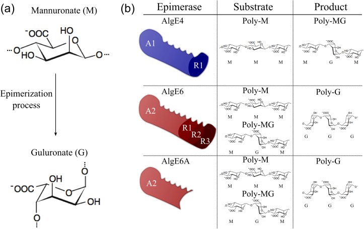
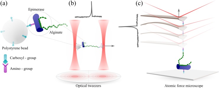
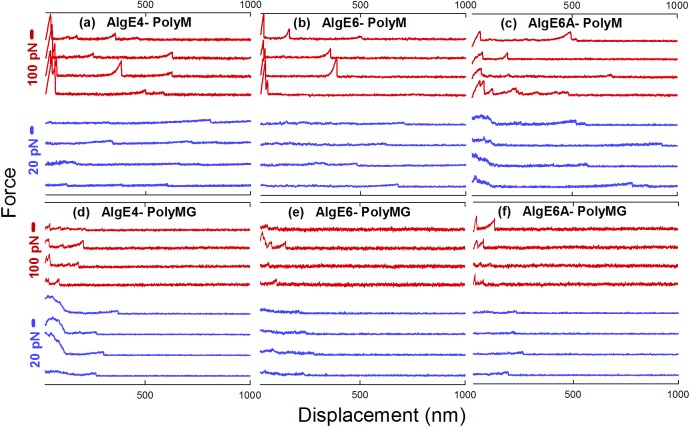
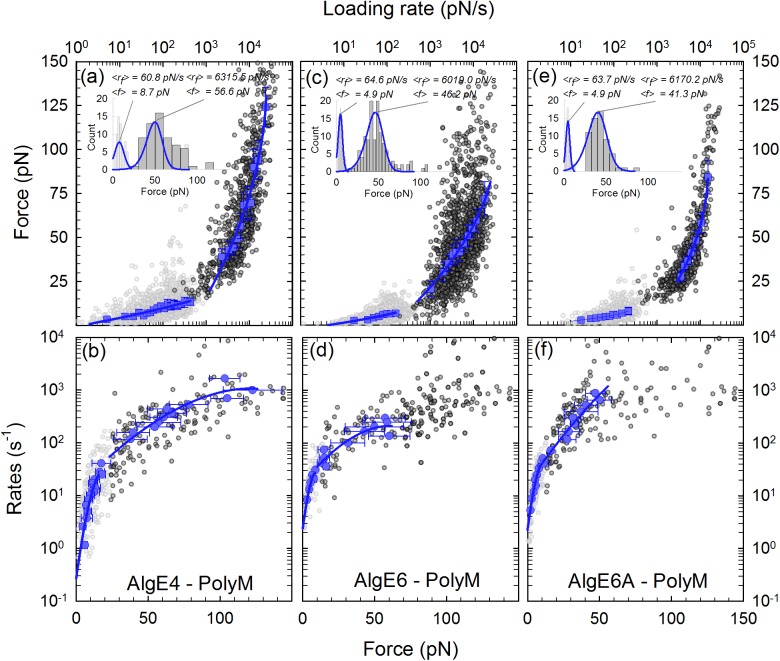
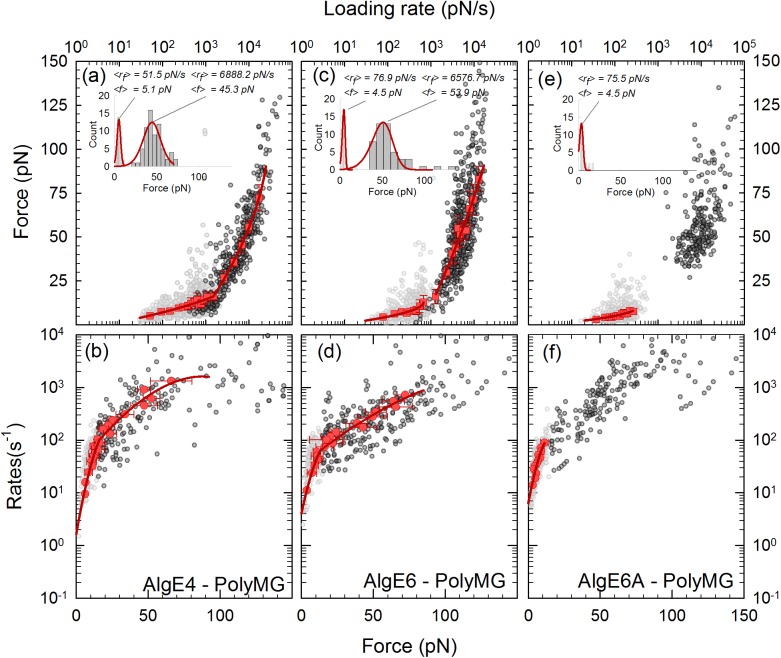
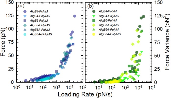
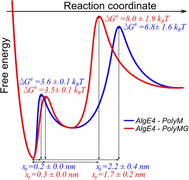
Mannuronan C-5 epimerases are a family of enzymes that catalyze epimerization of alginates at the polymer level. This group of enzymes thus enables the tailor-making of various alginate residue sequences to attain various functional properties, e.g. viscosity, gelation and ion binding. Here, the interactions between epimerases AlgE4 and AlgE6 and alginate substrates as well as epimerization products were determined. The interactions of the various epimerase-polysaccharide pairs were determined over an extended range of force loading rates by the combined use of optical tweezers and atomic force microscopy. When studying systems that in nature are not subjected to external forces the access to observations obtained at low loading rates, as provided by optical tweezers, is a great advantage since the low loading rate region for these systems reflect the properties of the rate limiting energy barrier. The AlgE epimerases have a modular structure comprising both A and R modules, and the role of each of these modules in the epimerization process were examined through studies of the A- module of AlgE6, AlgE6A. Dynamic strength spectra obtained through combination of atomic force microscopy and the optical tweezers revealed the existence of two energy barriers in the alginate-epimerase complexes, of which one was not revealed in previous AFM based studies of these complexes. Furthermore, based on these spectra estimates of the locations of energy transition states (xβ), lifetimes in the absence of external perturbation (τ0) and free energies (ΔG#) were determined for the different epimerase-alginate complexes. This is the first determination of ΔG# for these complexes. The values determined were up to 8 kBT for the outer barrier, and smaller values for the inner barriers. The size of the free energies determined are consistent with the interpretation that the enzyme and substrate are thus not tightly locked at all times but are able to relocate. Together with the observed different affinities determined for AlgE4-polymannuronic acid (poly-M) and AlgE4-polyalternating alginate (poly-MG) macromolecular pairs these data give important contribution to the growing understanding of the mechanisms underlying the processive mode of these enzymes.


Conflict of interest statement

Competing Interests: The authors have declared that no competing interests exist.

Figures

Fig 1
Fig 1. The epimerization process, epimerases and prevailing alginate residue sequences of the various epimerase substrates and resulting products.
(a) The mannuronan C-5 epimerases possess the ability to epimerize β-d-mannuronate residues (M) to its epimer form; α-l-guluronate residue (G). (b) The naturally occurring epimerases are known to form long stretches of systematically epimerized alginates. While AlgE4 can produce polyalternating structures from polymannuronic alginates, AlgE6 can epimerize both polymannuronic and polyalternating structures to form polyguluronic alginates.
Fig 2
Fig 2. Schematic of the techniques used to study alginate-epimerase interactions.
(a) Epimerase protein immobilized onto a polystyrene bead. The two force probes optical tweezers (b) and atomic force microscopy (c) display different force ranges associated to the difference in the spring constant of the optical traps and the AFM cantilever. Typical force-displacement curves displaying a force jump, as obtained when separating two functionalized and trapped polystyrene beads (b) or functionalized mica surface and AFM cantilever (c), are included. The force jumps reflect that the two surfaces were interconnected through an alginate-epimerase interaction, which was disrupted when increasing the separation distance between the two surfaces.
Fig 3
Fig 3. Gallery of rupture events of the various AlgE-poly-M (a,b,c) and AlgE-poly-MG (d,e,f) molecular pairs.
The red curves are recorded with AFM, while the blue curves represent forced ruptures recorded with OT. The displacement scale corresponds to the z-piezo translation distance, and bead separation for the data collected employing AFM and OT, respectively. Some of the AFM curves display interactions at short displacement distances (~ 0–50 nm) that may reflect non-specific interaction between the AFM-tip and the mica slide (e.g. red curves in a,b, and c). Bead-bead interactions are in some recordings present at the adhesion region of the OT experiments, such as the ones in (c) and (d). Interactions in the adhesion region either due to AFM-tip mica slide contact in the AFM experiments or due to strong bead-bead interactions in the OT were not included in the analysis as single-bond rupture events.
Fig 4
Fig 4. Interactions of AlgE–poly-M complexes determined by direct unbinding.
(a, c, e) Constant-speed rupture-force representation of the interactions i.e. mean forces versus mean loading rates (symbols) and fits of Eq 3 using ν = 1/2 (lines) to the experimental data. The estimates of the molecular interaction parameters are shown in Table 2. The light grey data points are forced ruptures recorded with the OT, while the dark grey data points are forced ruptures recorded with the AFM. By combining the two techniques we can access a larger range of loading rates as can be seen in (a, c, e). The inserts show two selected histograms of the unbinding forces, within the low and high loading rate regions, respectively, indicate typical distributions of the data within the loading rate intervals. The distribution of unbinding force P(f) (Eq 2) for ν = 1/2 (lines) is included on top of the histograms. (b, d, f) Constant-force rupture-rate representation with fits using ν = 1/2 of the interactions for the poly-M-complexes. The data are presented as mean rates versus mean forces obtained analytically from the constant-speed rupture-force experiments (Eq 6). The resulting estimates of the molecular interaction parameters are shown in Table 3.
Fig 5
Fig 5. Interactions of AlgE–poly-MG complexes determined by direct unbinding.
(a, c, e) Constant-speed rupture-force representation of the interactions i.e. mean forces versus mean loading rates (symbols) and fits of Eq 3 with ν = 1/2(lines) to the experimental data. The estimates of the molecular interaction parameters are shown in Table 2. The light grey data points are forced ruptures recorded with the OT, while the dark grey data points are forced ruptures recorded with the AFM. By combining the two techniques we can access a larger range of loading rates as can be seen in (a, c, e). The inserts show two selected histograms of the unbinding forces, within the low and high loading rate regions, respectively. The histogram plots, one for each energy barrier, exhibit the distribution of unbinding forces for which Eq 2 with ν = 1/2(lines) was fitted. (b, d, f) Constant-force rupture-rate representation with ν = 1/2 of the interactions for the poly-M-complexes. The data are presented as mean rates versus mean forces obtained analytically from the constant-speed rupture-force experiments (Eq 6). The resulting estimates of the molecular interaction parameters are shown in Table 3.
Fig 6
Fig 6. Comparison of mean force (a) and force variance (b) versus force loading rate for the alginate-epimerase pairwise interactions.
Fig 7
Fig 7. Schematic illustration of reconstructed energy landscapes for the AlgE4-poly-M and AlgE4 –poly-MG pairwise interactions based on the obtained parameters (Table 1).

References

    1. Hoare TR, Kohane DS. Hydrogels in drug delivery: Progress and challenges. Polymer. 2008;49(8):1993–2007. 10.1016/j.polymer.2008.01.027 - DOI
    1. Strand BL, Mørch YA, Syvertsen KR, Espevik T, SkjåkBræk G. Microcapsules made by enzymatically tailored alginate. Journal of Biomedical Materials Research Part A. 2003;64A(3):540–50. 10.1002/jbm.a.10337 - DOI - PubMed
    1. Zekorn T, Siebers U, Horcher A, Schnettler R, Zimmermann U, Bretzel RG, et al. Alginate coating of islets of Langerhans: in vitro studies on a new method for microencapsulation for immuno-isolated transplantation. Acta Diabetol. 1992;29(1):41–5. 10.1007/BF00572829 - DOI - PubMed
    1. Lim F, Sun AM. Microencapsulated islets as a bioartificial endocrine pancreas. Science. 1980;210:908–10. - PubMed
    1. Rozeboom HJ, Bjerkan TM, Kalk KH, Ertesvag H, Holtan S, Aachmann FL, et al. Structural and mutational characterization of the catalytic A-module of the mannuronan C-5-epimerase AlgE4 from Azotobacter vinelandii. Journal of Biological Chemistry. 2008;283(35):23819–28. 10.1074/jbc.M804119200 . - DOI - PMC - PubMed

LinkOut - more resources