

Otopathology of Vasculitis in Granulomatosis With Polyangitis
- PMID: 26496669
- PMCID: PMC4659735
- DOI: 10.1097/MAO.0000000000000868
Otopathology of Vasculitis in Granulomatosis With Polyangitis
Abstract
Objective: To describe the temporal bone histopathology of vasculitis in granulomatosis with polyangitis.
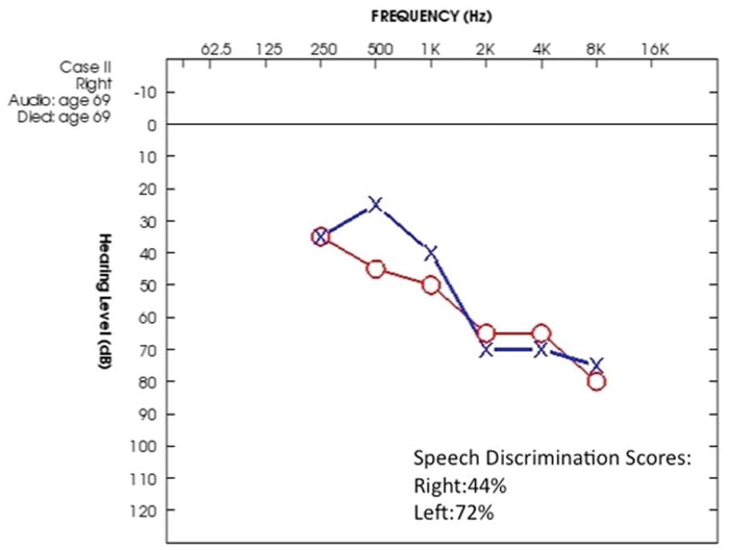
Background: Granulomatosis with polyangitis is an autoimmune disease that presents as granulomatosis and vasculitis. Otologic findings, including otitis media, hearing loss, vertigo, and facial paralysis are common in this condition.
Material and methods: The temporal bones of four subjects with manifestations of vasculitis attributed to granulomatosis with polyangitis were studied under light microscopy.
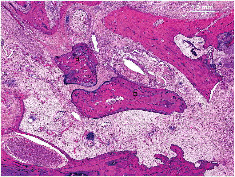
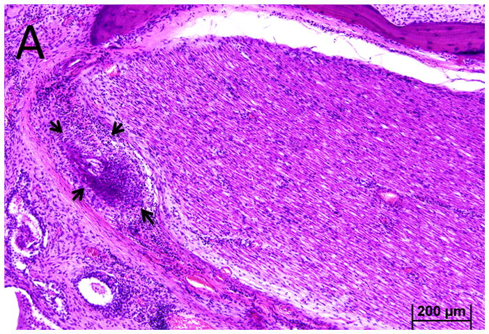
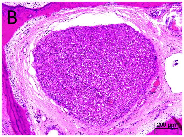
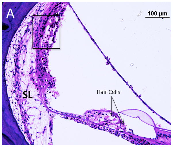
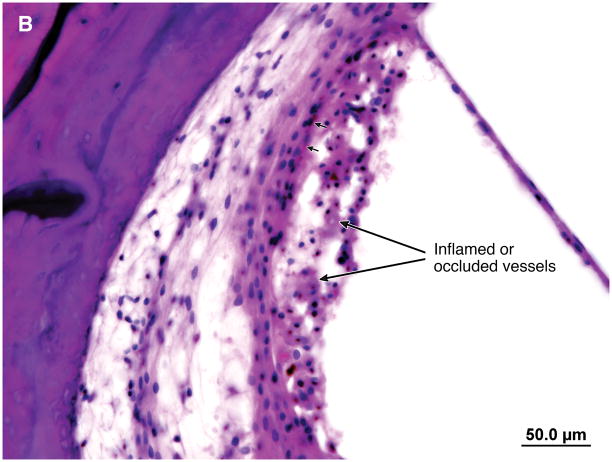
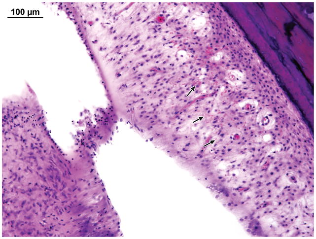
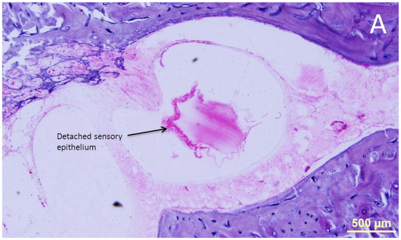
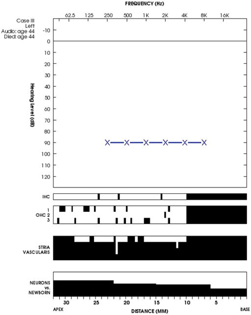
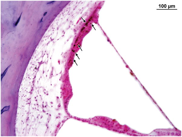
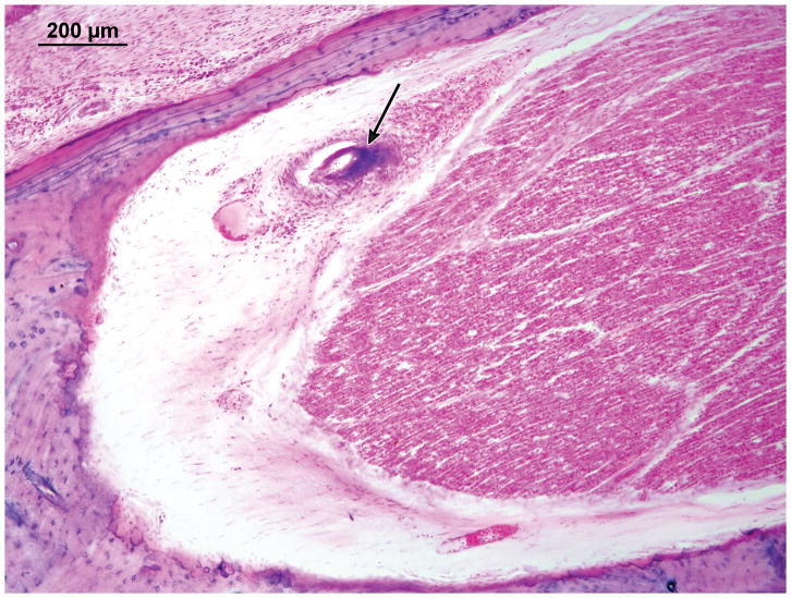
Results: The four subjects had manifestations of vasculitis including hemorrhage within the cochlea and vestibule, and inflammation and occlusion of vessels in the lateral cochlear wall and the vasa nervorum of the facial nerve.
Conclusions: We infer that sensorineural hearing loss, vestibulopathy, and facial nerve paresis in granulomatosis with polyangitis can be the results of vasculitis.
Figures

Similar articles
-
Otologic Wegener's granulomatosis with facial nerve palsy.Ann Otol Rhinol Laryngol. 1998 Jul;107(7):555-9. doi: 10.1177/000348949810700702. Ann Otol Rhinol Laryngol. 1998. PMID: 9682848
-
Subtotal Petrosectomy and Cochlear Implant Placement in Otologic Presentation of "Wegener's Granulomatosis".Kathmandu Univ Med J (KUMJ). 2017 Jan.-Mar.;15(57):94-98. Kathmandu Univ Med J (KUMJ). 2017. PMID: 29446374
-
Systemic vasculitis: a temporal bone histopathologic study.Laryngoscope. 1989 Jun;99(6 Pt 1):600-9. doi: 10.1288/00005537-198906000-00007. Laryngoscope. 1989. PMID: 2566880
-
Imaging Findings in Sensorineural Hearing Loss: A Pictorial Essay.Can Assoc Radiol J. 2017 May;68(2):106-115. doi: 10.1016/j.carj.2015.12.001. Epub 2016 May 18. Can Assoc Radiol J. 2017. PMID: 27209216 Review. No abstract available.
-
Granulomatosis with polyangiitis and facial palsy: Literature review and insight in the autoimmune pathogenesis.Autoimmun Rev. 2016 Jul;15(7):621-31. doi: 10.1016/j.autrev.2016.02.005. Epub 2016 Feb 4. Autoimmun Rev. 2016. PMID: 26851550 Review.
Cited by
-
Facial nerve paresis in the course of masked mastoiditis as a revelator of GPA.Eur Arch Otorhinolaryngol. 2022 Sep;279(9):4271-4278. doi: 10.1007/s00405-021-07166-w. Epub 2021 Nov 19. Eur Arch Otorhinolaryngol. 2022. PMID: 34797403 Free PMC article.
-
Enhanced fallopian canal as a potential marker for temporal bone vasculitis.Laryngoscope Investig Otolaryngol. 2020 Nov 4;5(6):1168-1175. doi: 10.1002/lio2.489. eCollection 2020 Dec. Laryngoscope Investig Otolaryngol. 2020. PMID: 33364409 Free PMC article.
-
Granulomatosis with Polyangiitis as a Cause of Sudden-Onset Bilateral Sensorineural Hearing Loss: Case Report and Recommendations for Initial Assessment.Case Rep Otolaryngol. 2021 Apr 20;2021:6632344. doi: 10.1155/2021/6632344. eCollection 2021. Case Rep Otolaryngol. 2021. PMID: 33968458 Free PMC article.
-
Quantitative assessment of vestibular otopathology in granulomatosis with polyangitis: A temporal bone study.Laryngoscope Investig Otolaryngol. 2018 Nov 5;3(6):473-477. doi: 10.1002/lio2.182. eCollection 2018 Dec. Laryngoscope Investig Otolaryngol. 2018. PMID: 30599032 Free PMC article.
-
Association Between Hearing Loss and Systemic Small-Vessel Vasculitis: Audiological Aspects Across Disease Types.Medicina (Kaunas). 2025 Jun 20;61(7):1117. doi: 10.3390/medicina61071117. Medicina (Kaunas). 2025. PMID: 40731747 Free PMC article.
References
-
- Takagi D, Nakamaru Y, Maguchi S, Furuta Y, Fukuda S. Otologic Manifestations of Wegener’s Granulomatosis. Laryngoscope. 2002;112:1684–1690. - PubMed
-
- Bakthavachalam S, Driver MS, Cox C, Spiegel JH, Grundfast KM, Merkel PA. Hearing loss in Wegener’s granulomatosis. Otol Neurotol. 2004;5:833–7. - PubMed
-
- Merchant SN, Nadol JB Jr, editors. Schuknecht’s Pathology of the Ear. 3. People’s Medical Publishing; House-USA, Shelton, Connecticut: 2010. p. 942.
-
- Travis WD, Hoffman GS, Leavitt RY, et al. Surgical pathology of the lung in Wegener’s granulomatosis. Review of 87 open lung biopsies from 67 patients. Am J Surg Pathol. 1991;15:315–33. - PubMed
-
- Yoon TH, Paparella MM, Schachern PA. Systemic vasculitis: a temporal bone histopathologic study. Laryngoscope. 1989 Jun;99(6 Pt 1):600–9. - PubMed
Publication types
MeSH terms
Grants and funding
LinkOut - more resources
Full Text Sources
Medical
