. 2015 Oct 23;11(10):e1005239.
doi: 10.1371/journal.ppat.1005239. eCollection 2015 Oct.

Identification of a Novel Lipoprotein Regulator of Clostridium difficile Spore Germination

Affiliations

Identification of a Novel Lipoprotein Regulator of Clostridium difficile Spore Germination

Kelly A Fimlaid et al. PLoS Pathog. .

Abstract

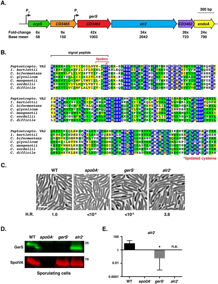
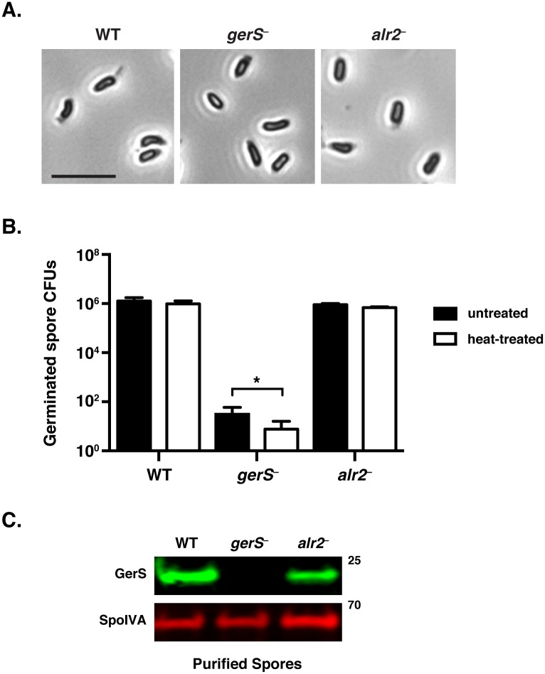
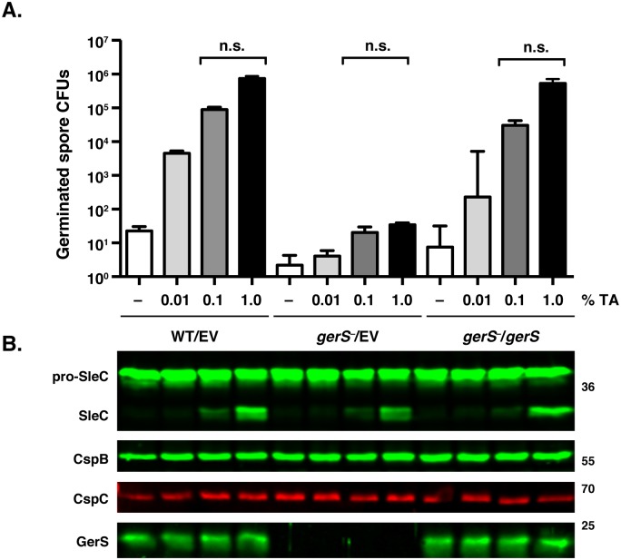
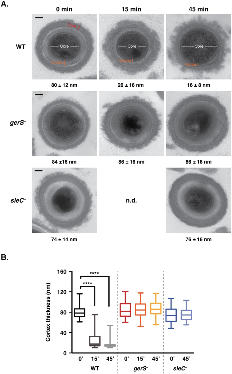
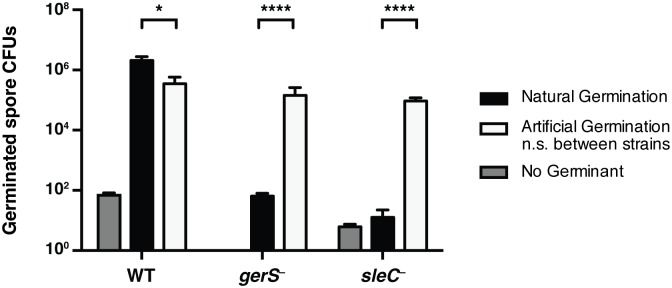
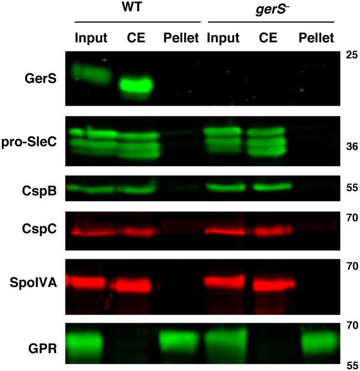
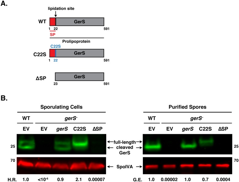
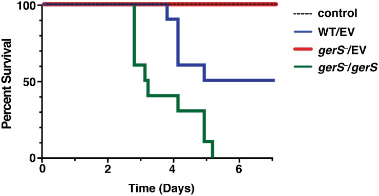
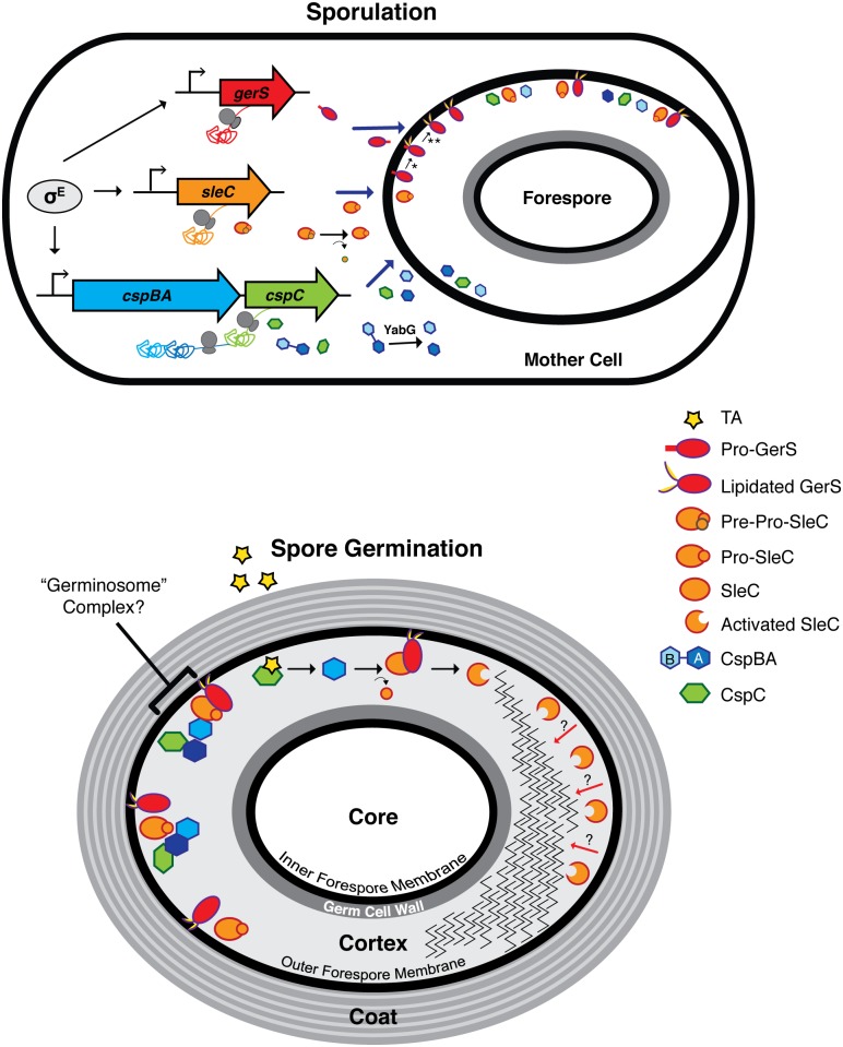
Clostridium difficile is a Gram-positive spore-forming pathogen and a leading cause of nosocomial diarrhea. C. difficile infections are transmitted when ingested spores germinate in the gastrointestinal tract and transform into vegetative cells. Germination begins when the germinant receptor CspC detects bile salts in the gut. CspC is a subtilisin-like serine pseudoprotease that activates the related CspB serine protease through an unknown mechanism. Activated CspB cleaves the pro-SleC zymogen, which allows the activated SleC cortex hydrolase to degrade the protective cortex layer. While these regulators are essential for C. difficile spores to outgrow and form toxin-secreting vegetative cells, the mechanisms controlling their function have only been partially characterized. In this study, we identify the lipoprotein GerS as a novel regulator of C. difficile spore germination using targeted mutagenesis. A gerS mutant has a severe germination defect and fails to degrade cortex even though it processes SleC at wildtype levels. Using complementation analyses, we demonstrate that GerS secretion, but not lipidation, is necessary for GerS to activate SleC. Importantly, loss of GerS attenuates the virulence of C. difficile in a hamster model of infection. Since GerS appears to be conserved exclusively in related Peptostreptococcaeace family members, our results contribute to a growing body of work indicating that C. difficile has evolved distinct mechanisms for controlling the exit from dormancy relative to B. subtilis and other spore-forming organisms.


Conflict of interest statement

The authors have declared that no competing interests exist.

Figures

Fig 1
Fig 1. C. difficile gerS is highly induced during sporulation and encodes a protein important for heat-resistant spore formation.
(A) Schematic of C. difficile gerS and alr2 genomic loci. gerS and alr2 are predicted to be part of an operon where transcription initiates from a P1 promoter immediately upstream of gerS (mapped by RNA-Seq transcriptional start site mapping, [42]) and potentially from the P2 promoter upstream of acpS. Fold-change represents the difference in gene expression between wild type and the sigE mutant [41]. Base mean refers to the number of transcripts detected for the respective gene normalized to the length of that gene. (B) ClustalW alignment of GerS. Completely conserved, identical residues are blocked in blue, conserved identical residues are blocked in green, and conserved similar residues in yellow. The predicted signal peptide is bracketed in black, and the lipobox is bracketed in red. The lipobox cysteine predicted to be lipidated is designated by a red asterisk. The following sequences from Peptostreptococcaceae family members [27] were analyzed: Peptostreptococcaceae bacterium VA2 (WP_026902346), Intestini bartlettii (WP_007287647), C. bifermentans (WP_021430359), C. glycolicum (WP_018591922), C. mangenotii (WP_027700975), C. sordellii (CEK32529), and C. difficile (YP_001089984). (C) Phase contrast microscopy of the indicated C. difficile strains grown on sporulation media for 20 hrs. The spo0A mutant cannot initiate sporulation [85]. The efficiency of heat-resistant spore formation (H.R.) was determined for each strain relative to wild type for three biological replicates. Scale bars represent 5 μm. (D) Western blot analysis of sporulating WT, spo0A , gerS , and alr2 cells. The mouse anti-SpoIVA antibody was used as a loading control [60]. (E) qRT-PCR analysis of alr2 transcription in the indicated mutants. RNA was isolated from the indicated strains after sporulation was induced for 18 hrs. Transcript levels were normalized to the housekeeping gene rpoB using the standard curve method. Data represents the average of three biological replicates. Error bars indicate the standard error of the mean. n.a. indicates not applicable, since the region amplified spans the disrupted alr2 gene. Statistical significance was determined using ANOVA and Tukey’s test (* p < 0.05).
Fig 2
Fig 2. C. difficile gerS mutant spores have a severe germination defect.
(A) Phase contrast microscopy of C. difficile spores isolated from wildtype, gerS , and alr2 strains. gerS and alr2 resemble wild type in size and their phase-bright appearance. Scale bars represent 5 μm. (B) Germination of wildtype, gerS , and alr2 spores following heat treatment. Heat-treated spores were incubated for 30 min at 60°C. Data represents the average of three biological replicates. Statistical significance was evaluated using ANOVA and Tukey’s test (* p < 0.05). (C) Western blot analysis of spores isolated from WT, gerS , and alr2 strains. Anti-SpoIVA was used as a loading control [60].
Fig 3
Fig 3. gerS spores process pro-SleC in response to germinant.
(A) in vitro germination of purified spores from wildtype carrying empty vector (WT/EV) and gerS carrying empty vector (gerS /EV) or the gerS complementation construct (gerS /gerS) spores in response to increasing amounts of taurocholate. Samples were plated on BHIS following taurocholate exposure. Data represents the average of three biological replicates. Statistical significance was evaluated using ANOVA and Tukey’s test. n.s. = no statistical significance. (B) Western blot analyses of samples from one representative replicate of the in vitro germination assay shown above. The zymogen pro-SleC is processed by CspB in response to taurocholate addition [23].
Fig 4
Fig 4. gerS spores are defective in cortex hydrolysis.
(A) TEM analyses of wildtype, gerS and sleC spores at time 0’, 15’, and 45’ post exposure to taurocholate. The 0’ timepoint was taken before taurocholate was added. sleC spores were used as a negative control, since they do not undergo cortex hydrolysis [36,37]. The average cortex thickness was determined for each sample from a minimum of 50 spores at each timepoint. n.d. designates not determined. Representative images are shown. Scale bars designate 100 nm. (B) Box and whiskers plot of cortex thickness of the in vitro germination assay shown in (A). Statistical significance was determined using ANOVA and Tukey’s test (**** p < 0.0001).
Fig 5
Fig 5. Artificial germination of gerS bypasses its germination defect.
Wildtype, gerS , and sleC spores were incubated with no germinant, taurocholate (natural germination) or thioglycollate and lysozyme (artificial germination). Mutants defective in cortex hydrolysis can be artificially germinated [58]. Data represents the average of 4 biological replicates. No statistical significance was observed between strains subjected to artificial germination, in contrast with natural germination. Statistical significance was determined using ANOVA and Tukey’s test (* p < 0.05; **** p < 0.001).
Fig 6
Fig 6. Germination proteins localize to a “coat-extractable” (CE) fraction.
Western blot analyses of “coat-extractable” (CE) and decoated spore lysate (pellet) fractions from wildtype and gerS spores. It should be noted that the CE fraction likely includes proteins localized to the cortex and outer-forespore membrane. Input represents the whole spore lysate without fractionation. SpoIVA is a coat morphogenetic protein [60]. GPR (germination protease) is localized to the core of spores [40,61,62].
Fig 7
Fig 7. The signal peptide of GerS, but not its lipidation site, is required for germination.
(A) Schematic of gerS complementation constructs. C22S designates a construct encoding a mutation of the invariant lipidation site cysteine to serine. ΔSP designates a construct encoding a truncated GerS lacking its signal peptide. (B) Western blot analyses of GerS in sporulating cells and purified spores from wild type carrying empty vector (EV) and the gerS mutant carrying either empty vector or the indicated complementation constructs. SpoIVA was used as a loading control [60]. The efficiency of heat-resistant spore formation (H.R.) during sporulation is shown, as is the germination efficiency (G.E.) of purified spores. Data is representative of at least 4 replicates.
Fig 8
Fig 8. GerS is required for virulence.
Kaplan-Meier survival curve of clindamycin-treated Syrian hamsters inoculated with 1,000 isolated spores of wildtype carrying empty vector (WT/EV), the gerS mutant carrying either empty vector (gerS /EV), or the wildtype complementation construct (gerS /gerS). The control designates antibiotic-treated hamsters that were not inoculated with spores.
Fig 9
Fig 9. Model of C. difficile germination regulator production and localization during sporulation and germination.
The sporulating cell depicts transcription of germination regulator genes and translation of the resulting transcripts. gerS, sleC, and cspBAC are all controlled by σE in the mother cell. σG may regulate the expression of gene products involved in germination, but these are unknown. In the mother cell, removal of the pre-domain is depicted by the curved arrow, while interdomain cleavage of CspBA is demarcated by the red X; these processing events occur during spore maturation [23]. Solid blue arrows designate transport of proteins from the mother cell into the forespore compartment. In mature spores, the germination regulators, GerS, SleC, CspC, and CspB, likely localize to the cortex region and/or to the outer forespore membrane. The possible association of these regulators into a “germinosome” complex [86] is designated by the bracket. The taurocholate (TA) germinant is depicted by the red star. Red arrows depict SleC-mediated cortex hydrolysis from the outer forespore membrane to the inner forespore membrane. * lipidation event; ** signal peptide cleavage event; dashed arrow designates an unknown event.

Similar articles

Cited by

References

    1. Carroll K, Bartlett J (2011) Biology of Clostridium difficile: implications for epidemiology and diagnosis. Annual review of microbiology 65: 501–521. 10.1146/annurev-micro-090110-102824 - DOI - PubMed
    1. Rupnik M, Wilcox M, Gerding D (2009) Clostridium difficile infection: new developments in epidemiology and pathogenesis. Nature reviews Microbiology 7: 526–536. 10.1038/nrmicro2164 - DOI - PubMed
    1. Warny M, Pepin J, Fang A, Killgore G, Thompson A, et al. (2005) Toxin production by an emerging strain of Clostridium difficile associated with outbreaks of severe disease in North America and Europe. Lancet 366: 1079–1084. - PubMed
    1. Badger VO, Ledeboer NA, Graham MB, Edmiston CE Jr. (2012) Clostridium difficile: epidemiology, pathogenesis, management, and prevention of a recalcitrant healthcare-associated pathogen. JPEN J Parenter Enteral Nutr 36: 645–662. 10.1177/0148607112446703 - DOI - PubMed
    1. Freeman J, Bauer M, Baines S, Corver J, Fawley W, et al. (2010) The changing epidemiology of Clostridium difficile infections. Clinical microbiology reviews 23: 529–549. 10.1128/CMR.00082-09 - DOI - PMC - PubMed

Publication types

LinkOut - more resources