. 2016 Jan:487:27-40.
doi: 10.1016/j.virol.2015.10.011. Epub 2015 Oct 20.

Biochemical analysis of the multifunctional vaccinia mRNA capping enzyme encoded by a temperature sensitive virus mutant

Affiliations

Biochemical analysis of the multifunctional vaccinia mRNA capping enzyme encoded by a temperature sensitive virus mutant

Jessica Tate et al. Virology. 2016 Jan.

Abstract

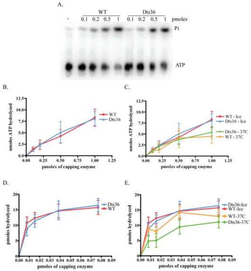
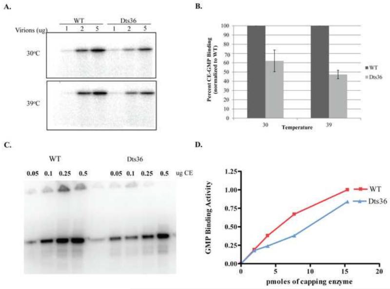
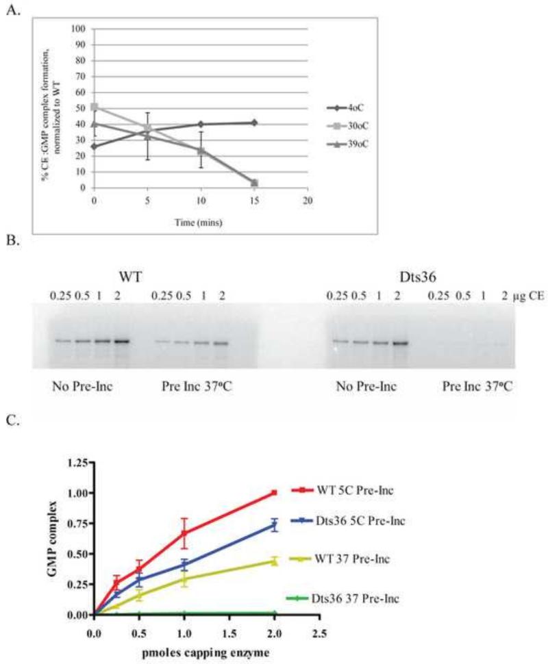
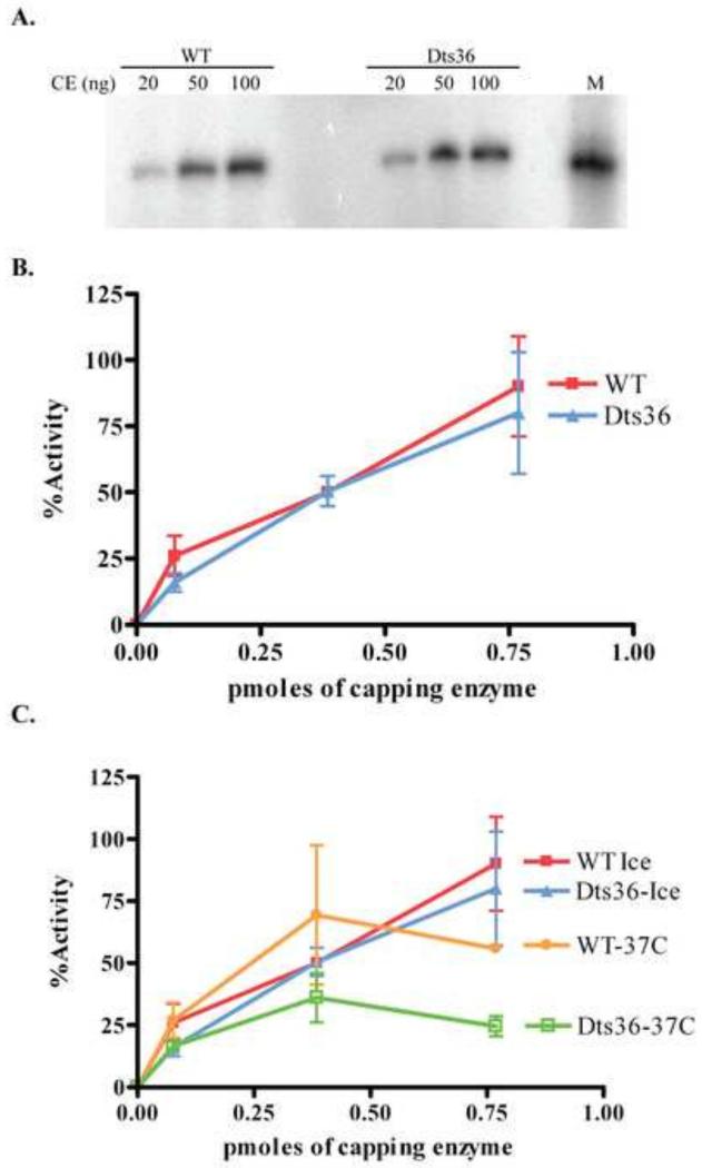
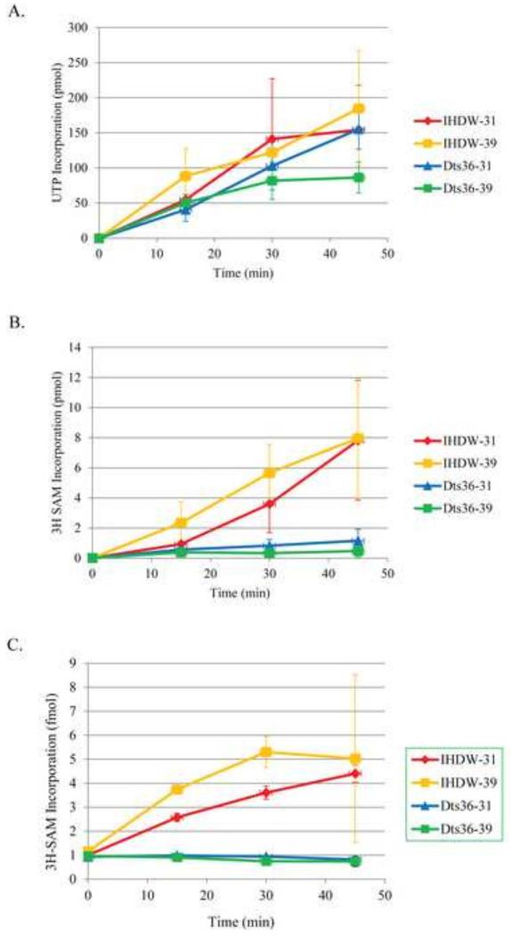
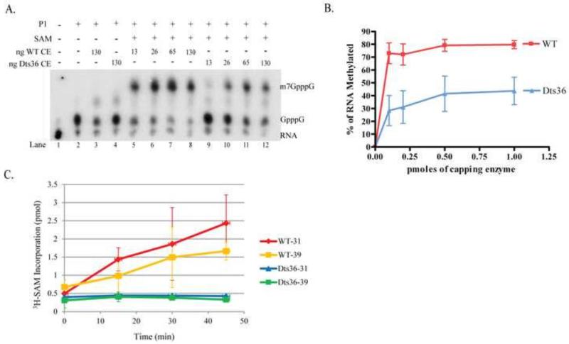
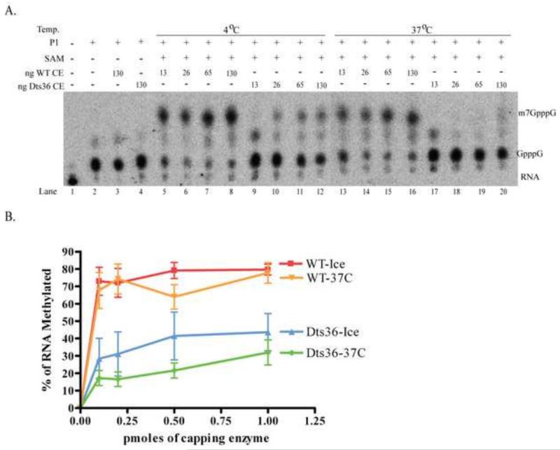
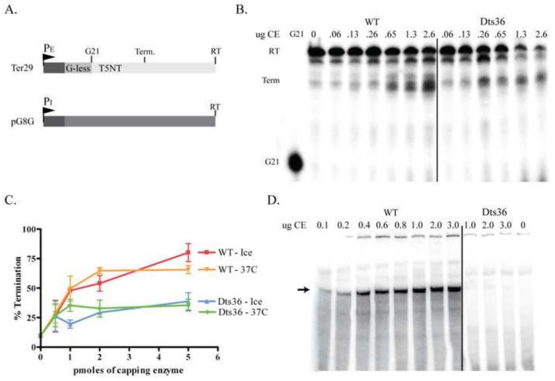
Prior biochemical analysis of the heterodimeric vaccinia virus mRNA capping enzyme suggests roles not only in mRNA capping but also in early viral gene transcription termination and intermediate viral gene transcription initiation. Prior phenotypic characterization of Dts36, a temperature sensitive virus mutant affecting the large subunit of the capping enzyme was consistent with the multifunctional roles of the capping enzyme in vivo. We report a biochemical analysis of the capping enzyme encoded by Dts36. Of the three enzymatic activities required for mRNA capping, the guanylyltransferase and methyltransferase activities are compromised while the triphosphatase activity and the D12 subunit interaction are unaffected. The mutant enzyme is also defective in stimulating early gene transcription termination and intermediate gene transcription initiation in vitro. These results confirm that the vaccinia virus mRNA capping enzyme functions not only in mRNA capping but also early gene transcription termination and intermediate gene transcription initiation in vivo.

Keywords: Capping enzyme; Transcription initiation; Transcription termination; Vaccinia virus; mRNA capping.


Figures

Fig. 1
Fig. 1. Structure of the vaccinia virus capping enzyme
Ribbon diagram representation of the structure of CE. The three domains of the large D1 subunit are represented in different colors. The TPase domain (residues 1-225) is blue, the GTase domain (residues 226-545) is green, and the MTase domain (residues 548-844) is red. The small D12 subunit is yellow. The bound S-adenosyl-L-homocysteine in the MTase active site is shown in stick models colored by element. Gly 705, which is replaced with Asp in the Dts36 mutant, is depicted in space-filling models colored light green.
Fig. 2
Fig. 2. SDS-PAGE and western blot analysis of virion-encapsidated and purified CE
Varying amounts of purified IHDW and Dts36 virions were denatured and their polypeptides separated by 11% SDS-PAGE. A) Polypeptides visualized by Coomassie stain. Protein standards in the first lane indicate the size of the polypeptides. B) Western blot of the CE subunits, D1 and D12. (Top panel) D1 was detected using anti-D1 polyclonal primary antibody followed by anti-rabbit-HRP monoclonal secondary antibody. (Bottom panel) D12 was probed using anti-D12 polyclonal primary antibody followed by anti-rabbit HRP monoclonal secondary antibody. Molecular weight markers are indicated to the right of the blot C) Recombinant wildtype and Dts36 CE proteins purified from E. coli analyzed on a 10% SDS-PAGE gel stained with Coomassie blue. Lanes 1 and 3 contain 3ug of each protein and lanes 2 and 4 contain 6ug of each protein. Molecular weight markers are indicated to the left of the gel, and the identities of the major bands are indicated at the right. D1 CT indicates a C-terminal proteolytic fragment of the D1 protein that is consistently observed when CE is expressed in and purified from E. coli.
Fig. 3
Fig. 3. Dts36 TPase activity
A) Autoradiogram of ATPase assay of WT and Dts36 CE. B) The moles of ATP hydrolyzed were determined by determining the fraction of Pi released from a known input mols of ATP. C) Analysis of the thermostability of the TPase activity of WT and Dts36 CE. Enzymes were pre-incubated for 15 min either on ice or at 37 °C after which hydrolysis of ATP was assayed as in (A) and plotted as in (B). D) TPase activity using an RNA substrate was determined by calculating the amount of Pi hydrolyzed from 5′-[32P] RNA. Pmoles hydrolyzed was plotted versus the amount of CE in the reaction. E) The thermostability of the TPase activity on an RNA substrate was analyzed by pre-incubating CE samples on ice or at 37°C for 15 minutes and then assaying TPase activity. Activity was calculated and plotted as is (D).
Fig. 4
Fig. 4. CE:GMP complex formation
A) Varying amounts of permeabilized IHDW or Dts36 virions were incubated with α32P-GTP at either 30°C or 39°C for 15 min. Reactions were analyzed on a 11% SDS-PAGE followed by autoradiography. B) The amount of CE:GMP present was calculate by dividing the band intensity by the amount of virions present in the reaction. Percent CE:GMP was then calculated by normalizing to the amount of CE:GMP present in the corresponding reactions containing IHDW virions. (C) Varying amounts of purified WT or Dts36 CE were incubated with α32P-GTP at 30 °C for 15 min and analyzed by SDS PAGE as described in (A). (D) Plot of the amount of CE-GMP complex formed with WT and Dts36 CE. The amount of CE-GMP complex formed with 0.5 μg WT CE was set at 100%
Fig. 5
Fig. 5. Thermolability of Dts36 CE:GMP complex formation
(A) Permeabilized IHDW and Dts36 virions were preincubated at 4, 30, or 39°C for 0, 5, 10, and 15 min. These virions were then incubated with α32P-GTP at 39°C for 15 min. Reactions were analyzed on an 11% SDS-PAGE followed by autoradiography. The amount of CE:GMP present was calculated by dividing the band intensity by the amount of virions present in the reaction. Percent CE:GMP was then calculated by normalizing to the amount of CE:GMP present in the corresponding reactions containing IHDW virions. (B) Thermolability of GMP complex formation with purified WT and Dts36 CE. Purified WT or Dts36 CE was pre-incubated at either 5°C or 37 °C for 15 min and then mixes with α32P-GTP and incubated at 30 °C for 15 min. The reactions were analyzed by SDS-PAGE as described in (A). (C) Plot of GMP complex formation from (B). The amount of complex formed with 2.0 μg WT CE pre-incubated at 5°C was set at 1.00 and the other samples normalized to this value.
Fig. 6
Fig. 6. Dts36 guanyltransferase activity
(A) Purified T7 synthesized RNA was incubated with α32P-GTP and either WT or Dts36 CE (20, 50, and 60 ng) at 37°C for 4 min. Samples were analyzed by denaturing PAGE and visualized by autoradiography. The marker lane (M) contains the same RNA used as substrate, however uniformly labeled during synthesis. (B) Plot of guanyltransferase activity from several assays as shown in (A). The activity obtained with 100 ng WT CE was set at 1.00 and the other samples normalized to this value. Values shown are the average of three independent assays each done in duplicate with error bars representing standard deviations. (C) Guanylyltransferase was assayed after preincubation of either WT or Dts36 CE for 10 min on ice or at 37°C. Reactions were analyzed and quantified as in (A) and (B).
Fig. 7
Fig. 7. Methyltransferase activity in IHDW and Dts36 virions
A) Transcription. Transcription was stimulated in permeabilized IHDW or Dts36 virions by the addition of all four nucleotides in the presence of α32P-UTP and SAM and incubation at either 30 or 39°C. A various times samples were TCA precipitated and quantified by scintillation counting. B) Transcript methylation. Transcription was stimulated in permeabilized IHDW or Dts36 virions by the addition of all four nucleotides in the presence 3H-SAM and incubation at either 30 or 39°C. A various times samples were TCA precipitated and quantified by scintillation counting. (C) GTP methylation. Permeabilized IHDW or Dts36 virions were incubated with GTP and 3H-SAM at either 31 or 39°C. A various times samples were spotted on DEAE-cellulose filters, washed to remove unincorporated 3H-SAM and quantified by scintillation counting.
Fig. 8
Fig. 8. Methyltransferase activity of purified Dts36 CE
A) Purified T7 synthesized RNA was guanylylated using α32P-GTP and WT CE and incubated with increasing amounts of either WT or Dts36 CE and in the presence or absence of SAM at 37°C for 15 min. The 5′ caps were cleaved off the RNA with P1 nuclease. Products were resolved by PEI cellulose TLC and visualized by autoradiography. B) Percent methylation was calculated as the amount of m7GpppG divided by the sum of m7GpppG and GpppG, and plotted versus the amount of CE present in the reaction. C) GTP methylation. Purified WT or Dts36 CE was incubated with GTP and 3H-SAM at either 30 or 39°C. A various times samples were captured on DEAE-cellulose filters and the amount of 3H-labeled GTP was measured by scintillation counting. The amount of 3H incorporation is plotted as a function of time.
Fig. 9
Fig. 9. Thermolability of methyltransferase activity of purified Dts36 CE
A) Purified T7 synthesized RNA was guanylylated using α32P-GTP and WT CE. These guanylated transcripts were incubated with increasing amounts of either WT or Dts36 CE preincubated at either 4 or 37°C, and in the presence or absence of SAM at 37°C for 15 min. The 5′ caps were cleaved from RNA with P1 nuclease and products were resolved by PEI cellulose TLC and visualized by autoradiography. B) Percent methylation was calculated as the amount of m7GpppG divided by the sum of m7GpppG and GpppG, and plotted against the amount of CE present in the reaction.
Fig. 10
Fig. 10. Dts36 activity in in vitro transcription termination and initiation
A) Diagram of the transcription templates Ter29 and pG8G. Ter29 contains an early gene promoter (PE) followed by a 20 bp G-less cassette and 169 bp of random sequence. Readthrough transcripts (RT) proceed to the end of the template to yield 189 nt transcript. The termination signal, T5NT, is located 9 bp downstream of the G-less cassette and stimulates termination to occur ~55 bp downstream of the G-less cassette to yield a ~75 nt terminated transcript (Term). Template pG8 contains an intermediate gene promoter (PI) followed by 380 bp G-less cassette (RT). B) Early transcription termination. Ter29 templates were incubated with ATP, CTP, α32P-UTP, 3′O-Methyl GTP, and virion extracts at 30°C for 15 min, which results in transcription elongation complexes stalled at G21 (leftmost lane). Stalled complexes were washed to remove endogenous CE and nucleotides. All four nucleotides and increasing amounts of either WT or Dts36 CE were added to the reactions, and transcription was allowed to proceed past G21 by incubating at 30°C for 15 min. Reactions were analyzed by denaturing PAGE and autoradiographed. Two predominant groups of transcripts, RT and Term, are observed. C) Percent termination was calculated by dividing the amount of Term transcripts by the sum of RT and Term transcripts. Percent termination was then plotted against the amount of CE. D) Intermediate transcription initiation. pG8 templates were incubated with ATP, GTP, CTP, α32P-UTP, purified His-A24 RNA Polymerase, VITF-3 and increasing amounts of either WT or Dts36 CE at 30°C for 30 min. Reactions were analyzed by denaturing PAGE and autoradiographed. The arrow indicates the expected 350 nt transcription product.

Similar articles

References

    1. Broyles SS. Vaccinia virus transcription. J.Gen.Virol. 2003;84:2293–2303. - PubMed
    1. Broyles SS, Moss B. Sedimentation of an RNA polymerase complex from vaccinia virus that specifically initiates and terminates transcription. Mol.Cell Biol. 1987;7:7–14. - PMC - PubMed
    1. Christen LA, Piacente S, Mohamed MR, Niles EG. Vaccinia virus early gene transcription termination factors VTF and Rap94 interact with the U9 termination motif in the nascent RNA in a transcription ternary complex. Virology. 2008;376:225–235. - PMC - PubMed
    1. Christen LM, Sanders M, Wiler C, Niles EG. Vaccinia virus nucleoside triphosphate phosphohydrolase I is an essential viral early gene transcription termination factor. Virology. 1998;245:360–371. - PubMed
    1. Condit RC, Lewis JI, Quinn M, Christen LM, Niles EG. Use of lysolecithin-permeabilized infected-cell extracts to investigate the in vitro biochemical phenotypes of poxvirus ts mutations altered in viral transcription activity. Virology. 1996;218:169–180. - PubMed

Publication types

MeSH terms

LinkOut - more resources