

A novel histopathologic finding in the Descemet's membrane of a patient with Peters Anomaly: a case-report and literature review
- PMID: 26496717
- PMCID: PMC4619091
- DOI: 10.1186/s12886-015-0131-y
A novel histopathologic finding in the Descemet's membrane of a patient with Peters Anomaly: a case-report and literature review
Abstract
Background: Peters anomaly is a rare developmental abnormality of the anterior segment of the eye and is one of the main causes of congenital corneal opacities. Typically, histopathology of Peters anomaly shows immature or absent Descemet's membrane and attenuated endothelial cells in the area of the corneal opacity, in addition to thinning or absence of Bowman's membrane and defects in the posterior stroma. In this report, we present a novel histopathological finding, which has not been previously reported, in the Descemet's membrane of a patient who is clinically diagnosed with Peters anomaly.
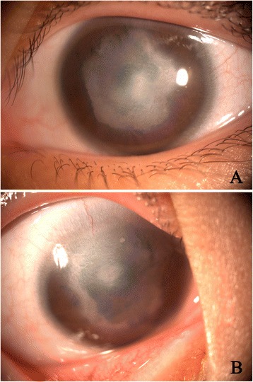
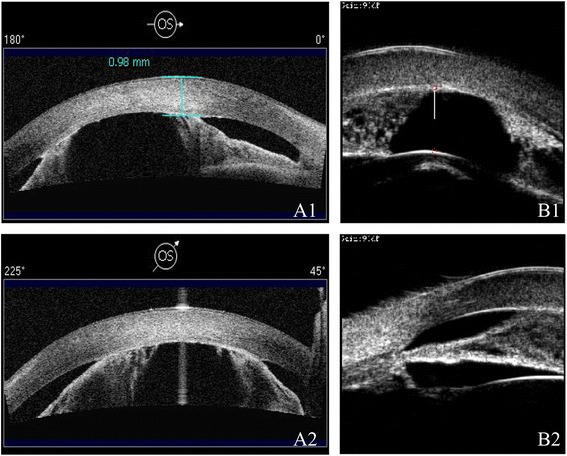
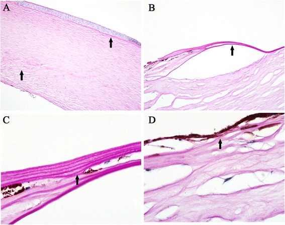
Case presentation: A 7-years old female child with developmentally delayed was born of a normal pregnancy, labor, and delivery. Apparent bilateral corneal opacifications were present at birth. On ophthalmologic examination, the child had a visual acuity of FC/20 cm in the right eye and that of FC/10 cm in the left one. Horizontal nystagmus and congenital cataract were found in both eyes. Slit-lamp examination revealed bilateral central corneal opacities which covered the iris and pupils. High-frequency UBM and AS-OCT both showed a shallow anterior chamber with multiple areas of iridocorneal adhesions and no corneal lenticular touch in each eye. A corneal specialist performed a penetrating keratoplasty with extra-capsular cataract extraction and intraocular lens implantation. Histopathologic procedures were conducted on the host corneal button, including Hematoxylin-Eosin stain and Periodic Acid-Schiff stain. All the sections were examined by light microscopy.
Conclusion: The "multiple-layer" structure of the Descemet's membrane described in our case has not been reported before as in association with abnormalities of the cornea tissues in Peters anomaly. Such pathological finding need to be reported to enhance further understanding of the special structure of Descemet's membrane as an abnormality during embryogenesis and neural crest cell differentiations.
Figures
Similar articles
-
Posterior lamellar keratoplasty (DSAEK) in Peters anomaly.Cornea. 2012 Oct;31(10):1201-5. doi: 10.1097/ICO.0b013e31825697a4. Cornea. 2012. PMID: 22790185
-
Surgical approach in type II Peters anomaly - case report.Rom J Ophthalmol. 2022 Jan-Mar;66(1):101-108. doi: 10.22336/rjo.2022.20. Rom J Ophthalmol. 2022. PMID: 35531449 Free PMC article.
-
Primary descemetorhexis without graft placement for type 1 Peters anomaly.Can J Ophthalmol. 2019 Apr;54(2):e52-e54. doi: 10.1016/j.jcjo.2018.05.014. Epub 2018 Aug 23. Can J Ophthalmol. 2019. PMID: 30975359 No abstract available.
-
Peters Anomaly: Novel Non-Invasive Alternatives to Penetrating Keratoplasty.Semin Ophthalmol. 2023 Apr;38(3):275-282. doi: 10.1080/08820538.2023.2176238. Epub 2023 Feb 14. Semin Ophthalmol. 2023. PMID: 36788651 Review.
-
Peters anomaly: review of the literature.Cornea. 2011 Aug;30(8):939-44. doi: 10.1097/ICO.0b013e31820156a9. Cornea. 2011. PMID: 21448066 Review.
Cited by
-
Terminology of Peters' anomaly variants: Summary of histopathological findings in 6 corneas and detailed clinicopathological correlation in 2 cases.Saudi J Ophthalmol. 2019 Jul-Sep;33(3):277-282. doi: 10.1016/j.sjopt.2018.02.015. Epub 2018 Mar 8. Saudi J Ophthalmol. 2019. PMID: 31686970 Free PMC article.
-
Bilateral anterior segment dysgenesis with the presumed Peters' anomaly in a cat.J Vet Med Sci. 2018 Feb 20;80(2):297-301. doi: 10.1292/jvms.17-0532. Epub 2017 Dec 15. J Vet Med Sci. 2018. PMID: 29249729 Free PMC article.
-
Axenfeld-Rieger syndrome: a novel histopathologic finding associated with corneal abnormalities.BMC Ophthalmol. 2022 Dec 28;22(1):514. doi: 10.1186/s12886-022-02754-8. BMC Ophthalmol. 2022. PMID: 36577962 Free PMC article. Review.
-
Novel Genetic Variants and Clinical Profiles in Peters Anomaly Spectrum Disorders.Int J Mol Sci. 2025 Jul 4;26(13):6454. doi: 10.3390/ijms26136454. Int J Mol Sci. 2025. PMID: 40650233 Free PMC article.
-
Selective endothelial removal: A case series of a phase I/II surgical trial with long-term follow up.Front Med (Lausanne). 2022 Jul 29;9:901187. doi: 10.3389/fmed.2022.901187. eCollection 2022. Front Med (Lausanne). 2022. PMID: 35966874 Free PMC article.
References
Publication types
MeSH terms
Supplementary concepts
LinkOut - more resources
Full Text Sources
Other Literature Sources
Research Materials
