. 2015 Dec;8(12):1543-53.
doi: 10.1242/dmm.023077. Epub 2015 Oct 23.

MAP3K1 function is essential for cytoarchitecture of the mouse organ of Corti and survival of auditory hair cells

Affiliations

MAP3K1 function is essential for cytoarchitecture of the mouse organ of Corti and survival of auditory hair cells

Rizwan Yousaf et al. Dis Model Mech. 2015 Dec.

Abstract

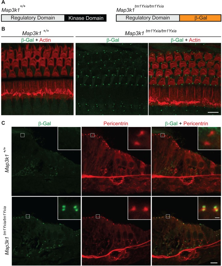
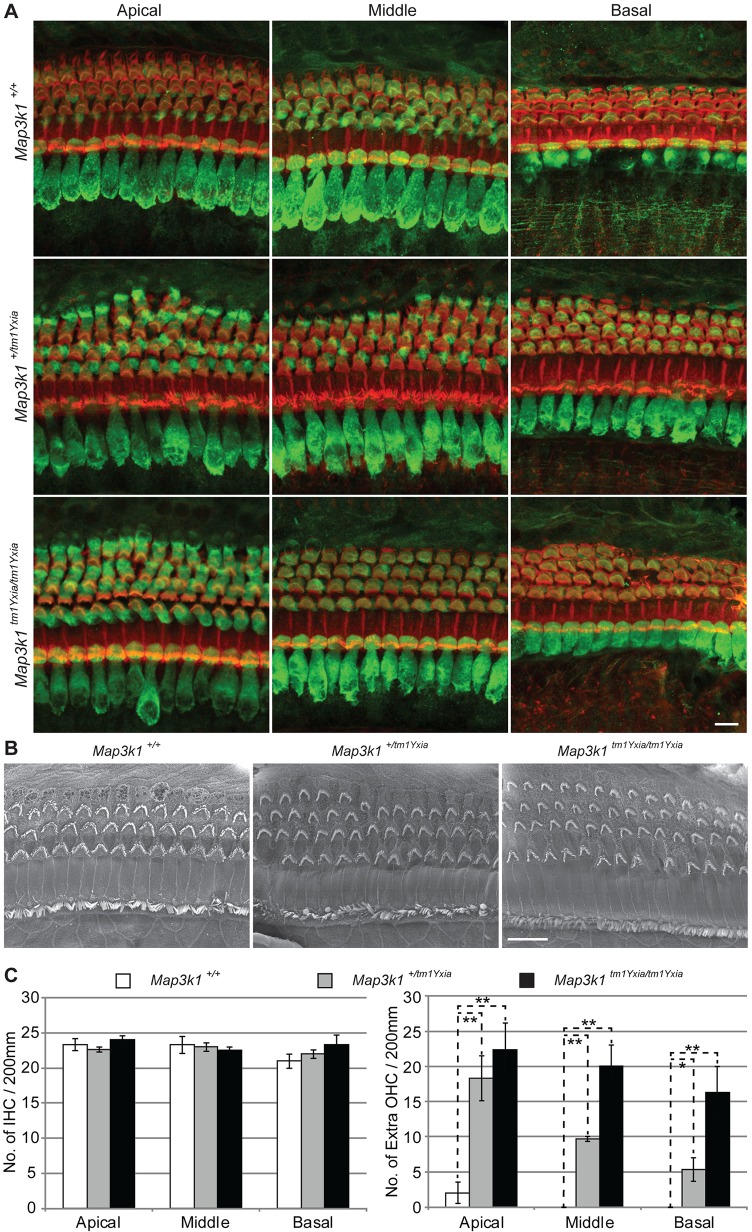
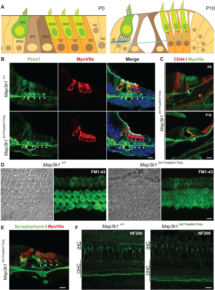
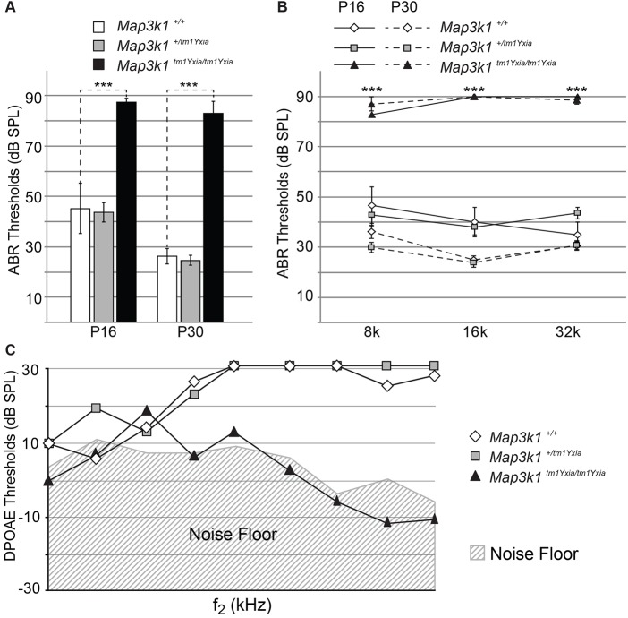
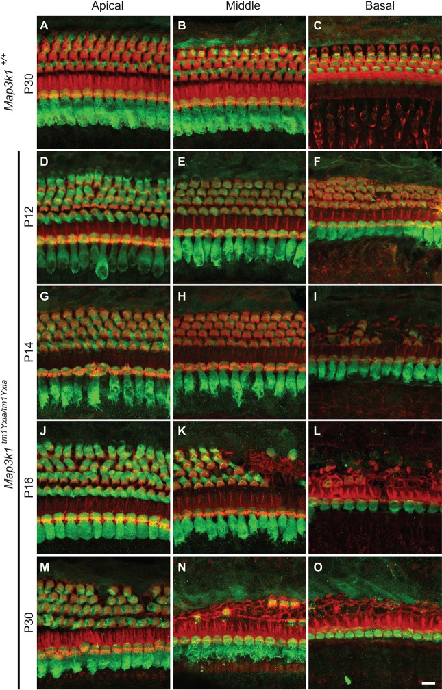
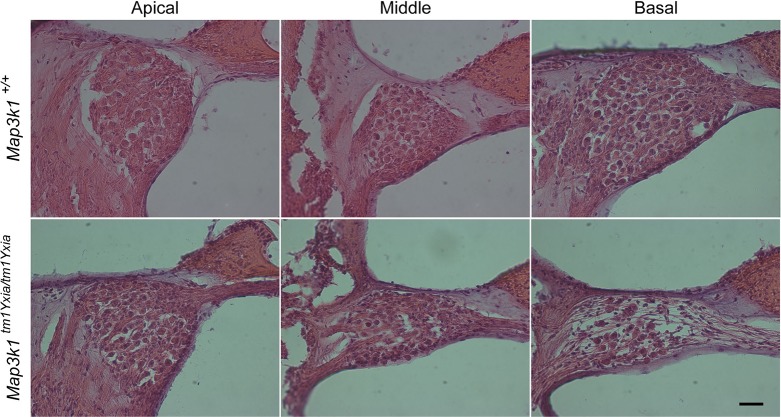
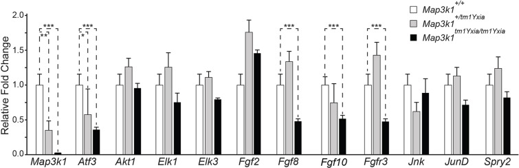
MAP3K1 is a serine/threonine kinase that is activated by a diverse set of stimuli and exerts its effect through various downstream effecter molecules, including JNK, ERK1/2 and p38. In humans, mutant alleles of MAP3K1 are associated with 46,XY sex reversal. Until recently, the only phenotype observed in Map3k1(tm1Yxia) mutant mice was open eyelids at birth. Here, we report that homozygous Map3k1(tm1Yxia) mice have early-onset profound hearing loss accompanied by the progressive degeneration of cochlear outer hair cells. In the mouse inner ear, MAP3K1 has punctate localization at the apical surface of the supporting cells in close proximity to basal bodies. Although the cytoarchitecture, neuronal wiring and synaptic junctions in the organ of Corti are grossly preserved, Map3k1(tm1Yxia) mutant mice have supernumerary functional outer hair cells (OHCs) and Deiters' cells. Loss of MAP3K1 function resulted in the downregulation of Fgfr3, Fgf8, Fgf10 and Atf3 expression in the inner ear. Fgfr3, Fgf8 and Fgf10 have a role in induction of the otic placode or in otic epithelium development in mice, and their functional deficits cause defects in cochlear morphogenesis and hearing loss. Our studies suggest that MAP3K1 has an essential role in the regulation of these key cochlear morphogenesis genes. Collectively, our data highlight the crucial role of MAP3K1 in the development and function of the mouse inner ear and hearing.

Keywords: FGF signaling pathway; Fgf10; Fgf8; Fgfr3; Hearing loss; MAPK pathway; Map3k1; Mekk1; Supernumerary outer hair cells.


Conflict of interest statement

Competing interests

The authors declare no competing or financial interests.

Figures

Fig. 1.
Fig. 1.
Localization of MAP3K1 in the inner ear. (A) MAP3K1 has two domains, a regulatory domain and a kinase domain, whereas, in Map3k1 mutant mice, the kinase domain has been replaced with a β-galactosidase reporter cassette, resulting in a MAP3K1–β-galactosidase fusion protein. (B) Confocal imaging of the whole-mount preparation of organ of Corti (OC) from P16 mice labeled with the anti-β-galactosidase antibody (green) and phalloidin (red). As anticipated, no β-galactosidase labeling was observed in control (Map3k1+/+) mice, whereas, in the Map3k1tm1Yxia mutant mice, a distinct expression of the MAP3K1–β-galactosidase fusion protein was observed at the apical surface of supporting cells of the OC. Scale bar: 10 μm. (C) In sections of P0 mice, the MAP3K1–β-galactosidase fusion protein (green) colocalizes with pericentrin (red), a marker for basal cell bodies. In addition, weak diffused cytoplasmic expression of fusion protein was also observed in inner and outer hair cells. Scale bar: 10 μm. Insets show higher magnification of β-galactosidase and pericentrin localization. Scale bar: 1 μm.
Fig. 2.
Fig. 2.
Map3k1tm1Yxia mutant mice have supernumerary outer hair cells (OHCs). (A) Whole-mount preparation of the OC from P12 mice labeled with myosin VIIa (green) and phalloidin (red). In contrast to three rows of OHCs observed in wild-type mice, Map3k1tm1Yxia mutant mice have an extra row of OHCs along the length of the cochlea, whereas sparse patches of a fourth row of OHCs was observed in Map3k1tm1Yxia heterozygous mice. Scale bar: 10 μm. (B) Scanning electron micrographs of P14 mice revealed characteristic polarized ‘V’-shaped stereocilia bundles at the apical surface of supernumerary OHCs present in Map3k1tm1Yxia heterozygous and homozygous mutant mice. Scale bar: 10 μm. (C) Quantitation of inner hair cells (IHCs) and OHCs in control and Map3k1tm1Yxia mice at P12. For quantification purposes, organ of Corti (OC) were isolated from four wild-type and Map3k1tm1Yxia mutant mice each and hair cells were counted in the apical middle and basal coil regions. No significant difference was observed in the IHC number. Statistically significant (*P<0.05, **P<0.01) increases in the OHC number were observed in the Map3k1tm1Yxia homozygous mutant and heterozygous mice, with a gradient from apex to base (mean±s.e.m.).
Fig. 3.
Fig. 3.
Cytoarchitecture of the organ of Corti (OC) is preserved in Map3k1tm1Yxia mutant mice. (A) Schematic representation of the developing mouse OC at P0 and P10. OHC, outer hair cell; IHC, inner hair cell; DC, Deiters' cell; OPC, outer pillar cell; IPC, inner pillar cell; HC, Hensen's cells; IPhC, inner phalangeal supporting cells. (B) Cross-section of Map3k1tm1Yxia mutant and wild-type control mice at P0, immunostained with Prox1 (green) and myosin VIIa (red). The arrows point to the pillar cells, whereas Deiters' cells are marked by the arrowheads. Map3k1tm1Yxia mutant mice have an extra row of OHCs accompanied by an extra row of Deiters' cells. Scale bar: 10 μm. (C) Immunostaining with the anti-CD44 antibody, a marker for OPCs, including Claudius cells, revealed an intact gross cytoarchitecture of the OC in Map3k1tm1Yxia mutant mice at P0 and P10. Scale bar: 10 μm. (D) No apparent difference in the FM1-43 dye uptake was observed among control and Map3k1tm1Yxia mutant explants. FM1-43 dye was also taken up by the supernumerary OHCs present in Map3k1tm1Yxia mutant mice. Scale bar: 10 μm. (E) Supernumerary OHCs in Map3k1tm1Yxia mutant mice are innervated and have synaptic junctions (arrowheads), immunolabeled with synaptophysin (green). Scale bar: 10 μm. (F) Map3k1tm1Yxia mutant mice have grossly intact neuronal wiring. Neurofilament (NF-200) protein immunostaining of wild-type control and Map3k1tm1Yxia mutant mice revealed grossly intact neuronal wiring of an extra row of OHCs. Scale bar: 10 μm.
Fig. 4.
Fig. 4.
Map3k1tm1Yxia mutant mice have elevated hearing thresholds. Hearing thresholds of 3 wild-type, 7 heterozygous and 8 Map3k1tm1Yxia homozygous mice were evaluated at P16, whereas 8 wild-type, 11 heterozygous and 7 Map3k1tm1Yxia homozygous mice were tested at P30. (A) Averaged auditory brainstem responses (ABR) thresholds of wild-type, and heterozygous and homozygous Map3k1tm1Yxia mice at P16 and P30 in response to click stimulus. The Map3k1tm1Yxia mutant mice showed significantly (***P<0.001) elevated thresholds compared with heterozygous and wild-type mice at both ages (mean±s.e.m.). (B) Averaged ABR thresholds of wild-type (white diamonds), heterozygous (gray squares) and homozygous Map3k1tm1Yxia mutant (black triangles) mice at P16 (solid lines) and P30 (dashed lines), in response to 8-kHz, 16-kHz and 32-kHz tone-bursts. At both developmental stages, Map3k1tm1Yxia mutant mice showed significantly (***P<0.001) elevated thresholds compared with the wild-type control and heterozygous mice at all frequencies tested (mean±s.e.m.). (C) Distortion product otoacoustic emissions (DPOAEs) of Map3k1tm1Yxia mutant (black triangles), heterozygous (gray squares) and wild-type control (white diamonds) mice at P30, represented as a function of f2 stimulus frequencies. Map3k1tm1Yxia mutant mice showed no responses, with values close to the noise floor, indicating that the residual OHCs were non-functional. SPL, sound pressure level.
Fig. 5.
Fig. 5.
Outer hair cells (OHCs) in Map3k1tm1Yxia mutant mice degenerate as early as P14. Maximum intensity projections of confocal Z-stacks of whole-mount cochleae labeled with the anti-myosin-VIIa antibody (green) and phalloidin (red) are shown. (A-C) Representative images from the apical, middle and basal turns of the organ of Corti (OC) of a wild-type control mouse at P30. (D-O) Images of the OC from the three turns of the cochlea of Map3k1tm1Yxia mutant mice at P12 (D-F), P14 (G-I), P16 (J-L) and P30 (M-O). The hair cells appear to have normal development and morphology at P12 in the apical and middle cochlear turns in Map3k1tm1Yxia mutant mice. Initial signs of OHC degeneration are evident in the basal turn (F). At P14, obvious degeneration of OHCs was observed in Map3k1tm1Yxia mutant mice. (K,L) Severe OHC degeneration can be observed by P16 in the middle (K) and basal (L) turns. The OHC loss progresses rapidly and, by P30, severe degeneration is evident in all three cochlear turns. In contrast, inner hair cells (IHCs) remained intact along the length of the cochlea. Scale bar: 10 μm.
Fig. 6.
Fig. 6.
Map3k1tm1Yxia mice at P90 displayed the degeneration of spiral ganglions in the basal turn. Cochlear sections from control and Map3k1tm1Yxia mutant mice were stained with hematoxylin and eosin. Scale bar: 20 μm.
Fig. 7.
Fig. 7.
Fgfr3, Fgf8, Fgf10 and Atf3 genes are downregulated in the organ of Corti (OC) of Map3k1tm1Yxia mutant mice. Semi-quantification expression analysis of various developmental and MAP3K1-mediated signaling pathway genes normalized against Gapdh and actin (endogenous controls) at P10. As anticipated, a dose-dependent, statistically significant reduction in the expression of Map3k1 was observed in heterozygous and homozygous mutant mice. Furthermore, the expression of Fgfr3, Fgf8, Fgf10 and Atf3 was also significantly abolished (Student's t-test, ***P<0.001), suggesting a role of Map3k1 in regulating their expression in the inner ear (mean±s.e.m.). **P<0.01; *P<0.05.

Similar articles

Cited by

References

    1. Bonvin C., Guillon A., van Bemmelen M. X., Gerwins P., Johnson G. L. and Widmann C. (2002). Role of the amino-terminal domains of MEKKs in the activation of NF kappa B and MAPK pathways and in the regulation of cell proliferation and apoptosis. Cell. Signal. 14, 123-131. 10.1016/S0898-6568(01)00219-4 - DOI - PubMed
    1. Cardone M. H., Salvesen G. S., Widmann C., Johnson G. and Frisch S. M. (1997). The regulation of anoikis: MEKK-1 activation requires cleavage by caspases. Cell 90, 315-323. 10.1016/S0092-8674(00)80339-6 - DOI - PubMed
    1. Christerson L. B., Vanderbilt C. A. and Cobb M. H. (1999). MEKK1 interacts with alpha-actinin and localizes to stress fibers and focal adhesions. Cell Motil. Cytoskeleton 43, 186-198. 10.1002/(SICI)1097-0169(1999)43:3<186::AID-CM2>3.0.CO;2-1 - DOI - PubMed
    1. Christerson L. B., Gallagher E., Vanderbilt C. A., Whitehurst A. W., Wells C., Kazempour R., Sternweis P. C. and Cobb M. H. (2002). p115 Rho GTPase activating protein interacts with MEKK1. J. Cell. Physiol. 192, 200-208. 10.1002/jcp.10125 - DOI - PubMed
    1. Cuevas B. D., Abell A. N., Witowsky J. A., Yujiri T., Johnson N. L., Kesavan K., Ware M., Jones P. L., Weed S. A., DeBiasi R. L. et al. (2003). MEKK1 regulates calpain-dependent proteolysis of focal adhesion proteins for rear-end detachment of migrating fibroblasts. EMBO J. 22, 3346-3355. 10.1093/emboj/cdg322 - DOI - PMC - PubMed

Publication types

MeSH terms

Substances