. 2015 Oct 26:6:8372.
doi: 10.1038/ncomms9372.

Inhibition of DYRK1A and GSK3B induces human β-cell proliferation

Affiliations

Inhibition of DYRK1A and GSK3B induces human β-cell proliferation

Weijun Shen et al. Nat Commun. .

Abstract

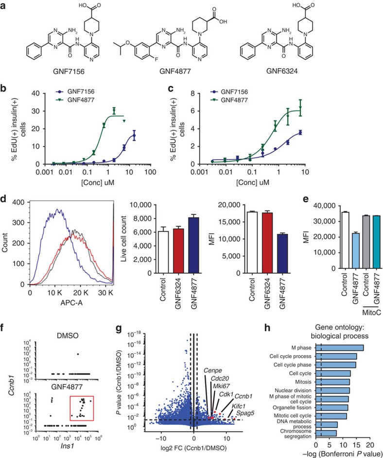
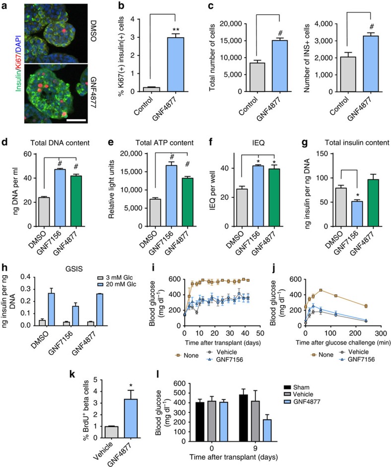
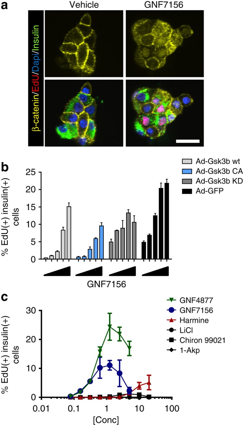
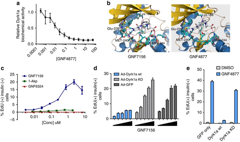
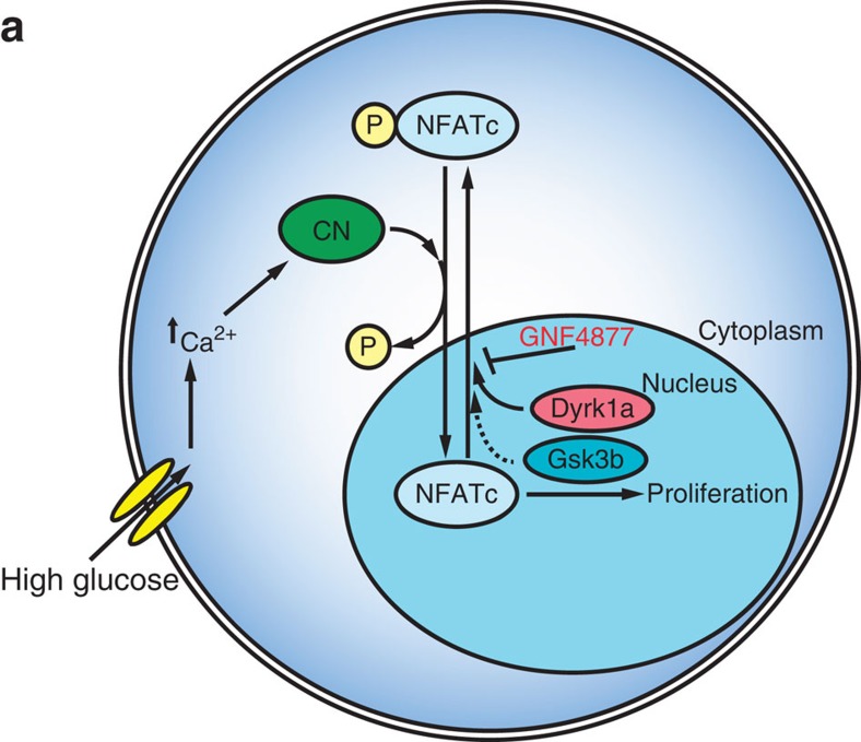
Insufficient pancreatic β-cell mass or function results in diabetes mellitus. While significant progress has been made in regulating insulin secretion from β-cells in diabetic patients, no pharmacological agents have been described that increase β-cell replication in humans. Here we report aminopyrazine compounds that stimulate robust β-cell proliferation in adult primary islets, most likely as a result of combined inhibition of DYRK1A and GSK3B. Aminopyrazine-treated human islets retain functionality in vitro and after transplantation into diabetic mice. Oral dosing of these compounds in diabetic mice induces β-cell proliferation, increases β-cell mass and insulin content, and improves glycaemic control. Biochemical, genetic and cell biology data point to Dyrk1a as the key molecular target. This study supports the feasibility of treating diabetes with an oral therapy to restore β-cell mass, and highlights a tractable pathway for future drug discovery efforts.


Conflict of interest statement

B.T., Q.J., V.N.-T., A.P., Y.-Q.Z., C.M.F., J.L., L.W., B.B., G.H., M.Q., Y.L., X.H., T.M., S.Y., J.L., T.W., P.M., R.G. and B.L. are employees of the Genomics Institute of the Novartis Research Foundation. A.H. is an employee of Genentech, Inc. W.S. is an employee of the California Institute for Biomedical Research. H.M.S. is an employee of the Novartis Institutes for BioMedical Research. B.H. serves as a consultant for Janssen Pharmaceuticals, Novartis Pharmaceuticals Corporation, and Sanofi-Aventis Group.

Figures

Figure 1
Figure 1. Aminopyrazine compounds induce primary rodent and human β-cell proliferation.
(a) Structures of the aminopyrazine (AP) compounds GNF7156, GNF4877 and GNF6324. (bc) Dose response of GNF7156 and GNF4877-induced EdU incorporation into β cells from rat (b) or human (c) islets. (de) Cell division induced by GNF4877. (d) Rat islets were loaded with eFluor670 and subsequently treated with GNF4877 or the inactive analogue GNF6324 for 5 days. Fluorescence was determined by FACS and represented as mean fluorescence intensity (MFI; mean±s.d.; n=5). (e) Dilution of eFluor670 dye was abrogated by cell cycle inhibitor Mitomycin C (MitoC). (fh) Single cell RNA sequencing from rat islets. (f) Single cell RNA sequencing of rat islets reveals a greater number of Insulin 1 (Ins1) and Ccnb1 co-positive cells with GNF4877 treatment (red box). (g) Volcano plot comparing gene expression of Ccnb1/ Ins1 positive cells from GNF4877 treatment (red box) to Ins1 expressing cells in DMSO reveals a significant increase in expression of cell cycle genes (g) and gene ontology biological processes (h) strongly associated with cell cycle progression.
Figure 2
Figure 2. AP compounds expand human islets ex vivo with retention of function after transplantation.
(ac) Treatment of intact primary human islets with GNF4877 for 8 days results in increased beta cell numbers relative to vehicle control–treated islets. (a) Immunofluorescence for insulin, Ki67 and DAPI on DMSO or GNF4877-treated human intact islets (scale bar, 50 μm). (b) Quantification of Ki67+ as a percent of total insulin+ cells (n=3). (c) Total number of islet cells and total number of insulin+ cells after 8 days of treatment of human islets with GNF4877 or vehicle (n=24 islets; bars represent mean±s.d.; **P<0.01, #P<0.0001, Student's t-test. (dj) Primary human islets were treated for 7 days with the AP compounds or vehicle control (6.7 μM GNF7156 or 2 μM GNF4877; n=9 per condition) and then tested for (d) total DNA content, (e) total ATP content, (f) islet equivalent units (IEQ), (g) total insulin content and (h) glucose stimulated insulin release (GSIS) (*P<0.05, #P<0.0001, Student's t-test). (ij) GNF7156 and DMSO-expanded human islets are functionally equivalent when equal numbers were transplanted into the kidney capsule of STZ-induced diabetic NOD–SCID mice: (i) Fed blood glucose and (j) oral glucose tolerance test. Data are shown as mean±s.d. (six mice per group; transplanted with islets from a single human donor). (kl) NSG mice were transplanted with a sub-optimal dose of human islets under the kidney capsule before treatment with GNF4877 (oral, 50 mg kg−1, twice daily) or vehicle control. GNF4877-treated NSG mice transplanted with human islets demonstrated increased BrdU incorporation into insulin-positive cells by immunohistochemistry (k) and display improved glucose control as measured by fed glucose levels (l). Data shown as mean±s.d.; (n=6 mice/group, *P<0.05, Student's t-test).
Figure 3
Figure 3. AP compounds improve glucose control and β-cell mass in the RIP-DTA diabetes model.
(ah) Diabetic RIP-DTA mice were treated with GNF4877 (oral, 50 mg kg−1 twice daily) after disease induction (when blood glucose reached 400 mg dl−1). (a) GNF4877 caused progressive reduction of hyperglycemia (fed glucose measurement) relative to vehicle-treated animals. (bc) GNF4877-treated animals showed improved oral glucose tolerance following 14 days of dosing. (c) Area under the curve (AUC) from oral glucose tolerance test (OGTT) data was significantly improved by GNF4877 treatment (n=8 mice/group; data shown as mean±s.d.; *P<0.05; **P<0.01, Student's t-test). (dh) Histological analysis for insulin and Ki67 from pancreata of non-diabetic and diabetic mice after 14 days of treatment with GNF4877 for 14 days reveals increased percentage of Ki67+ Ins+ cells (d,e), increased β-cell mass (f), increased insulin content (g), and mean fluorescent intensity of insulin (h) compared with vehicle-treated control mice. (n=8 mice/group; data shown as mean±s.d.; *P<0.05; **P<0.01, Student's t-test). Scale bar, 100 μm.
Figure 4
Figure 4. Aminopyrazines inhibit Gsk3b.
(a) GNF7156 induces nuclear localization of β-catenin in EdU and insulin co-positive cells from primary rat islets. (b) Adenoviral overexpression of wild type (wt), constitutively active (CA) and kinase-dead (KD) Gsk3b partially inhibited GNF7156-induced β-cell proliferation in primary dispersed rat islets as judged by EdU incorporation (n=8/group; mean±s.d.). (c) Gsk3b inhibitors LiCl, 1-Akp and Chiron99021 induce minimal or no proliferation of rat β-cells from dispersed islets, whereas the Dyrk1a inhibitor harmine induces EdU incorporation into insulin-positive cells (n=3 per data point; mean±s.d.). Scale bar, 20 μm.
Figure 5
Figure 5. Dyrk1a inhibition is a critical mechanism of AP-stimulated β-cell proliferation.
(a) GNF4877 potently inhibits Dyrk1a kinase activity in a biochemical assay (data point is mean±s.d., n=3). (b) Molecular modelling of GNF7156 and GNF4877 in the Dyrk1a crystal structure. (c) GNF6324, a Gsk3b inhibitor compound closely related to GNF7156, but lacking Dyrk1a activity, does not induce proliferation of primary rat β-cells. Dispersed primary rat islets were treated with the indicated compounds and β-cell proliferation was measured by EdU incorporation into insulin-positive cells (n=3 per data point; mean±s.d.). (de) Adenoviral overexpression of wild type (wt) but not kinase-dead (KD) Dyrk1a blocked GNF7156 (d) or GNF4877 (e) induced β-cell proliferation in dispersed rat islets. Proliferation of β-cells was measured by EdU incorporation into insulin-positive cells (n=3; mean±s.d.).
Figure 6
Figure 6. AP compounds induce β-cell proliferation through activation of the calcineurin–NFAT pathway.
(a) AP compounds stimulate NFATc1-GFP nuclear localization. Two days after infection of Ins1 cells with adenoviral NFATc1-GFP, cells were treated with GNF4877, GNF7156 or vehicle control for the indicated time before high content imaging for NFATc1-GFP cellular localization. Scale bar, 50 μm. (b) AP compounds inhibit NFATc1 nuclear export after induction of NFATc1 nuclear localization with ionophore. Following ionophore withdrawal, cells were treated with GNF4877, GNF7156 or DMSO for the indicated times and NFATc1-GFP localization was monitored by high content imaging. (c) Primary human β-cell proliferation induced by GNF4877 (2 μM) was inhibited by FK506 (12 nM to 1 μM) in the ex vivo human dissociated islet assay (n=3; mean±s.d.; **P<0.001, #P<0.0001, ANOVA). (de) Endogenous NFATc1 and NFATc3 are nuclear localized by treatment with GNF4877. Primary rat islets were treated with GNF4877 or DMSO control before immunofluorescent staining for endogenous NFATc1 (d) or NFATc3 (e). Scale bar, 50 μm.
Figure 7
Figure 7. Calcium signaling regulates AP-induced β-cell proliferation.
(ad) Elevation of intracellular Ca2+ levels in β cells augments the response to AP compounds. (a) Primary human islets were treated with glucokinase activators (Ro28-1675 or GKA50), high glucose (16.7 mM glucose) or the sulfonylurea (Glibenclamide)±a sub-optimal concentration (0.1 μM) of GNF4877. β-cell proliferation was measured by EdU incorporation into insulin-positive cells (n=3 per data point; mean±s.d.). (bd) Combination of GNF4877 with agents that elevate intracellular calcium increases proliferation of rat β-cells. Concentrations of GNF4877, below the EC50 for rat or human β-cell proliferation, are effective in inducing proliferation in the presence of agents that elevate intracellular calcium: (b) high glucose; (c) the glucokinase activator GKA50 or (d) glibenclamide (a sulfonylurea receptor 1 inhibitor) or Bay K8644 (an L-type Ca2+ channel activator).
Figure 8
Figure 8. Model of AP-stimulated β-cell proliferation.
(a) In the presense of high glucose, beta cells increase intracellular Ca2+ levels leading to activation of calcineurin (CN). CN dephosphorylates NFATc proteins such as NFATc1 leading to nuclear import. Previous studies demonstrated a critical role for the CN–NFATc pathway in the regulation of β-cell proliferation. Nuclear NFATc kinases Dyrk1a and Gsk3b phosphorylate NFATc proteins leading to nuclear export. AP compounds, such as GNF4877, inhibit Dyrk1a and Gsk3b leading to blockade of NFATc nuclear export and increased β-cell proliferation.

Similar articles

Cited by

References

    1. Matveyenko A. V. & Butler P. C. Relationship between beta-cell mass and diabetes onset. Diabetes Obes. Metab. 10, (Suppl 4): 23–31 (2008). - PMC - PubMed
    1. Butler A. E. et al.. Beta-cell deficit and increased beta-cell apoptosis in humans with type 2 diabetes. Diabetes 52, 102–110 (2003). - PubMed
    1. Stumvoll M., Goldstein B. J. & van Haeften T. W. Type 2 diabetes: pathogenesis and treatment. Lancet 371, 2153–2156 (2008). - PubMed
    1. Schafer S. A., Machicao F., Fritsche A., Haring H. U. & Kantartzis K. New type 2 diabetes risk genes provide new insights in insulin secretion mechanisms. Diabetes Res. Clin. Pract. 93, (Suppl 1): S9–S24 (2011). - PubMed
    1. Dor Y., Brown J., Martinez O. I. & Melton D. A. Adult pancreatic beta-cells are formed by self-duplication rather than stem-cell differentiation. Nature 429, 41–46 (2004). - PubMed

Publication types

MeSH terms