

Agreement between transperineal ultrasound measurements and digital examinations of cervical dilatation during labor
- PMID: 26496894
- PMCID: PMC4619348
- DOI: 10.1186/s12884-015-0704-z
Agreement between transperineal ultrasound measurements and digital examinations of cervical dilatation during labor
Abstract
Background: To compare 2D transperineal ultrasound assessment of cervical dilatation with vaginal examination and to investigate intra-observer variability of the ultrasound method.
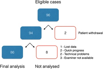
Methods: A prospective observational study was performed at Skane University Hospital, Lund, Sweden between October 2013 and June 2014. Women with one fetus in cephalic presentation at term had the cervical dilatation assessed with ultrasound and digital vaginal examinations during labor. Inter-method agreement between ultrasound and digital examinations and intra-observer repeatability of ultrasound examinations were tested.
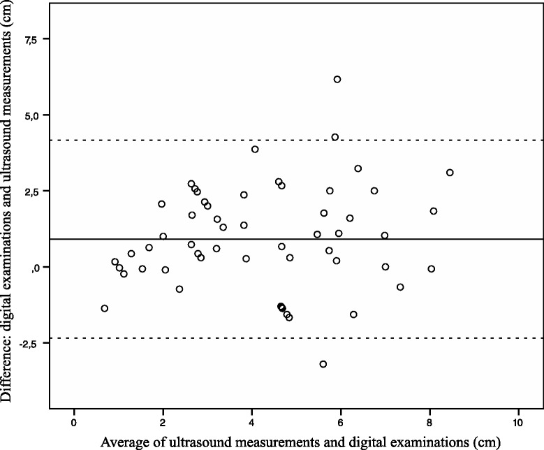
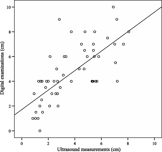
Results: Cervical dilatation was successfully assessed with ultrasound in 61/86 (71 %) women. The mean difference between cervical dilatation and ultrasound measurement was 0.9 cm (95 % CI 0.47-1.34). Interclass correlation coefficient (ICC) was 0.83 (95 % CI 0.72-0.90). Intra-observer repeatability was analysed in 26 women. The intra-observer ICC was 0.99 (95 % CI 0.97-0.99). The repeatability coefficient was ± 0.68 (95 % CI 0.45-0.91).
Conclusion: The mean ultrasound measurement of cervical dilatation was approximately 1 cm less than clinical assessment. The intra-observer repeatability of ultrasound measurements was high.
Figures
References
-
- Sherer DM, Miodovnik M, Bradley KS, Langer O. Intrapartum fetal head position I: comparison between transvaginal digital examination and transabdominal ultrasound assessment during the active stage of labor. Ultrasound Obstet Gynecol. 2002;19:258–63. doi: 10.1046/j.1469-0705.2002.00656.x. - DOI - PubMed
Publication types
MeSH terms
LinkOut - more resources
Full Text Sources
Other Literature Sources
Medical
