Review
. 2015;4(6):399-410.
doi: 10.2217/cns.15.20. Epub 2015 Oct 26.

Assessing and monitoring intratumor heterogeneity in glioblastoma: how far has multimodal imaging come?

Affiliations
Review

Assessing and monitoring intratumor heterogeneity in glioblastoma: how far has multimodal imaging come?

Natalie R Boonzaier et al. CNS Oncol. 2015.

Abstract

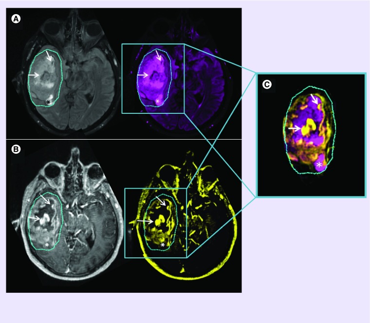
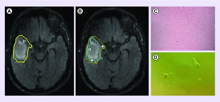
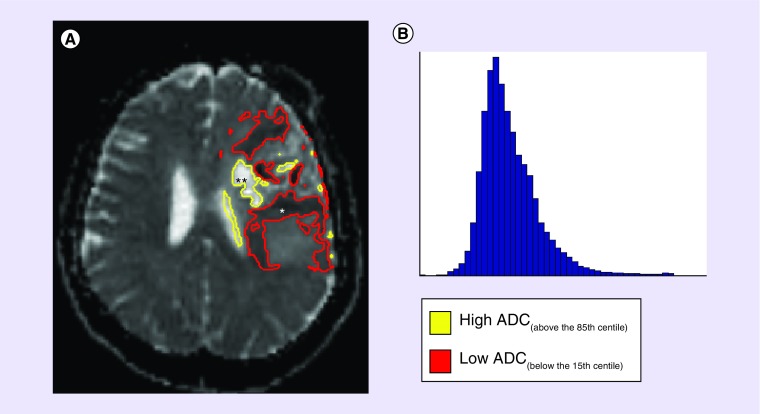
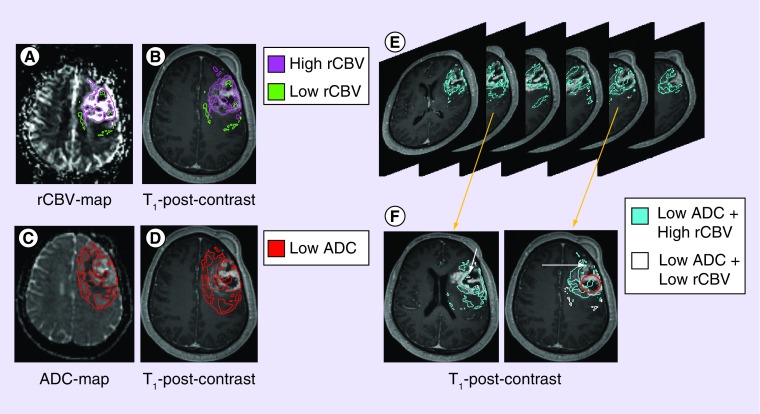
Glioblastoma demonstrates imaging features of intratumor heterogeneity that result from underlying heterogeneous biological properties. This stems from variations in cellular behavior that result from genetic mutations that either drive, or are driven by, heterogeneous microenvironment conditions. Among all imaging methods available, only T1-weighted contrast-enhancing and T2-weighted fluid-attenuated inversion recovery are used in standard clinical glioblastoma assessment and monitoring. Advanced imaging modalities are still considered emerging techniques as appropriate end points and robust methodologies are missing from clinical trials. Discovering how these images specifically relate to the underlying tumor biology may aid in improving quality of clinical trials and understanding the factors involved in regional responses to treatment, including variable drug uptake and effect of radiotherapy. Upon validation and standardization of emerging MR techniques, providing information based on the underlying tumor biology, these images may allow for clinical decision-making that is tailored to an individual's response to treatment.

Keywords: advanced imaging; glioblastoma; intratumor heterogeneity; subregional assessment; underlying biology.


Conflict of interest statement

Financial & competing interests disclosure

SJ Price is funded by a Clinician Scientist Award from the National Institute for Health Research. Research conducted by Stephen J Price is supported by the Cambridge Biomedical Research Centre and Cancer Research UK. The authors have no other relevant affiliations or financial involvement with any organization or entity with a financial interest in or financial conflict with the subject matter or materials discussed in the manuscript apart from those disclosed.

No writing assistance was utilized in the production of this manuscript.

Figures

<b>Figure 1.</b>
Figure 1.. Conventional MRI.
T2-weighted FLAIR images (A) are used to non-specifically assess edema surrounding the T1-weighted contrast-enhancement (B). Though signal hyper- or hypo-intensities may indicate certain biological scenarios, one cannot be certain of the eco-biology within ROIs. Combined ROIs (C) of low T2-FLAIR with high T1-post-contrast signal (arrows) can imply absence of edema, as a result of tumor and/or inflammatory cell presence, with compromised BBB integrity and leaky vasculature; ROIs of increased T2-FLAIR with low T1-post contrast signal (*) can imply regional edema, resulting from an adjacent inflammatory response or from an absence of tumor cells (possible necrosis), with a probable intact BBB and preserved vascular integrity. Contrary to what may be expected, a low signal on a T1-post-contrast image does not necessarily imply a reduced or compromised blood supply- information of this nature would only be obtainable with perfusion imaging. BBB: Blood–brain barrier; FLAIR: Fluid attenuated inversion recovery; ROI: Region of interest.
<b>Figure 2.</b>
Figure 2.. T2-weighted FLAIR image of a GBM lesion with a superimposed delineation of the p-abnormality, which extends beyond FLAIR enhancement (A).
This, in conjunction with the q-abnormality (B), can be used to differentiate the tumor bulk (q) from the margin (*), which has a normal histological appearance (C), but which harbors tumor ††cells (D). Regions of p- and q- abnormality were obtained using the p- and q- components of the diffusion tensor, as described by [30]. Tissue samples obtained using 5-ALA fluorescence-guided resection, allowing access to normal-appearing tissue, stained with H&E, 10× magnification. ††Margin cells were plated in vitro and observed 4DIV. 5-ALA: 5-aminolevulinic acid; DIV: Days in vitro; FLAIR: Fluid-attenuated inversion recovery; GBM: Glioblastoma; H&E: Hematoxylin and eosin stain.
<b>Figure 3.</b>
Figure 3.. A pre-treatment ADC scan of GBM (A) can show marked variation of low (*) and high (**) ADC values, which can regionally represent various biological scenarios- low ADC: high tumor or inflammatory cell presence, ischemia or a combination of both; high ADC: vasogenic edema, limited tumor and/or inflammatory cells, necrosis or a combination of any.
Studies assessing distribution of ADC with histogram analyses (B), although providing interesting quantitative data, give no information pertaining to the spatial distribution of ADC throughout the lesion. ADC: Apparent diffusion coefficient; GBM: Glioblastoma.
<b>Figure 4.</b>
Figure 4.. ADC and rCBV can give better indications of what may be happening within sub-regions of GBM.
rCBV-maps (A) provide clues pertaining to the microenvironment that surrounds cells. Superimposing these sub-regions to T1-post-contrast (B) images provides details pertaining to where these ecosystems may be in relation to the tumor ‘bulk’ (T1-enhancing region)- either within the ‘necrotic core’, within the ‘bulk’, or just outside. Combining this with regional low ADC (C) and assessing its location on T1-post-contrast images (D) can also give spatial information relative to tumor ‘bulk’. As one scans through images of combined ADC and rCBV ROIs (E), further scenarios can be presumed (F): in the ‘necrotic core’, cellular regions with adequate perfusion suggest a population residing in regions of adequate oxygen and nutrient supply (O); cellular regions with lowered blood volume (arrow), however, suggest a population adapted to a hypoxic and acidic microenvironment conditions. Note how these advanced methods also suggest that the conventional ‘necrotic core’ may harbor cells and possess regional blood supply. ADC: Apparent diffusion coefficient; GBM: Glioblastoma; rCBV: Relative cerebral blood volume.

References

    1. Stupp R, Mason WP, van den Bent MJ, et al. Radiotherapy plus concomitant and adjuvant temozolomide for glioblastoma. N. Engl. J. Med. 2005;352(10):987–996. - PubMed
    2. • The major single advancement in GBM treatment over the past years, incorporating RT with concomitant and adjuvant temozolomide.

    1. Jackson RJ, Fuller GN, Abi-Said D, et al. Limitations of stereotactic biopsy in the initial management of gliomas. Neuro Oncol. 2001;3(3):193–200. - PMC - PubMed
    1. Stummer W, Pichlmeier U, Meinel T, Wiestler OD, Zanella F, Reulen H-J. Fluorescence-guided surgery with 5-aminolevulinic acid for resection of malignant glioma: a randomised controlled multicentre Phase III trial. Lancet Oncol. 2006;7(5):392–401. - PubMed
    1. Deacon J, Peckham MJ, Steel GG. The radioresponsiveness of human tumours and the initial slope of the cell survival curve. Radiother. Oncol. 1984;2(4):317–323. - PubMed
    1. Diehn M, Nardini C, Wang DS, et al. Identification of noninvasive imaging surrogates for brain tumor gene-expression modules. Proc. Natl Acad. Sci. USA. 2008;105(13):5213–5218. - PMC - PubMed

Publication types

LinkOut - more resources