. 2015 Oct 23;82(1):211-7.
doi: 10.1128/AEM.02302-15. Print 2016 Jan 1.

A Dual Microscopy-Based Assay To Assess Listeria monocytogenes Cellular Entry and Vacuolar Escape

Affiliations

A Dual Microscopy-Based Assay To Assess Listeria monocytogenes Cellular Entry and Vacuolar Escape

Juan J Quereda et al. Appl Environ Microbiol. .

Abstract

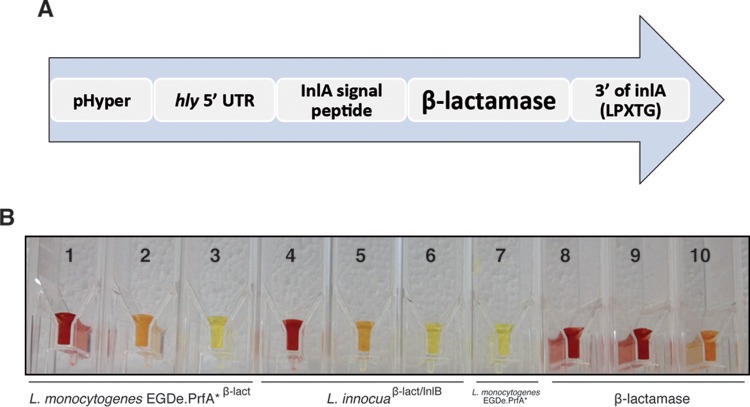
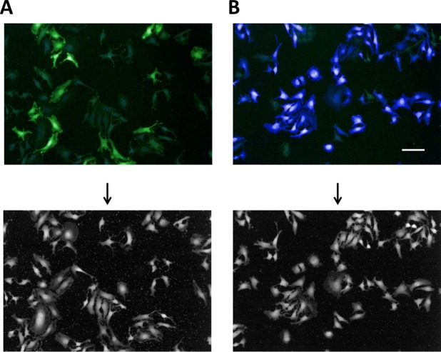
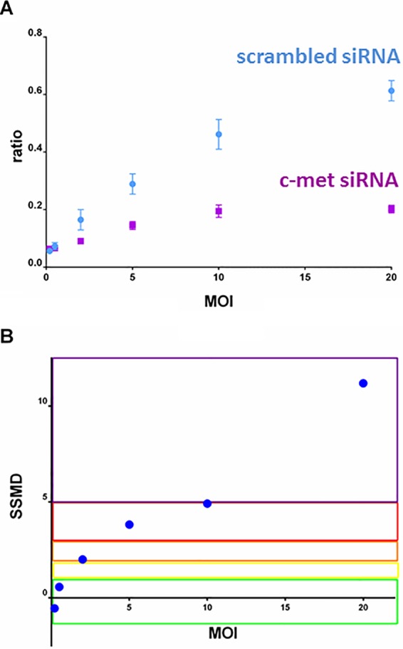
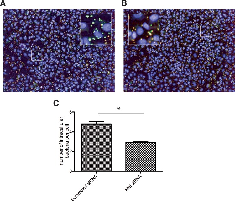
Listeria monocytogenes is a Gram-positive bacterium and a facultative intracellular pathogen that invades mammalian cells, disrupts its internalization vacuole, and proliferates in the host cell cytoplasm. Here, we describe a novel image-based microscopy assay that allows discrimination between cellular entry and vacuolar escape, enabling high-content screening to identify factors specifically involved in these two steps. We first generated L. monocytogenes and Listeria innocua strains expressing a β-lactamase covalently attached to the bacterial cell wall. These strains were then incubated with HeLa cells containing the Förster resonance energy transfer (FRET) probe CCF4 in their cytoplasm. The CCF4 probe was cleaved by the bacterial surface β-lactamase only in cells inoculated with L. monocytogenes but not those inoculated with L. innocua, thereby demonstrating bacterial access to the host cytoplasm. Subsequently, we performed differential immunofluorescence staining to distinguish extracellular versus total bacterial populations in samples that were also analyzed by the FRET-based assay. With this two-step analysis, bacterial entry can be distinguished from vacuolar rupture in a single experiment. Our novel approach represents a powerful tool for identifying factors that determine the intracellular niche of L. monocytogenes.


Figures

FIG 1
FIG 1
Construction of a β-lactamase constitutively expressed at the surface of Listeria spp. (A) Modular domain construction of the gene for constitutive expression of a surface β-lactamase in Listeria. The construction consists of the constitutive promoter Phyper fused to the hly 5′-untranslated region and the β-lactamase gene (codon usage optimized for L. monocytogenes) with the signal peptide of InlA. Finally, the region encoding the last 284 amino acids of InlA, which code for an LPXTG motif to anchor the protein to the peptidoglycan, was added at the 3′ end. (B) A chromogenic cephalosporin test based on nitrocefin confirms β-lactamase activity on the surface of L. monocytogenes EGDe PrfA*β-lact and L. innocuaβ-lact/InlB strains. Hydrolysis of nitrocefin produces a shift in the visible light spectrum from intact (yellow) to degraded (red) nitrocefin. Tubes 1, 2, and 3, L. monocytogenes EGDe PrfA*β-lact and 10-fold dilutions 10−1 and 10−2; tubes 4, 5, and 6, L. innocuaβ-lact/InlB and 10-fold dilutions 10−1 and 10−2; tube 7, L. monocytogenes EGDe PrfA*; tubes 8, 9, and 10, β-lactamase diluted to 1, 0.1, and 0.01 mg/ml, respectively. The volume in all tubes was 105 μl. Bacterial concentration in tubes 1, 4, and 7 was ≈1.4 × 10⁸ bacteria/ml.
FIG 2
FIG 2
Tracking of L. monocytogenes vacuolar escape using a FRET-based microscopic assay. Cellular infection with L. innocuaβ-lact/InlB (A) or L. monocytogenes EGDe PrfA*β-lact (B). HeLa cells were loaded with CCF4-AM for 2.30 h and infected with bacterial strains for 1 h. After paraformaldehyde fixation for 10 min, nuclei were stained with Draq5, and cells were imaged using an Opera QEHS confocal microscope with a 10× objective. Pictures were obtained with the following merged channels: the intact CCF4 probe peaks at 535 nm (green), and the cleaved CCF4 probe peaks at 450 nm (blue). As shown, L. monocytogenes EGDe PrfA*β-lact can escape its vacuolar compartments and induce the cleavage of the CCF4 probe (B). Bar = 10 μm. Panels at the bottom show Draq5 staining of images A and B.
FIG 3
FIG 3
Quality control by MOI for the efficiency of the CCF4 assay. HeLa cells transfected with scrambled or InlB cellular receptor c-Met siRNAs were infected using a dynamic range of MOIs (0.1, 0.2, 2, 5, 10, and 20). Seven wells per MOI were automatically acquired and analyzed to extract the 450 nm/535 nm ratio for each condition. (A) The 450 nm/535 nm ratio as a function of MOI for c-Met (purple) and scrambled (blue) siRNA-treated cells. Each plot corresponds to the mean ratio between the intensity in the 450- and 535-nm channels on 405 nm excitation (n = 7 for each experimental condition). Vertical bars represent the standard deviation. (B) Quality control for the efficiency of the CCF4 assay for each MOI. For quality control, SSMD statistical tests were used to quantify the magnitude of difference between negative and positive controls for each MOI. This test yields a score for each pair of controls per assay condition. The SSMD scores to qualify the robustness of an assay were defined by Zhang (30, 31) as follows:  SSMD  ≥ 5 for extremely strong (purple), 5 >  SSMD  ≥ 3 for very strong (red), 3 >  SSMD  ≥ 2 for strong (orange), 2 >  SSMD  ≥ 1.645 for fairly strong (yellow), 1.645 >  SSMD  ≥ 1.28 for moderate (green), and  SSMD  < 1.28 for weak or no effects.
FIG 4
FIG 4
Differential immunofluorescence staining for quantification of extracellular versus total L. monocytogenes numbers. HeLa cells were previously transfected with control (A) or c-Met-specific (B) siRNAs in 96-well plates and infected with L. monocytogenes EGDe PrfA*β-lact. Indirect immunofluorescence was performed 1 day after CCF4 cleavage acquisition, and plates were kept at 4°C without probenecid for 4 days in order to decrease the CCF4 cell load. Extracellular bacteria were labeled with a secondary goat anti-rabbit antibody–Alexa Fluor 647 stain (red), and total bacteria were labeled with a secondary goat anti-rabbit antibody–Alexa Fluor 488 stain (green) after cell permeabilization. Nuclei were stained with Hoechst 33342 (blue). Intracellular and extracellular bacteria are seen in green and red, respectively. Inactivation of c-Met reduces the number of intracellular bacteria displaying a very distinct green signal. Bars = 100 μm; inset bars = 8 μm. (C) Measurement of the number of intracellular bacteria per cell from images displayed in panels A and B. The experiment was repeated three times (4 wells per condition in each experiment). The results from one representative experiment are shown and were statistically analyzed using a t test. *, P < 0.05.

References

    1. Allerberger F, Wagner M. 2010. Listeriosis: a resurgent foodborne infection. Clin Microbiol Infect 16:16–23. doi:10.1111/j.1469-0691.2009.03109.x. - DOI - PubMed
    1. Cossart P. 2011. Illuminating the landscape of host-pathogen interactions with the bacterium Listeria monocytogenes.. Proc Natl Acad Sci U S A 108:19484–19491. doi:10.1073/pnas.1112371108. - DOI - PMC - PubMed
    1. Pizarro-Cerda J, Bonazzi M, Cossart P. 2010. Clathrin-mediated endocytosis: what works for small, also works for big. Bioessays 32:496–504. doi:10.1002/bies.200900172. - DOI - PubMed
    1. Pizarro-Cerda J, Cossart P. 2009. Listeria monocytogenes membrane trafficking and lifestyle: the exception or the rule? Annu Rev Cell Dev Biol 25:649–670. doi:10.1146/annurev.cellbio.042308.113331. - DOI - PubMed
    1. Kuhbacher A, Dambournet D, Echard A, Cossart P, Pizarro-Cerda J. 2012. Phosphatidylinositol 5-phosphatase oculocerebrorenal syndrome of Lowe protein (OCRL) controls actin dynamics during early steps of Listeria monocytogenes infection. J Biol Chem 287:13128–13136. doi:10.1074/jbc.M111.315788. - DOI - PMC - PubMed

Publication types

MeSH terms

LinkOut - more resources