Review
. 2015 Nov;268(1):139-59.
doi: 10.1111/imr.12349.

Human IgG4: a structural perspective

Affiliations
Review

Human IgG4: a structural perspective

Anna M Davies et al. Immunol Rev. 2015 Nov.

Abstract

IgG4, the least represented human IgG subclass in serum, is an intriguing antibody with unique biological properties, such as the ability to undergo Fab-arm exchange and limit immune complex formation. The lack of effector functions, such as antibody-dependent cell-mediated cytotoxicity and complement-dependent cytotoxicity, is desirable for therapeutic purposes. IgG4 plays a protective role in allergy by acting as a blocking antibody, and inhibiting mast cell degranulation, but a deleterious role in malignant melanoma, by impeding IgG1-mediated anti-tumor immunity. These findings highlight the importance of understanding the interaction between IgG4 and Fcγ receptors. Despite a wealth of structural information for the IgG1 subclass, including complexes with Fcγ receptors, and structures for intact antibodies, high-resolution crystal structures were not reported for IgG4-Fc until recently. Here, we highlight some of the biological properties of human IgG4, and review the recent crystal structures of IgG4-Fc. We discuss the unexpected conformations adopted by functionally important Cγ2 domain loops, and speculate about potential implications for the interaction between IgG4 and FcγRs.

Keywords: Fc receptor; IgG1; IgG4; antibody; immunoglobulin.


Figures

Fig. 1
Fig. 1
Overall IgG architecture. (A) Schematic of an IgG antibody. The Fc fragment, comprising Cγ2 and Cγ3 domains from the heavy chain, is connected to the Cγ1 domain of each Fab fragment through a hinge region. The sequence composition of the hinge, and number of inter-heavy chain disulfide bonds varies between the four human IgG subclasses. In this figure, two inter-heavy chain disulfide bonds are indicated for IgG1. The variable regions of the Fab fragment (VH and VL) are responsible for antigen recognition. (B) Crystal structure of an intact human IgG1 antibody, solved at 2.7 Å resolution, reveals an asymmetric conformation (36). (C) Cartoon representation of the IgG-Fc fragment (50), showing the internal oligosaccharide moiety. The figure was prepared with PyMOL (166).
Fig. 2
Fig. 2
IgG utilizes the Cγ2–Cγ3 domain interface to interact with a variety of different proteins. (A) The neonatal receptor, FcRn (63). (B) HSV-1 (herpes simplex virus type I) gE-gI receptor (53). (C) Streptococcal protein G (52). (D) TRIM21 (tripartite motif-containing 21) (61). (E) Fab fragment from an IgM rheumatoid factor (74). (F) Fc–Fc interactions revealed by crystal packing (18). (G) Staphylococcal protein A (49). (H) Fc–Fc-mediated hexamer involved in complement activation (19,36,90). The figure was prepared with PyMOL (167).
Fig. 3
Fig. 3
Overall structure of IgG1-Fc/FcγR complexes. (A) Crystal structure of the IgG1-Fc/FcγRIIIa complex (57). The domain arrangement is similar in the IgG1-Fc/FcγRIIa (60), FcγRIIb (65) and FcγRIIIb (56,59) complexes. (B) Crystal structure of the IgG1-Fc/FcγRI complex (55). In both panels, receptor domains are labeled D1-D3 and IgG-Fc domains Cγ2–Cγ3. The figure was prepared with PyMOL (167).
Fig. 4
Fig. 4
Sites of interaction in IgG1-Fc/FcγR complexes. A crystal structure for IgG1-Fc in complex with FcγRIIIa (58) is shown, although the interface is similar in all FcγRs. One IgG1-Fc Cγ2 domain (blue) interacts with the receptor (yellow) D1-D2 domain linker and D2 domain BC loop. The second IgG1-Fc Cγ2 domain (pink) interacts with the D2 domain C and C′ strands. The lower hinge contacts the D2 domain BC and FG loops. The figure was prepared with PyMOL (167).
Fig. 5
Fig. 5
IgG1-Fc interactions with FcγRs. (A) The hydrophobic proline sandwich interaction, in which Pro329 from the Cγ2 FG loop interacts with two conserved tryptophan residues from the receptor. A position adjacent to the proline sandwich is a site of sequence variation, in which structurally equivalent residues are Arg102 in FcγRI (white) (54), Ser88 in FcγRII (pink) (65), and Ile88 in FcγRIII (beige) (58). In the FcγRI complex, Arg102 forms a hydrogen bond with the Pro329 backbone carbonyl group. (B) A second site of interaction involves the IgG1 Cγ2 domain BC and DE loops. In one IgG1-Fc/FcγRI complex (54), Lys142 from the receptor packs against Tyr296 (Cγ2 DE loop) while Lys145 from the receptor forms a hydrogen bond with Glu269 (Cγ2 BC loop). (C) In the IgG1-Fc/FcγRIIIa complex (58), the lower hinge from one IgG1 chain rests above a shallow groove created by His119, Lys120, His134, and His135 from the receptor. (D) In the IgG1-Fc/FcγRIIIa complex (58), the lower hinge from the second IgG1 chain is positioned above a depression created by Thr116, Ala117, Val158, and Lys161 from the receptor. (E) The position of the lower hinge differs in the two IgG1-Fc/FcγRI complexes. In one structure (white) (54), the lower hinge adopts a conformation akin to that in FcγRII and FcγRIII complexes, while in another structure (pink) (55), the hinge points away from the Fc region. (F) In one IgG1-Fc/FcγRI complex (55), Leu235 from the lower hinge occupies a hydrophobic pocket on the receptor. The figure was prepared with PyMOL (167).
Fig. 6
Fig. 6
Fab-arm exchange. (A) Two intact IgG4 antibodies with different specificities are indicated by different colors for the variable domains. (B) Antibodies separate into ‘half-molecules’, each comprising one heavy and one light chain. (C) Half-molecules recombine to form bi-specific antibodies. (D) Amino acid sequence of the IgG1 and IgG4 hinges (168). In IgG4, position 228 is serine, compared with proline in IgG1. Inter-chain disulfide bonds form between Cys226 and Cys229 in IgG1, while intra-chain disulfide bonds can form in IgG4.
Fig. 7
Fig. 7
IgG4-Fc structure. (A) Crystal structure of the IgG4 Cγ3 domain dimer (17). The two Cγ3 domains are colored in light and dark gray, and the position of Arg409 at the Cγ3–Cγ3 interface is colored pink. (B) Arg409 adopts two conformations at the Cγ3–Cγ3 interface. One conformation (pink) is compatible with a conserved network of four water molecules, of which one is shown. The second conformation (white) disrupts the conserved network. IgG1-Fc, in which residue 409 is lysine, is colored beige. Residues from the second Cγ3 domain are indicated by a prime symbol. (C) Overall structure of IgG4-Fc (18). The two chains are colored in light and dark gray. The oligosaccharide moiety from one chain is colored as follows: N-acetylglucosamine, yellow; mannose, pink; fucose, blue; galactose, green. The figure was prepared with PyMOL (167).
Fig. 8
Fig. 8
Conformational differences between IgG1 and IgG4 Cγ2 domain loops. (A) Overall structure of the IgG1 (yellow) (58) and IgG4 (green) (18) Cγ2 domain. While the overall domain structure is conserved, the conformation of BC and FG loops is different, and in IgG4, the FG loop folds away from the Cγ2 domain. (B) In IgG4 (blue) (18), Cα atoms for residues 327 (Gly in IgG4, Ala in IgG1) and Pro329 from the FG loop differ from their positions in IgG1 (white) (55) by approximately 6.7 and 9.9 Å, respectively. The positions of Asp270 and Pro271 from the BC loop are also significantly altered. (C) In IgG1 (55), the Asn325 side chain is able to form hydrogen bonds, indicated by black lines, with carbonyl oxygen atoms of Asp270 and Glu272 from the BC loop. (D) In IgG4 (18), Asn325 could instead form a hydrogen with the carbonyl oxygen atom of Pro271. The figure was prepared with PyMOL (167).
Fig. 9
Fig. 9
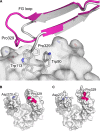
Disrupted FcγR and C1q binding sites in IgG4. (A) In all IgG1-Fc/FcγR complex structures, a hydrophobic ‘proline sandwich’ interaction forms between Pro329 from the IgG Cγ2 domain FG loop and two tryptophan residues from the receptor. The interaction between IgG1-Fc and FcγRIIIa is shown in gray (58). In IgG4-Fc (pink), the unique Cγ2 FG loop conformation would disrupt this conserved interaction (18). (B) Residues from IgG1-Fc (55) which are important for C1q binding are colored according to a model for the interaction between IgG1-Fc and C1q (164). The positions of Asp270 and Pro329 are indicated. (C) In IgG4-Fc (18), the positions of C1q binding residues are altered. The figure was prepared with PyMOL (167).
Fig. 10
Fig. 10
The IgG4 Cγ2 FG loop conformation is unique. The IgG1 Cγ2 FG loop (white) (50), IgE Cε3 FG loop (pink) (82), IgY Cυ3 FG loop (salmon) (153), IgM Cμ3 FG loop (yellow) (160), and IgA Cα2 FG loop (light green) (156) adopt a conserved conformation. The IgG4 Cγ2 FG loop conformation (dark green) (18), which contains a single proline residue at position 329, is unique. Residue numbering is according to the Protein Data Bank entry for each structure. The figure was prepared with PyMOL (167).
Fig. 11
Fig. 11
Deglycosylated IgG4-Fc crystal structure. (A) Two IgG4-Fc molecules (blue/yellow and green/pink) form an interlocked arrangement. (B) The Cγ2 domain FG loop forms crystal packing interactions and in the molecule colored pink, adopts the conserved conformation found in IgG1. The figure was prepared with PyMOL (167).
Fig. 12
Fig. 12
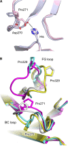
Cγ2 domain loop conformations. (A) In the IgG1-Fc/FcγRIIIa complex (light blue) (58), the conserved Cγ2 BC loop conformation precludes hydrogen bond formation between Asp270 (IgG1 Cγ2 BC loop) and a histidine residue from the receptor. In one IgG1-Fc/FcγRI complex (pink) (55), Pro271 isomerization alters the conformation of the BC loop, permitting hydrogen bond formation. (B) Four different combinations of Cγ2 BC and FG loop conformations are possible: yellow – conserved BC and FG loop, found in non-receptor-bound human IgG1 (e.g. (50)), blue – conserved BC loop and unique FG loop, found in deglycosylated IgG4-Fc (19), gray – non-conserved BC loop and conserved FG loop, found in one IgG1-Fc/FcγRI complex (55), purple – non-conserved BC loop and unique FG loop, found in IgG4-Fc (18). The figure was prepared with PyMOL (167).
Fig. 13
Fig. 13
Potential interactions between IgG4-Fc and FcγRs. (A) The IgG4 Cγ2 domain FG loop (pink) (18) disrupts the hydrophobic proline sandwich. The Pro329 carbonyl oxygen atom and the Ser330 side chain could form hydrogen bonds, indicated by black lines, with Arg102 from FcγRI (white) (55). Of the human IgG subclasses, Ser330 is unique to IgG4. (B) The conserved Cγ2 BC loop conformation found in IgG1 (light blue) (55) precludes hydrogen bond formation between Asp270 and His148 from FcγRI. The Cγ2 BC loop conformation in IgG4 (pink) (18), in which Pro271 undergoes a cis/trans isomerization, would enable Asp270 to form a hydrogen bond with His148 from FcγRI (white) (55). In one FcγRI complex structure, Pro271 from the IgG1 Cγ2 BC loop also undergoes a cis/trans isomerization (55). (C) The same cis/trans isomerization would enable Asp270 from the IgG4 Cγ2 BC loop (pink) (18) to form a salt bridge with Arg134 from FcγRIIb (white) (65). (D) In the IgG1-Fc/FcγRIIIa complex (white) (58), Glu269 from the Cγ2 BC loop forms a hydrogen bond with Lys131. Gln268 from the IgG4 Cγ2 BC loop (pink) (18) would also be able to form a hydrogen bond with Lys131. The figure was prepared with PyMOL (167).

References

    1. Aalberse RC, Stapel SO, Schuurman J, Rispens T. Immunoglobulin G4: an odd antibody. Clin Exp Allergy. 2009;39:469–477. - PubMed
    1. Aalberse RC, Schuurman J. IgG4 breaking the rules. Immunology. 2002;105:9–19. - PMC - PubMed
    1. van der Zee JS, van Swieten P, Aalberse RC. Inhibition of complement activation by IgG4 antibodies. Clin Exp Immunol. 1986;64:415–422. - PMC - PubMed
    1. van der Zee JS, van Swieten P, Aalberse RC. Serologic aspects of IgG4 antibodies. II. IgG4 antibodies form small, nonprecipitating immune complexes due to functional monovalency. J Immunol. 1986;137:3566–3571. - PubMed
    1. Salfeld JG. Isotype selection in antibody engineering. Nat Biotechnol. 2007;25:1369–1372. - PubMed

Publication types

MeSH terms