Multicenter Study
. 2015 Dec 29;6(42):44254-73.
doi: 10.18632/oncotarget.5471.

FN14 and GRP94 expression are prognostic/predictive biomarkers of brain metastasis outcome that open up new therapeutic strategies

Affiliations
Multicenter Study

FN14 and GRP94 expression are prognostic/predictive biomarkers of brain metastasis outcome that open up new therapeutic strategies

Antonio Martínez-Aranda et al. Oncotarget. .

Abstract

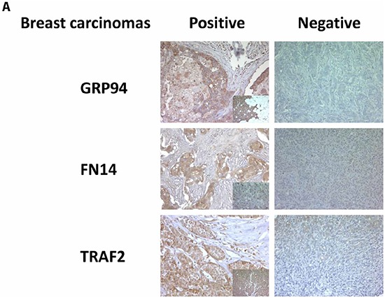
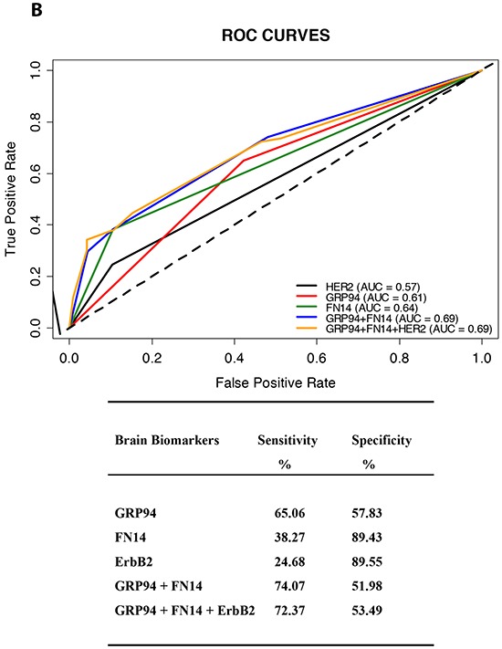
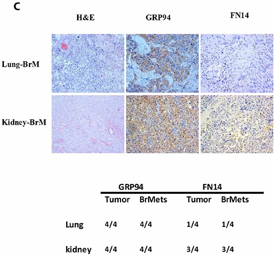
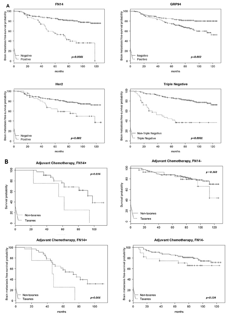
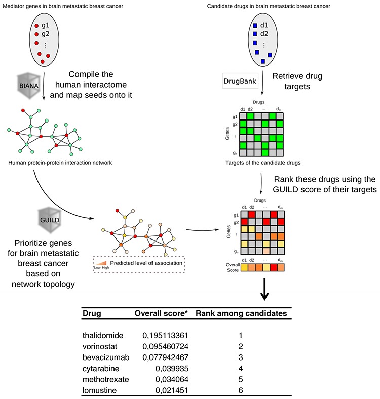
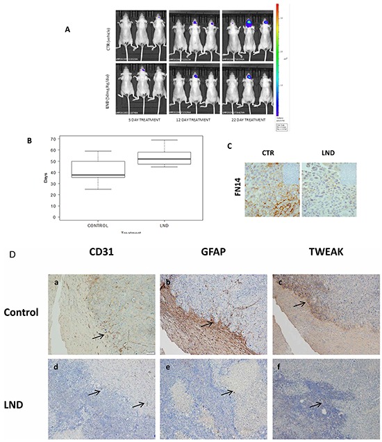
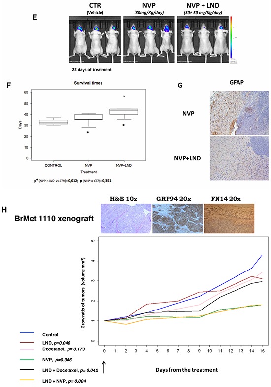
Brain metastasis is a devastating problem in patients with breast, lung and melanoma tumors. GRP94 and FN14 are predictive biomarkers over-expressed in primary breast carcinomas that metastasized in brain. To further validate these brain metastasis biomarkers, we performed a multicenter study including 318 patients with breast carcinomas. Among these patients, there were 138 patients with metastasis, of whom 84 had brain metastasis. The likelihood of developing brain metastasis increased by 5.24-fold (95%CI 2.83-9.71) and 2.55- (95%CI 1.52-4.3) in the presence of FN14 and GRP94, respectively. Moreover, FN14 was more sensitive than ErbB2 (38.27 vs. 24.68) with similar specificity (89.43 vs. 89.55) to predict brain metastasis and had identical prognostic value than triple negative patients (p < 0.0001). Furthermore, we used GRP94 and FN14 pathways and GUILD, a network-based disease-gene prioritization program, to pinpoint the genes likely to be therapeutic targets, which resulted in FN14 as the main modulator and thalidomide as the best scored drug. The treatment of mice with brain metastasis improves survival decreasing reactive astrocytes and angiogenesis, and down-regulate FN14 and its ligand TWEAK. In conclusion our results indicate that FN14 and GRP94 are prediction/prognosis markers which open up new possibilities for preventing/treating brain metastasis.

Keywords: FN14; GRP94; biomarkers; brain metastasis; breast cancer.


Conflict of interest statement

CONFLICTS OF INTEREST

Angels Sierra has received research funding from Celgene Research, S. L.

Figures

Figure 1
Figure 1. GRP94 and FN14 expression predict brain metastasis progression in breast cancer patients
A. TMAs were used to identify the indicated proteins by IHC analysis in paraffin-embedded primary breast carcinomas (x 20). To score the positivity of the three proteins we considered samples with more than 70% of tumor positive cells with high levels of staining to represent positive control samples (small squares in the left column), ignoring samples with less intense staining or fewer positive cells. B. The area under the ROC curve obtained with the integrated predictive indexes. Markers were assessed in a multivariate logistic regression model using a forward stepwise procedure to identify the best combination for predicting brain metastasis. The area under the ROC curve obtained for ErbB2 alone (AUC = 0.57), for GRP94 (AUC = 0.61), FN14 (AUC = 0.64) and the combination of GRP94 and FN14 (AUC = 0.69) and for ErbB2, GRP94 and FN14 (AUC = 0.69), is represented in the upper part of the figure. The sensitivity and specificity of the markers are shown in the lower part of the figure, indicating the most specific GRP94 and the most sensitive FN14, which was similar to ErbB2 in terms of sensitivity and specificity. C. Expression of GRP94 and FN14 in four pairs tumor/BrM of lung and clear cell kidney carcinomas. Representative IHC of BrM are showed in the upper part of the figure and at the bottom the relation of positive samples of each protein.
Figure 2
Figure 2. Kaplan-Meier survival estimates of brain metastasis-free survival among patients
A. According to the expression of FN14 (total N = 308, events N = 81), GRP94 (total N = 313, events N = 83), ErbB2 (total N = 297, events N = 77) and TNBC (total N = 301, events N = 77). The p values were obtained from the log-rank test. B. Kaplan-Meier survival estimates of overall free survival (both upper panels) and brain metastasis-free survival (both bottom panels) among patients who received chemoadjuvant therapy, with or without taxanes, according to the FN14 expression in tumors.
Figure 3
Figure 3. Modeling personalized therapies to prevent brain metastases using GRP94 and FN14 pathways as targets
Workflow chart describing the process of prioritizing genes involved in breast cancer brain metastasis based on network topology, retrieval of drugs from the DrugBank and ranking of these drugs using the GUILD score to determine the best treatment. The list of targets prioritized is shown at the bottom, indicating that thalidomide is the best drug to target FN14.
Figure 4
Figure 4. Experimental brain metastasis models to validate the therapeutic effect of a thalidomide derivative
Lenalidomide, 50 mg/Kg/day, alone or in combination with Docetaxel 15 mg/Kg/day and NVP-AUY922 (NVP) 30 mg/Kg/day. Docetaxel was administered every 4 days for 2 weeks and NVP-AUY922 every 2 days for 2 weeks. A. Representative images show bioluminescence in animals at day 5, 12 and 22 from the start of LND treatment 50 mg/Kg/day every day (Celgene Corporation, Summit, NJ). B. The survival time (box plot) of mice treated with LND (N = 8) or with vehicle (Control, N = 8) was evaluated using the log-rank test (p = 0.0802) and the differences observed in the survival rate between the two groups (treated or not with LND) are represented. C. IHC analysis in paraffin-embedded experimental brain metastasis (x 20) shows the downregulation of FN14 in mice treated with LND. D. IHC analysis in paraffin-embedded experimental brain metastasis (x 20). Representative figures showing the upfront of the metastasis invading the brain tissue in controls (upper part of the figure). Each protein expressed is indicated. CD31 a. GFAP b. and TWEAK c. Decreased angiogenesis d. few reactive astrocytes e. and downregulation of TWEAK f. indicated with arrows in tissues from mice treated with LND with regard to controls (lower part of the figure). E. Representative images show bioluminescence in animals at day 22 from the start of indicated treatments. The bioluminescence data were transformed using the log(1+x) function (where x = AvR), in order to obtain a more regular and positive distribution. Subsequently, these data were normalized by subtracting the first observation (day 14) from each of the following observations. F. The survival time (box plot) of mice treated with NVP-AUY922 (N = 6) alone or in combination with LND (N = 6) are shown. The differences in mouse survival were statistically significant in NVP-AUY922 + LND vs. CTR (p = 0.012) and NVP-AUY922 alone vs NVP-AUY922 + LND (p = 0.033); but the survival of mice treated only with NVP-AUY922 and controls was similar (p = 0.351). G. IHC of brain metastasis from mice treated with NVP-AUY922 alone and in convination with LND. The expression of GFAP indicative of reactive astrocytes is higher in NVP-AUY922 + LDN group than in brain metastasis from mice treated with NVP-AUY922 alone. H. Athymic Nude-Foxn1nu female mice. The xenograft expression of GRP94 and FN14 is shown in the upper part, in tumoral cells (GRP94) and in tumor and estromal cells (FN14). The statistical differences between groups of treatment (*): control treated with vehicle (N=17), Docetaxel (N=10), LND (N=16), NVP (N=15), NVP+LND (N=9) and Docetaxel+LND (N=20) according the volume slope are indicated at the bottom of the figure.

References

    1. Tham YL, Sexton K, Kramer R, Hilsenbeck S, Elledge R. Primary breast cancer phenotypes associated with propensity for central nervous system metastases. Cancer. 2006;107:696–704. - PubMed
    1. Stemmler HJ, Heinemann V. Central nervous system metastases in HER-2-overexpressing metastatic breast cancer: a treatment challenge. Oncologist. 2008;13:739–750. - PubMed
    1. Matthias P, Capper D, Ilhan-Mutlu AI, Berghoff AS, Birner P, Bartsch R, Marosi C, Zielinski C, Mehta MP, Winkler F, Wick W, von Deimling A. Brain metastases: pathobiology and emerging targeted therapies. Acta Neuropathol. 2012;123:205–222. - PubMed
    1. Soffietti R, Trevisan E, Ruda R. Targeted therapy in brain metastasis. Curr. Opin. Oncol. 2012;24:679–686. - PubMed
    1. Lockman PR, Mittapalli RK, Taskar KS, Rudraraju V, Gril B, Bohn KA, Adkins CE, Roberts A, Thorsheim HR, Gaasch JA, Huang S, Palmieri D, Steeg PS, et al. Heterogeneous blood-tumor barrier permeability determines drug efficacy in experimental brain metastases of breast cancer. Clin Cancer Res. 2010;16:5664–5678. - PMC - PubMed

Publication types

MeSH terms