. 2015 Oct 24:14:182.
doi: 10.1186/s12943-015-0456-4.

Activin and TGFβ use diverging mitogenic signaling in advanced colon cancer

Affiliations

Activin and TGFβ use diverging mitogenic signaling in advanced colon cancer

Jessica Bauer et al. Mol Cancer. .

Abstract

Background: Understanding cell signaling pathways that contribute to metastatic colon cancer is critical to risk stratification in the era of personalized therapeutics. Here, we dissect the unique involvement of mitogenic pathways in a TGFβ or activin-induced metastatic phenotype of colon cancer.

Method: Mitogenic signaling/growth factor receptor status and p21 localization were correlated in primary colon cancers and intestinal tumors from either AOM/DSS treated ACVR2A (activin receptor 2) -/- or wild type mice. Colon cancer cell lines (+/- SMAD4) were interrogated for ligand-induced PI3K and MEK/ERK pathway activation and downstream protein/phospho-isoform expression/association after knockdown and pharmacologic inhibition of pathway members. EMT was assessed using epithelial/mesenchymal markers and migration assays.

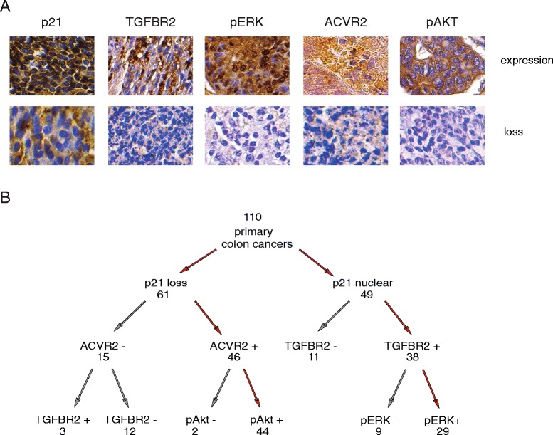
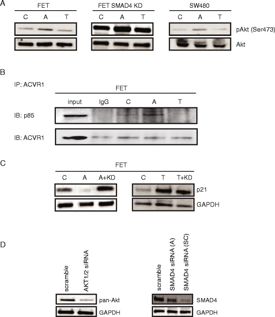
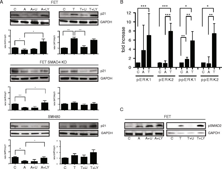
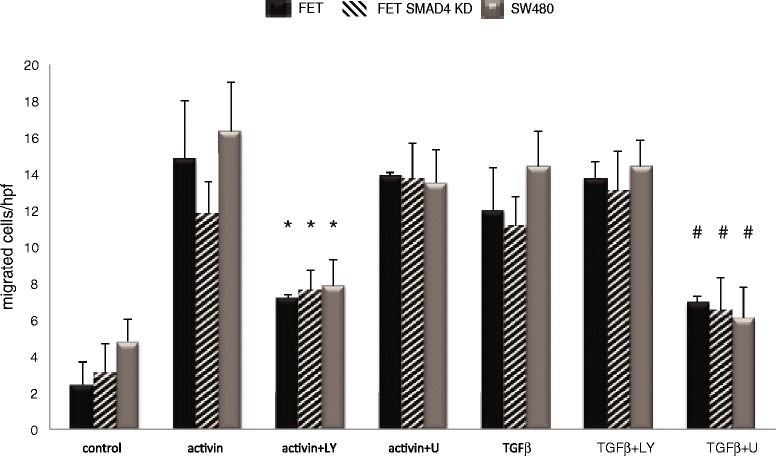
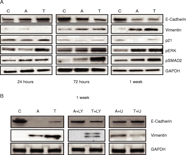
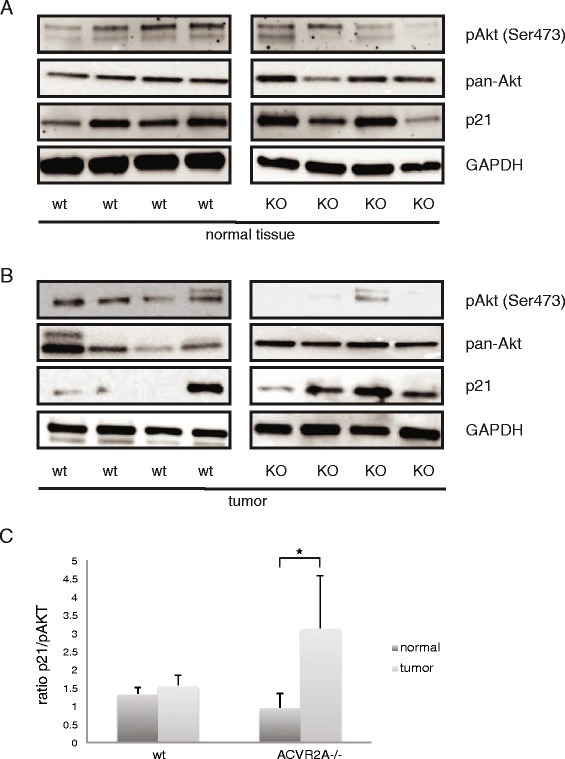
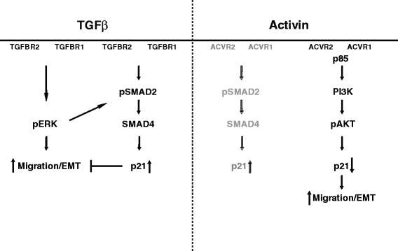
Results: In primary colon cancers, loss of nuclear p21 correlated with upstream activation of activin/PI3K while nuclear p21 expression was associated with TGFβ/MEK/ERK pathway activation. Activin, but not TGFβ, led to PI3K activation via interaction of ACVR1B and p85 independent of SMAD4, resulting in p21 downregulation. In contrast, TGFβ increased p21 via MEK/ERK pathway through a SMAD4-dependent mechanism. While activin induced EMT via PI3K, TGFβ induced EMT via MEK/ERK activation. In vivo, loss of ACVR2A resulted in loss of pAkt, consistent with activin-dependent PI3K signaling.

Conclusion: Although activin and TGFβ share growth suppressive SMAD signaling in colon cancer, they diverge in their SMAD4-independent pro-migratory signaling utilizing distinct mitogenic signaling pathways that affect EMT. p21 localization in colon cancer may determine a dominant activin versus TGFβ ligand signaling phenotype warranting further validation as a therapeutic biomarker prior to targeting TGFβ family receptors.


Figures

Fig. 1
Fig. 1
Loss of nuclear p21 is indicative of activin/pAkt activation, while nuclear p21 is associated with TGFβ/pERK activation in primary colon cancer tissues. In human colon cancer tissue, loss of nuclear p21 is associated with ACVR2A/pAkt expression while nuclear p21 is associated with TGFBR2/pERK expression. A total of 110 primary human colon cancer tissues were stained for ACVR2A/TGFBR2/p21/pERK/pAkt expression and pathway expression associations were determined. a Representative colon cancer tissues showing nuclear (p21/pERK) or cytosolic (TGFBR2/ACVR2A/pAkt) expression (upper panel) versus loss (lower panel). b Schematic of colon cancer signaling pathways based on signal component staining. Dominant pathways with p21 expression as diverging point drawn in red (p21 loss/ACVR2A+/pAkt + and p21 expression/TGFBR2+/pERK+, respectively). For statistical analysis, see Table 1
Fig. 2
Fig. 2
Activin but not TGFβ utilizes PI3K/Akt to downregulate p21 in a SMAD4-independent manner. a Activin but not TGFβ leads to PI3K activation in a SMAD4-independent manner. ACVR2A/TGFBR2 wild type FET cells, FET with SMAD4 knockdown, and the SMAD4-null colon cancer cell line SW480 were stimulated with activin or TGFβ for 24 h following serum starvation. pAkt level was determined by Western Blot. Akt was used as a loading control. pAkt increased after cells were treated with activin but not TGFβ. b ACVR1B, ACVR2A’s primary binding partner, interacts with p85, the regulatory subunit of PI3K in an activin-dependent manner. Co-immunoprecipitation (Co-IP) with ACVR2A/TGFBR2 wild type FET cells were used to detect a protein-protein interaction between ACVR1B and p85. c p21 downregulation is dependent on Akt, a PI3K downstream target. ACVR2A/TGFBR2 wild type FET cells were transfected with siRNA to Akt1/2 (KD) and treated with activin or TGFβ for 24 h following serum starvation. Activin-induced downregulation of p21 was abrogated after Akt1/2 knockdown implicating Akt in activin-induced p21 regulation. d Knock down of downstream target in FET cell. ACVR2A/TGFBR2 wild type FET cells were transfected with siRNA Akt1/2 and siRNA SMAD4. Resulting loss of respective protein expression is shown using Western blotting. For siRNA SMAD4 we tested two different siRNA from Ambion (A: middle panel) and Santa Cruz (SC: right panel) and the latter was used in all our experiments. (C control; A Activin; T TGFβ; KD siRNA Akt1/2; IP immunoprecipitation)
Fig. 3
Fig. 3
TGFβ but not activin stabilizes p21 via SMAD4 and MEK/ERK. a TGFβ-induced p21 upregulation is MEK/ERK dependent and not influenced by PI3K inhibition. In contrast, activin-induced p21 downregulation is dependent on PI3K signaling, but not dependent on SMAD4. ACVR2A/TGFBR2 wild type FET cells, FET with SMAD4 knockdown, and the SMAD4-null colon cancer cell line SW480 were treated with activin or TGFβ for 24 h 30 min after pharmacologic inhibition of PI3K (LY 290042) or MEK1/2 inhibition (U0126) and p21 levels determined by Western blot. GAPDH was used as a loading control. Three independent experiments entered the densitometric analysis shown below the representative blots (*p < 0.05, **p < 0.01, ***p < 0.001). For simplicity, we only show level of significance between control versus activin; control versus TGFβ; and activin versus LY and TGFβ versus U. b TGFβ treatment prominently increases the phosphorylation of ERK1/2 in colon cancer cells. Isoelectric point immunoassay was performed in FET cells treated with activin or TGFβ and pERK1/2 and ppERK1/2 compared to vehicle control. Five independent experiments were performed (*p < 0.05, **p < 0.01, ***p < 0.001). c TGFβ utilizes MEK/ERK to induce pSMAD2. ACVR2A/TGFBR2 wild type FET cells were treated with activin, TGFβ or vehicle control for 24 h following pretreatment with PI3K and MEK/ERK inhibition. pSMAD2 was determined by Western blot analysis and GAPDH was used as a loading control. Activin-induced pSMAD2 increase is independent from PI3K and MEK/ERK, but inhibition of MEK/ERK abolishes TGFβ-induced upregulation of pSMAD2 (A + LY Activin + LY; A + U Activin + U0126; T + LY TGFβ + LY; T + U TGFβ + U0126)
Fig. 4
Fig. 4
Activin and TGFβ use distinct mitogenic signaling to affect SMAD4- independent migration. Activin and TGFβ are both inducers of migration independent of SMAD4. ACVR2A/TGFBR2 wild type FET cells, FET with SMAD4 knockdown, and the SMAD4-null colon cancer cell line SW480 were seeded in transwell plates, serum starved, and pretreated with pharmacological inhibitors of PI3K (LY 290042) or MEK (U0126). Activin and TGFβ-induced migration is SMAD4-independent. Activin-induced migration is decreased in the absence of PI3K signaling and TGFβ-induced migration is MEK/ERK dependent. Inserts show representative fields with migrated FET cells. Graph shows data from 4 independently performed experiments (*p < 0.05 Activin versus Activin + LY; #p < 0.05 TGFβ versus TGFβ + U0126)
Fig. 5
Fig. 5
Activin and TGFβ induce epithelial to mesenchymal transition via distinct mitogenic signaling and downregulation of p21. a Both TGFβ and activin treatment induce a decrease of E-Cadherin and an increase in vimentin, indicative of EMT while p21 is downregulated. TGFβ-induced p21 upregulation normalizes over time while activin induced downregulation of p21 persists. ACVR2AsTGFBR2 wild type FET colon cancer cells were treated with activin or TGFβ and lysed after 24 h, 72 h or 1 week, respectively. EMT was determined using E-Cadherin as an epithelial and vimentin as a mesenchymal marker. p21, pERK, pSMAD2 during activin and TGFβ-induced EMT were also interrogated. Phosphorylation of SMAD2 and ERK increased over time after TGFβ treatment. b Inhibition of PI3K following activin and inhibition of MEK following TGFβ treatment leads to a decrease in EMT. FET cells were pretreated with PI3K (LY 290042) or MEK (U0126) prior to stimulation with activin or TGFβ and lysed after one week. EMT was determined using E-Cadherin as an epithelial marker. Representative blots of 3 independent experiments are shown
Fig. 6
Fig. 6
Loss of ACVR2A in vivo is associated with pAkt downregulation in intestinal tumors. Loss of ACVR2A leads to pAkt downregulation and p21 upregulation in ACVR2A knockout KO mice. ACVR2A wild type (wt) and ACVR2A KO mice were used in a DSS/AOM intestinal cancer model. Mice were injected intraperitoneally with AOM and after 5 days were given 3 cycles of DSS. Lysates from normal (a) and 10.1186/s12943-015-0456-4 intestinal tumor tissue (b) were probed for p21, pan-Akt, and pAkt expression via Western blot. Tumor and normal tissue from four different mice from each group of ACVR2A wt and ACVR2A KO mice is shown. c Loss of ACVR2A is associated with a decrease in pAkt and an increase of p21, which is most pronounced in intestinal tumor tissue. Immunobloy signal was quantified by densitometry and the ratio of p21 to pAKT expression was determined
Fig. 7
Fig. 7
Parallel activin and TGFβ signaling in advanced colon cancer. TGFβ and activin both share SMAD4 signaling to upregulate p21. However, in colon cancer, there is ligand-specific SMAD4-independent signaling utilizing distinct mitogenic signaling. Moreover, activin dominantly induces downregulation of p21 via PI3K/Akt signaling over early SMAD4-dependent p21 upregulation in colon cancer (non-dominant pathway indicated in grey). Net nuclear p21 expression in colon cancer may be a functional surrogate of intact TGFβ/SMAD growth suppression and a negative possible predictor of growth enhancing response to TGFβ pathway inhibition. In contrast, colon cancers with loss of nuclear p21 may benefit from activin, TGFβ or combination inhibitory therapy. In summary, there is complex parallel signaling with feedback loops operative in colon cancer downstream of activin and TGFβ. In order to predict net functional effects of targeted pathway disruption on tumor behavior, it is crucial that the interplay of pathways is fully appreciated to minimize unwanted side effects

Similar articles

Cited by

References

    1. SEER Stat Fact Sheets: Colon and Rectum Cancer 2014. Available from: (http://seer.cancer.gov/statfacts/html/colorect.html).
    1. Vogelstein B, Papadopoulos N, Velculescu VE, Zhou S, Diaz LA, Jr, Kinzler KW. Cancer genome landscapes. Science. 2013;339(6127):1546–58. doi: 10.1126/science.1235122. - DOI - PMC - PubMed
    1. Massague J. TGFbeta signalling in context. Nat Rev Mol Cell Biol. 2012;13(10):616–30. doi: 10.1038/nrm3434. - DOI - PMC - PubMed
    1. Loomans HA, Andl CD. Intertwining of activin a and TGFb signaling: dual roles in cancer progression and cancer cell invasion. Cancers. 2014;7:70–91. doi: 10.3390/cancers7010070. - DOI - PMC - PubMed
    1. Principe DR, Doll JA, Bauer J, Jung B, Munshi HG, Bartholin L, et al. TGF-beta: duality of function between tumor prevention and carcinogenesis. J Natl Cancer Inst. 2014;106(2):djt369. doi: 10.1093/jnci/djt369. - DOI - PMC - PubMed

Publication types

MeSH terms

Substances