Review
. 2016 Jan;49(1):26-36.
doi: 10.5483/BMBRep.2016.49.1.165.

Myocardial tissue engineering using electrospun nanofiber composites

Affiliations
Review

Myocardial tissue engineering using electrospun nanofiber composites

Pyung-Hwan Kim et al. BMB Rep. 2016 Jan.

Abstract

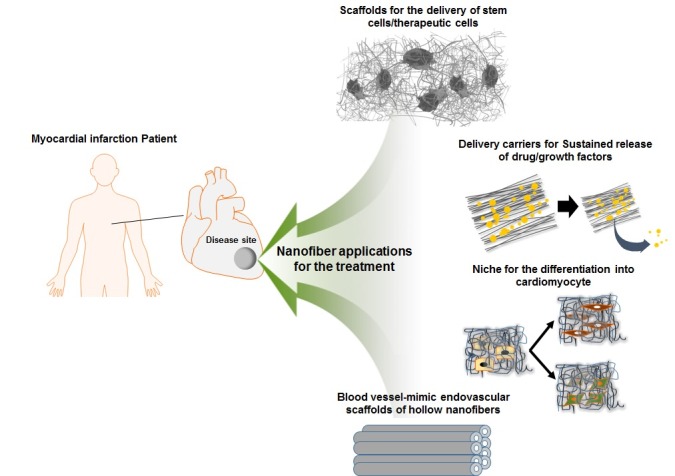
Emerging trends for cardiac tissue engineering are focused on increasing the biocompatibility and tissue regeneration ability of artificial heart tissue by incorporating various cell sources and bioactive molecules. Although primary cardiomyocytes can be successfully implanted, clinical applications are restricted due to their low survival rates and poor proliferation. To develop successful cardiovascular tissue regeneration systems, new technologies must be introduced to improve myocardial regeneration. Electrospinning is a simple, versatile technique for fabricating nanofibers. Here, we discuss various biodegradable polymers (natural, synthetic, and combinatorial polymers) that can be used for fiber fabrication. We also describe a series of fiber modification methods that can increase cell survival, proliferation, and migration and provide supporting mechanical properties by mimicking micro-environment structures, such as the extracellular matrix (ECM). In addition, the applications and types of nanofiber-based scaffolds for myocardial regeneration are described. Finally, fusion research methods combined with stem cells and scaffolds to improve biocompatibility are discussed.


Figures

Fig. 1.
Fig. 1.. Schematic representation of applicable strategies of multi-functional fiber-based scaffolds for myocardial regeneration.
Fig. 2.
Fig. 2.. Scheme of the electrospinning composed of major four parts diameter (A), the change of Taylor cone (B) and fiber diameter according to a broad range of voltage. The diameter of fiber fabricated by electrospinning is variously changed depending on the polymer concentration, voltage, flow rate, distance between capillary and collector, and solution conductivity (C).
Fig. 3.
Fig. 3.. Fiber surface modification for the generation of multi-functional nanofibers. (A) physical adsorption, (B) blend electrospinning, (C) coaxial electrospinning, and (D) surface-grafted modification by radiation, plasma, and chemical treatment for the formation of functional groups.

Similar articles

Cited by

References

    1. Ovize M, Thibault H, Przyklenk K. Myocardial conditioning: opportunities for clinical translation. Circ Res. (2013);113:439–450. doi: 10.1161/CIRCRESAHA.113.300764. - DOI - PubMed
    1. Jawad H, Ali NN, Lyon AR, Chen QZ, Harding SE, Boccaccini AR. Myocardial tissue engineering: a review. J Tissue Eng Regen Med. (2007);1:327–342. doi: 10.1002/term.46. - DOI - PubMed
    1. Finosh GT, Jayabalan M. Regenerative therapy and tissue engineering for the treatment of end-stage cardiac failure: new developments and challenges. Biomatter. (2012);2:1–14. doi: 10.4161/biom.19429. - DOI - PMC - PubMed
    1. Jugdutt BI. Ventricular remodeling after infarction and the extracellular collagen matrix: when is enough enough? Circulation. (2003);108:1395–1403. doi: 10.1161/01.CIR.0000085658.98621.49. - DOI - PubMed
    1. Kim HN, Jiao A, Hwang NS, et al. Nanotopographyguided tissue engineering and regenerative medicine. Adv Drug Deliv Rev. (2013);65:536–558. doi: 10.1016/j.addr.2012.07.014. - DOI - PMC - PubMed

Publication types