. 2015 Dec;89(Pt B):268-79.
doi: 10.1016/j.yjmcc.2015.10.022. Epub 2015 Oct 20.

A critical role of cardiac fibroblast-derived exosomes in activating renin angiotensin system in cardiomyocytes

Affiliations

A critical role of cardiac fibroblast-derived exosomes in activating renin angiotensin system in cardiomyocytes

Linmao Lyu et al. J Mol Cell Cardiol. 2015 Dec.

Abstract

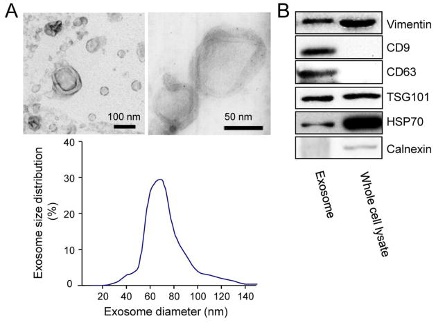
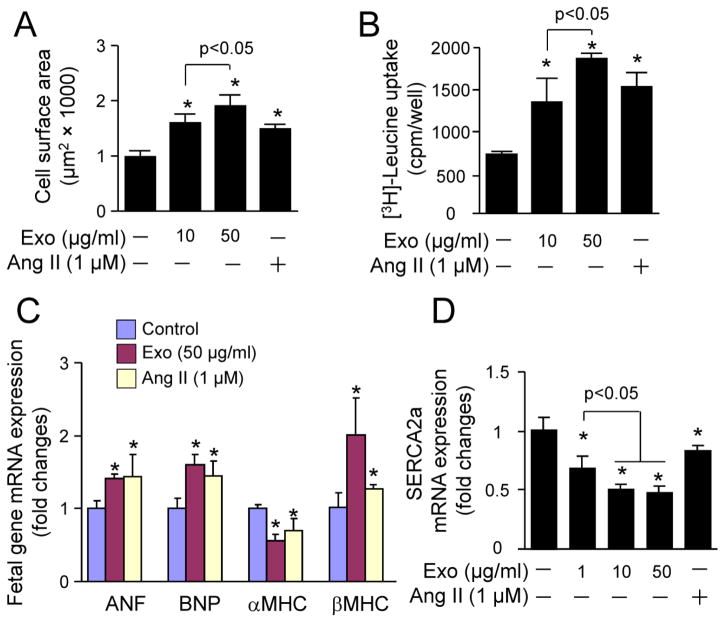
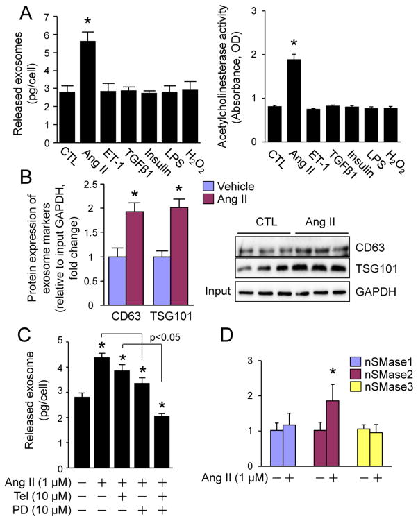
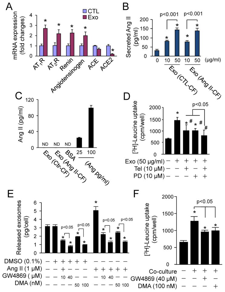
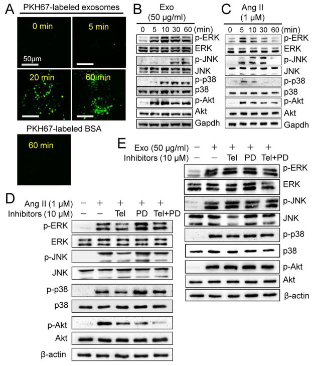
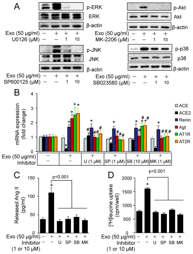
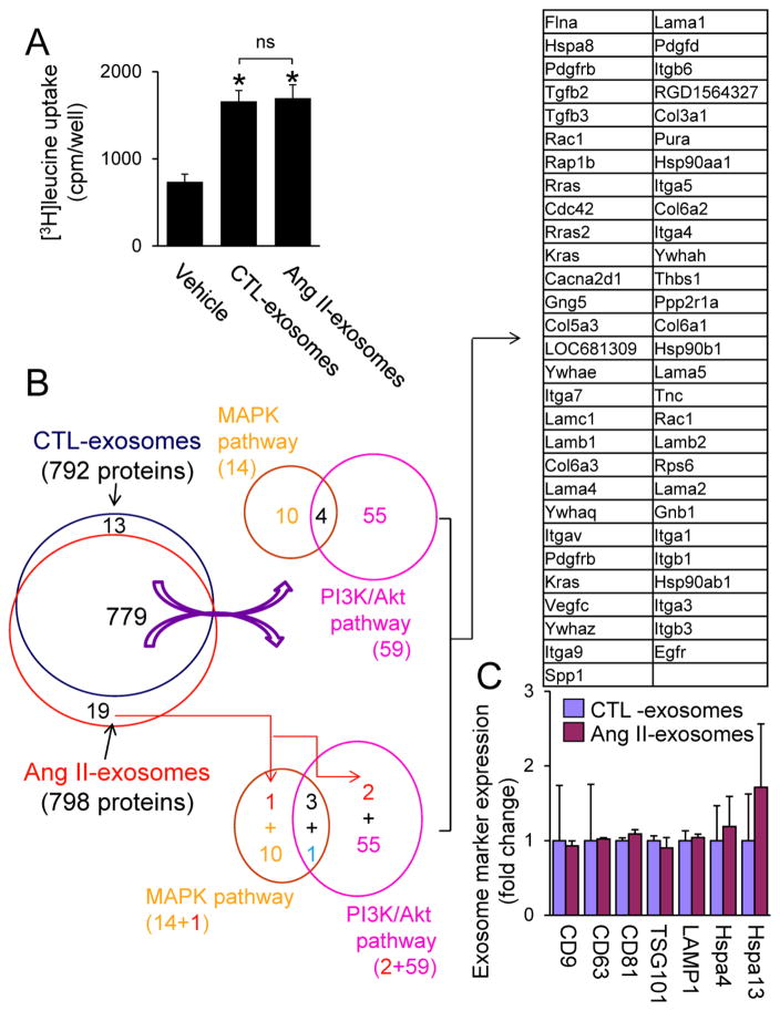
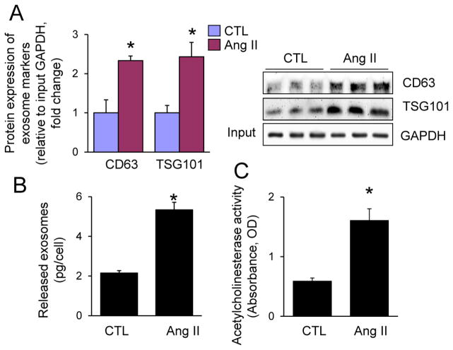
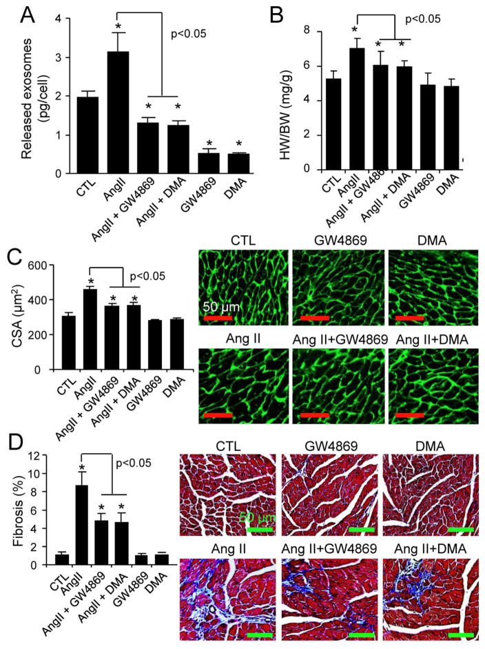
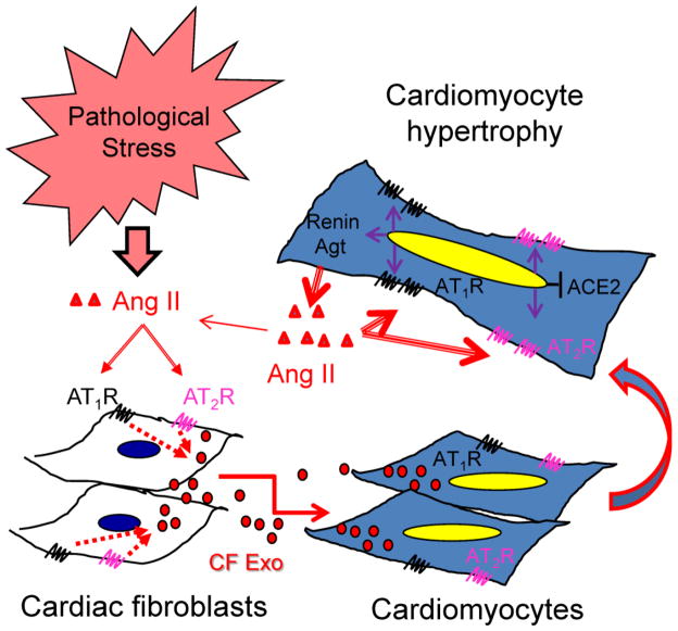
Chronic activation of the myocardial renin angiotensin system (RAS) elevates the local level of angiotensin II (Ang II) thereby inducing pathological cardiac hypertrophy, which contributes to heart failure. However, the precise underlying mechanisms have not been fully delineated. Herein we report a novel paracrine mechanism between cardiac fibroblasts (CF)s and cardiomyocytes whereby Ang II induces pathological cardiac hypertrophy. In cultured CFs, Ang II treatment enhanced exosome release via the activation of Ang II receptor types 1 (AT1R) and 2 (AT2R), whereas lipopolysaccharide, insulin, endothelin (ET)-1, transforming growth factor beta (TGFβ)1 or hydrogen peroxide did not. The CF-derived exosomes upregulated the expression of renin, angiotensinogen, AT1R, and AT2R, downregulated angiotensin-converting enzyme 2, and enhanced Ang II production in cultured cardiomyocytes. In addition, the CF exosome-induced cardiomyocyte hypertrophy was blocked by both AT1R and AT2R antagonists. Exosome inhibitors, GW4869 and dimethyl amiloride (DMA), inhibited CF-induced cardiomyocyte hypertrophy with little effect on Ang II-induced cardiomyocyte hypertrophy. Mechanistically, CF exosomes upregulated RAS in cardiomyocytes via the activation of mitogen-activated protein kinases (MAPKs) and Akt. Finally, Ang II-induced exosome release from cardiac fibroblasts and pathological cardiac hypertrophy were dramatically inhibited by GW4869 and DMA in mice. These findings demonstrate that Ang II stimulates CFs to release exosomes, which in turn increase Ang II production and its receptor expression in cardiomyocytes, thereby intensifying Ang II-induced pathological cardiac hypertrophy. Accordingly, specific targeting of Ang II-induced exosome release from CFs may serve as a novel therapeutic approach to treat cardiac pathological hypertrophy and heart failure.

Keywords: Angiotensin II; Cardiac hypertrophy; Exosomes; Heart failure.


Figures

Fig. 1
Fig. 1
Characterization of cardiac fibroblasts-derived exosomes. (A) Upper panel: Transmission electron microscopic images of neonatal rat cardiac fibroblast-derived (CF) exosomes; Lower panel: Distribution of CF exosome sizes. (B) Western blot analysis of several exosome biomarkers in CF exosomes. Immunoblots are representative of six separate experiments.
Fig. 2
Fig. 2
The effect of cardiac fibroblast-derived exosomes on cardiomyocyte hypertrophy. Neonatal rat cardiomyocytes were treated with neonatal rat cardiac fibroblasts-derived exosomes (Exo) or angiotensin II (Ang II) as indicated for 48 h and then subjected to (A) cell surface area measurement, (B) [3H]-Leucine uptake, (C and D) qPCR analysis of gene expression. n=4, *p<0.05 vs. vehicle treated control (−).
Fig. 3
Fig. 3
Ang II-induced release of exosomes from cardiac fibroblasts. (A, B) The effect of pathological stress on the release of exosomes from cardiac fibroblasts. (A) The amount of proteins and the activity of acetylcholinesterase in total released exosomes from the same number of neonatal rat cardiac fibroblasts that were stimulated with vehicle, Ang II (1 μM), ET-1 (0.1 μM), TGFβ1 (10 ng/ml), insulin (0.1 μM), LPS (0.1 μg/ml), H2O2 (500 μM) as indicated for 48 h. n=3, *p<0.05 vs. vehicle-treated control (CTL). (B) Western blot analysis of CD63 and TSG101 in total exosomes from the same number of neonatal rat cardiac fibroblasts that were stimulated with vehicle or Ang II for 48 h. Left panel shows semi-quantified results. n=3, *p<0.05 vs. vehicle-treated control (CTL). Right panel is the immunoblots (n=3). Input: 10 μl of the neonatal rat cardiac fibroblast lysates. (C) The effect of Telmisartan and PD123319 on Ang II-induced secretion of exosomes from cardiac fibroblasts. Neonatal rat cardiac fibroblasts were treated with Ang II (1 μM), Telmisartan (Tel, 10 μM), PD123319 (PD, 10 μM) as indicated for 48 h. The total proteins of exosomes secreted were measured by BCA assay and normalized by cell number. n=3, *p<0.05 vs. vehicle treated control (−). (D) The effect of Ang II on mRNA expression of neutral sphingomyelinase (nSMase) in cardiac fibroblasts. Neonatal rat cardiac fibroblasts were treated with vehicle of Ang II (1 M) for 48 h and subjected to qPCR analysis of nSMase1, 2, and 3 expression. n=4, *p<0.05 vs. vehicle-treated control (−).
Fig. 4
Fig. 4
The effects of cardiac fibroblast-derived exosomes on activation of renin angiotensin system (RAS) in cardiomyocytes. (A) Cardiac fibroblast-derived exosomes (Exo)-induced expression of RAS components. Neonatal rat cardiomyocytes were treated with or without Exo (50 μg/ml) for 48 h and then subjected to qPCR analysis of mRNA expression of AT1R, AT2R, ACE, ACE2, and angiotensinogen. n=4, *p<0.05 vs. vehicle-treated control (CTL). (B) Measurement of Ang II in culture medium of cardiomyocytes treated with Exo. Neonatal cardiomyocytes were treated with or without exosomes derived from neonatal rat cardiac fibroblasts treated with (Ang II-CF) or without Ang II (CTL-CF) for 48 h and then the culture medium were subjected to enzyme immunoassay (EIA) of Ang II. n=4, *p<0.05 vs. vehicle treated control (0). (C) Measurement of Ang II in cardiac fibroblast-derived exosomes (Exo). Lysates (50 μg) of exosomes derived from neonatal rat cardiac fibroblasts treated with (Ang II-CF) or without Ang II (CTL-CF) were subjected to EIA analysis of Ang II as indicated, and 25–100 pg/ml Ang II was used as a positive control. Bovine serum albumin (BSA, 50 μg) was used as a negative control; ND - non-detectable. n=4. (D) The effects of Telmisartan (Tel) and PD123319 (PD) on cardiac fibroblast-derived exosomes (Exo)-induced [3H]-Leucine uptake in cardiomyocytes. Neonatal rat cardiomyocytes were treated with Exo (50 μg/ml), Tel (10 μM), and PD (10 μM) as indicated for 48 h. n=4, *p<0.05 vs. control (−); #p<0.05 vs. Exo (+). (E) The effect of GW4869 and DMA on exosome release from cardiac fibroblasts. Neonatal rat cardiac fibroblasts were treated with GW4869 and DMA as indicated for 48 h and the culture medium was subjected to exosome isolation and quantification. n=4, *p<0.05 vs. vehicle-treated control (−). (F) The effect of GW4869 and DMA on cardiac fibroblast-induced [3H]-Leucine uptake in cardiomyocytes. Neonatal rat cardiac myocytes and fibroblasts were co-cultured with GW4869 and DMA as indicated for 48 h. n=4, *p<0.05 vs. vehicle-treated control (−).
Fig. 5
Fig. 5
The effects of cardiac fibroblast-derived exosomes on pro-hypertrophic signaling in cardiomyocytes. (A) Exosomes uptake assay. Cardiac fibroblast-derived exosomes were labeled with green membrane dye PKH67 and incubated with neonatal rat cardiomyocytes as indicated and visualized using a confocal microscopy. The results are representatives of 3 separated experiments. Scale bar = 50 μm. (B) Exosome (Exo)-induced activation of mitogen-activated protein kinases (MAPKs) and Akt. Neonatal rat cardiomyocytes were treated with (5, 10, 30 and 60 min) and without (0 min) Exo (50 μg/ml) derived from neonatal rat cardiac fibroblasts as indicated and then subjected to Western blot analysis of phosphorylated and total ERK1/2, JNK, p38, and Akt. The results are representatives of 4 separated experiments. (C) Ang II-induced activation of MAPKs and Akt. Neonatal rat cardiomyocytes were treated with (5, 10, 30 and 60 min) or without (0 min) Ang II (1 μM) as indicated and then subjected to Western blot analysis of phosphorylated and total ERK1/2, JNK, p38, and Akt. The results are representatives of 4 separated experiments. (D) The effects of Telmisartan (Tel) and PD123319 (PD) on Exo-induced activation of MAPKs and Akt. Neonatal rat cardiomyocytes were treated with Exo, Tel (10 μM), and PD (10 μM) as indicated for 20 min and subjected to Western blot analysis. The immunoblots are representatives of 4 separated experiments. (E) The effects of Tel and PD on Ang II-induced activation of MAPKs and Akt. Neonatal rat cardiomyocytes were treated with Ang II, Tel (10 μM), and PD (10 μM) as indicated for 5 min and subjected to Western blot analysis. The immunoblots are representatives of 4 separate experiments.
Fig. 6
Fig. 6
The effects of MAPK and Akt inhibitors on cardiac fibroblast-derived exosomes-induced activation of RAS in cardiomyocytes. (A) Dose response of MAPK and Akt inhibitors on cardiac fibroblast-derived exosomes (Exo)-induced activation of MAPKs and Akt. Neonatal rat cardiomyocytes were treated with or without Exo (50 μg/ml), U0126, SP600125, MK-2206, and SB023580 for 20 min and subjected to Western blot analysis. The results are from 4 separate experiments. (B) The effects of MAPK and Akt inhibitors on Exo-induced mRNA expression of RAS components. Neonatal rat cardiomyocytes were treated with or without Exo (50 μg/ml), U0126 (U, 1 μM), SP600125 (SP, 1 μM), SB023580 (SB, 10 μM), and MK-2206 (MK, 1 μM) for 48 h and then subjected to qPCR analysis of mRNA expression of ACE, ACE2, renin, angiotensinogen (Agt), AT1R, and AT2R. n=4, *p<0.05 vs. Control (−). #p<0.05 vs. Exo (+) group. (C) The effects of MAPK and Akt inhibitors on Exo-induced Ang II release. Neonatal rat cardiomyocytes were treated with or without Exo (50 μg/ml), U0126 (U, 1 μM), SP600125 (SP, 1 μM), SB023580 (SB, 10 μM), and MK-2206 (MK, 1 μM) for 48 h. Ang II was measured in the culture medium. n=4, *p<0.05 vs. Control (−). (D) The effects of MAPK and Akt inhibitors on Exo-induced [3H]-Leucine uptake. Neonatal rat cardiomyocytes were treated with or without Exo (50 μg/ml), U0126 (U, 1 μM), SP600125 (SP, 1 μM), SB023580 (SB, 10 μM), and MK-2206 (MK, 1 μM) for 48 h. n=4, *p<0.05 vs. Control (−).
Fig. 7
Fig. 7
Protein array analysis of cardiac fibroblast (CF)-derived exosomes. A, The effect of released exosomes of control vehicle-treated (CTL-exosomes) and Ang II-treated neonatal cardiac fibroblasts (CFs) (Ang II-exosomes) on cardiomyocyte hypertrophy. Neonatal rat cardiomyocytes were treated with or without CTL-exosomes (50 μg/ml) and Ang II-exosomes (50 μg/ml) for 48 h and then subjected to [3H]leucine uptake assay. n=4, *p<0.05 vs. vehicle control. B, C, Exosome proteins extracted from CTL-exosomes (20 μg) and Ang II-exosomes (20 μg) (n=3) were subjected to proteomics analysis as described in Online Supplement. B, The effect of Ang II on overall distribution of CF exosome proteins which potentially regulate PI3K/Akt and MAPK pathways. Ang II downregulates one protein (blue) and upregulates 2 proteins (red) in CF exosomes (Supplementary tables 6–8). C, The effect of Ang II on CF exosome marker expression in CTL- and Ang II-exosomes. There is no difference in the expression of exosome marker proteins in CTL- and Ang II-exosomes. Hspa4; Heat shock 70 kDa protein 4, Hspa13; Heat shock 70 kDa protein 13.
Fig. 8
Fig. 8
Ang II-induced exosome release from mouse adult cardiac fibroblasts. Mouse adult cardiac fibroblasts were treated with vehicle control (CTL) or Ang II (1 μM) for 48 h and then subjected to (A) Western blot analysis of CD63 and TSG101, (B) protein quantity measurement, and (C) acetylcholinesterase activity assay. n=3, *p<0.05 vs. CTL.
Fig. 9
Fig. 9
The effects of GW4869 and DMA on Ang II-induced cardiac hypertrophy. A, Adult male C57BL/6N mice (n=10) at 8 weeks of age were treated with Ang II, GW4869, and DMA for 5 days as described in “Methods”, and then cardiac fibroblasts were isolated for measuring exosome release. *p<0.05 vs. vehicle-treated control (CTL). B–D, Adult male C57BL/6N mice (n=6~10) at 8 weeks of age were treated with Ang II, GW4869, and DMA for 14 days as described in “Methods”, and then (B) heart weight (HW)/body weight (BW) ratio, (C) cardiomyocyte cross sectional area (CSA), and (D) cardiac fibrosis were quantified. n=6~10. *p<0.05 vs. vehicle-treated control (CTL).
Fig. 10
Fig. 10
Schematic working hypothesis. Under pathological stress, myocardial Ang II stimulates exosome release from cardiac fibroblasts via activation of both AT1R and AT2R and then the cardiac fibroblast-derived exosomes upregulate expression of renin, angiotensinogen, AT1R, and AT2R while downregulating ACE2 in cardiomyocytes in a paracrine manner. Thus, cardiomyocytes produce and release more Ang II, which in turn activates the upregulated AT1R and AT2R to induce hypertrophic growth in an autocrine fashion. As a result, Ang II intensifies its own pro-hypertrophic signaling in cardiomyocytes by upregulation of RAS via increasing paracrine release of exosomes from cardiac fibroblasts.

Similar articles

Cited by

References

    1. Wollert KC, Drexler H. The renin-angiotensin system and experimental heart failure. Cardiovascular research. 1999;43:838–49. - PubMed
    1. Balakumar P, Jagadeesh G. A century old renin-angiotensin system still grows with endless possibilities: AT1 receptor signaling cascades in cardiovascular physiopathology. Cell Signal. 2014;26:2147–60. - PubMed
    1. Aoki H, Richmond M, Izumo S, Sadoshima J. Specific role of the extracellular signal-regulated kinase pathway in angiotensin II-induced cardiac hypertrophy in vitro. Biochem J. 2000;347(Pt 1):275–84. - PMC - PubMed
    1. Gul R, Shawl AI, Kim SH, Kim UH. Cooperative interaction between reactive oxygen species and Ca2+ signals contributes to angiotensin II-induced hypertrophy in adult rat cardiomyocytes. Am J Physiol Heart Circ Physiol. 2012;302:H901–9. - PubMed
    1. Mathivanan S, Ji H, Simpson RJ. Exosomes: extracellular organelles important in intercellular communication. Journal of proteomics. 2010;73:1907–20. - PubMed

Publication types

MeSH terms