A novel prohibitin-binding compound induces the mitochondrial apoptotic pathway through NOXA and BIM upregulation
- PMID: 26497683
- PMCID: PMC4747186
- DOI: 10.18632/oncotarget.6154
A novel prohibitin-binding compound induces the mitochondrial apoptotic pathway through NOXA and BIM upregulation
Abstract
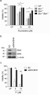
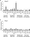
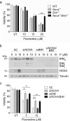
We previously described diaryl trifluorothiazoline compound 1a (hereafter referred to as fluorizoline) as a first-in-class small molecule that induces p53-independent apoptosis in a wide range of tumor cell lines. Fluorizoline directly binds to prohibitin 1 and 2 (PHBs), two proteins involved in the regulation of several cellular processes, including apoptosis. Here we demonstrate that fluorizoline-induced apoptosis is mediated by PHBs, as cells depleted of these proteins are highly resistant to fluorizoline treatment. In addition, BAX and BAK are necessary for fluorizoline-induced cytotoxic effects, thereby proving that apoptosis occurs through the intrinsic pathway. Expression analysis revealed that fluorizoline induced the upregulation of Noxa and Bim mRNA levels, which was not observed in PHB-depleted MEFs. Finally, Noxa(-/-)/Bim(-/-) MEFs and NOXA-downregulated HeLa cells were resistant to fluorizoline-induced apoptosis. All together, these findings show that fluorizoline requires PHBs to execute the mitochondrial apoptotic pathway.
Keywords: BCL-2 family members; apoptosis; cancer; mitochondria; prohibitins.
Conflict of interest statement
A.P-P., D.M.G-G., S.P., F.A., R.L., D.I-S., and J.G. patented the use of fluorinated thiazolines in the treatment of cancer. The remaining authors declare no conflict of interest.
Figures


Similar articles
-
AICAR induces Bax/Bak-dependent apoptosis through upregulation of the BH3-only proteins Bim and Noxa in mouse embryonic fibroblasts.Apoptosis. 2013 Aug;18(8):1008-16. doi: 10.1007/s10495-013-0850-6. Apoptosis. 2013. PMID: 23605481
-
NOXA upregulation by the prohibitin-binding compound fluorizoline is transcriptionally regulated by integrated stress response-induced ATF3 and ATF4.FEBS J. 2021 Feb;288(4):1271-1285. doi: 10.1111/febs.15480. Epub 2020 Jul 26. FEBS J. 2021. PMID: 32648994
-
The BCL-2 family members NOXA and BIM mediate fluorizoline-induced apoptosis in multiple myeloma cells.Biochem Pharmacol. 2020 Oct;180:114198. doi: 10.1016/j.bcp.2020.114198. Epub 2020 Aug 13. Biochem Pharmacol. 2020. PMID: 32798467
-
A review of the role of Puma, Noxa and Bim in the tumorigenesis, therapy and drug resistance of chronic lymphocytic leukemia.Cancer Gene Ther. 2013 Jan;20(1):1-7. doi: 10.1038/cgt.2012.84. Epub 2012 Nov 23. Cancer Gene Ther. 2013. PMID: 23175245 Review.
-
The BCL2 Family: Key Mediators of the Apoptotic Response to Targeted Anticancer Therapeutics.Cancer Discov. 2015 May;5(5):475-87. doi: 10.1158/2159-8290.CD-15-0011. Epub 2015 Apr 20. Cancer Discov. 2015. PMID: 25895919 Free PMC article. Review.
Cited by
-
Development of artesunate intelligent prodrug liposomes based on mitochondrial targeting strategy.J Nanobiotechnology. 2022 Aug 13;20(1):376. doi: 10.1186/s12951-022-01569-5. J Nanobiotechnology. 2022. PMID: 35964052 Free PMC article.
-
The prohibitin protein complex promotes mitochondrial stabilization and cell survival in hematologic malignancies.Oncotarget. 2017 Jul 1;8(39):65445-65456. doi: 10.18632/oncotarget.18920. eCollection 2017 Sep 12. Oncotarget. 2017. PMID: 29029444 Free PMC article.
-
The Prohibitin-Binding Compound Fluorizoline Activates the Integrated Stress Response through the eIF2α Kinase HRI.Int J Mol Sci. 2023 Apr 29;24(9):8064. doi: 10.3390/ijms24098064. Int J Mol Sci. 2023. PMID: 37175767 Free PMC article.
-
Loss of prohibitin 2 in Schwann cells dysregulates key transcription factors controlling developmental myelination.Glia. 2024 Dec;72(12):2247-2267. doi: 10.1002/glia.24610. Epub 2024 Aug 31. Glia. 2024. PMID: 39215540
-
Prohibitin ligands: a growing armamentarium to tackle cancers, osteoporosis, inflammatory, cardiac and neurological diseases.Cell Mol Life Sci. 2020 Sep;77(18):3525-3546. doi: 10.1007/s00018-020-03475-1. Epub 2020 Feb 15. Cell Mol Life Sci. 2020. PMID: 32062751 Free PMC article. Review.
References
-
- Hanahan D, Weinberg RA. Hallmarks of cancer: the next generation. Cell. 2011;144:646–74. - PubMed
-
- Pérez-Perarnau A, Preciado S, Palmeri CM, Moncunill-Massaguer C, Iglesias-Serret D, González-Gironès DM, Miguel M, Karasawa S, Sakamoto S, Cosialls AM, Rubio-Patiño C, Saura-Esteller J, Ramón R, Caja L, Fabregat I, et al. A Trifluorinated Thiazoline Scaffold Leading to Pro-apoptotic Agents Targeting Prohibitins. Angew Chemie Int Ed. 2014;53:10150–4. - PubMed
-
- Vousden KH, Lane DP. p53 in health and disease. Nat Rev Mol Cell Biol. 2007;8:275–83. - PubMed
-
- Artal-Sanz M, Tavernarakis N. Prohibitin and mitochondrial biology. Trends Endocrinol Metab. 2009;20:394–401. - PubMed
-
- Artal-Sanz M, Tavernarakis N. Prohibitin couples diapause signalling to mitochondrial metabolism during ageing in C. elegans. Nature. 2009;461:793–7. - PubMed
Publication types
MeSH terms
Substances
LinkOut - more resources
Full Text Sources
Other Literature Sources
Research Materials
Miscellaneous

