

Case Reports
doi: 10.5144/0256-4947.2015.321.
Multifocal giant cell reparative granuloma involving maxilla and mandible: a rare entity
Affiliations
- PMID: 26497714
- PMCID: PMC6074220
- DOI: 10.5144/0256-4947.2015.321
Item in Clipboard
Case Reports
Multifocal giant cell reparative granuloma involving maxilla and mandible: a rare entity
Ann Saudi Med.
2015 Jul-Aug.
Abstract
Giant cell reparative granuloma (GCRG) is a rare lesion that is a reactive process, not a true neoplasm. It was originally coined by Jaffe to describe lesions, which he believed were a response to intraosseous hemorrhage from jaw trauma. Regardless, GCRG is much more distinct from giant cell tumor (GCT) of bone, both histologically and clinically. We report a patient who presented with multiple facial swelling involving the facial skeleton that showed a multiloculated cystic appearance on CT involving the maxilla and mandible. The patient refused surgery, but after 6 months of follow up there was no progression.
Conflict of interest statement
The authors report no conflict of interest and no funding was received for this purpose.
Figures

Orthopantomogram showing a well-defined, multiloculated expansile, lytic lesion in left paramedian aspect of the body of the mandible with evidence of displacement of roots of adjacent teeth. There was no evidence of sclerosis, internal calcification or resorption of tooth, and the matrix appeared clear.

Contrast enhanced axial and coronal-CT findings shows an expansile multiloculated lesion involving the body and adjacent part of ramus of mandible on the left side with a heterogeneous pattern of enhancement.

Bone algorithm-correlating image demonstrates cortical thinning with a breech at the anterolateral aspect of the mandibular lesion with displacement of the roots of an adjacent tooth. No evidence of internal calcification/mineralization.

Contrast-enhanced axial and coronal-CT findings shows similar appearing lesions involving the superior alveolar arch of maxilla on either side of midline, with heterogeneous pattern of enhancement.
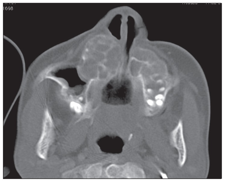
Bony algorithm of the maxillary lesions demonstrates cortical thinning giving a soap bubble appearance.

Photomicrograph shows solid, cellular proliferation of oval to spindle fibroblasts with no cellular pleomorphism and multinucleated giant cells scattered throughout these stromal cells (hematoxylin and eosin, ×10).

Photomicrograph shows spindle-shaped fibroblasts without atypia and scattered multinucleated giant cells (hematoxylin and eosin, ×40).
References
-
- Jaffe HL. Giant-cell reparative granuloma, traumatic bone cyst, and fibrous (fibrooseous) dysplasia of the jawbones. Oral Surg Oral Med Oral Pathol. 1953;6:159–75. - PubMed
-
- Ackerman LV, Spjut HJ. Giant cell reaction in tumors of bones and cartilage. In: Spjut HJ, Dorfman HD, Fechner RE, Ackerman LV, editors. Atlas of Tumor Pathology. Washington: Armed Forces Institute of Pathology; 1962. p. 82.
-
- Rosenthal L, Lisbona R. Radionuclide bone and joint imaging. In: Wilner D, editor. Radiology of bone tumors and allied disorders. Philadelphia, Pa: Saunders; 1982. pp. 4161–4220.
-
- Austin LT, Jr, Dahlin DC, Royer RQ. Giant cell reparative granuloma and related conditions affecting the jaws. Oral Surg. 1959;12:1285–1295. - PubMed
-
- Caskey PM, Wolf MD, Fechner RE. Multicentric giant cell reparative granuloma of the small bones of the hand. Clin Orthop. 1985;193:199–205. - PubMed
Publication types
MeSH terms
LinkOut - more resources
Full Text Sources
Other Literature Sources