

Preparation of Stabilizer-Free Silver Nanoparticle-Coated Micropipettes as Surface-Enhanced Raman Scattering Substrate for Single Cell Detection
- PMID: 26497732
- PMCID: PMC4620108
- DOI: 10.1186/s11671-015-1122-x
Preparation of Stabilizer-Free Silver Nanoparticle-Coated Micropipettes as Surface-Enhanced Raman Scattering Substrate for Single Cell Detection
Abstract
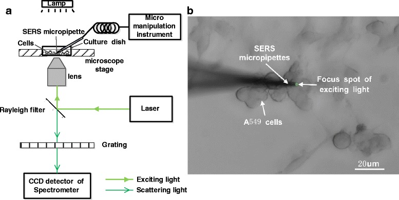
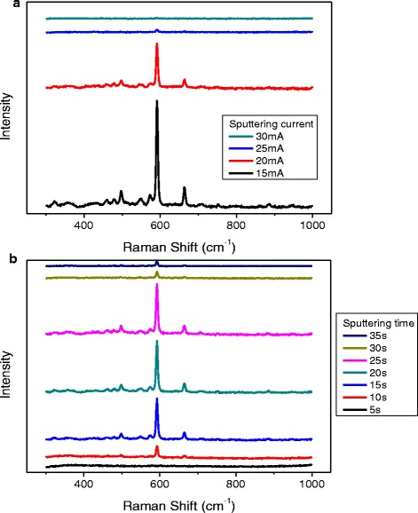
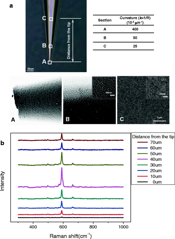
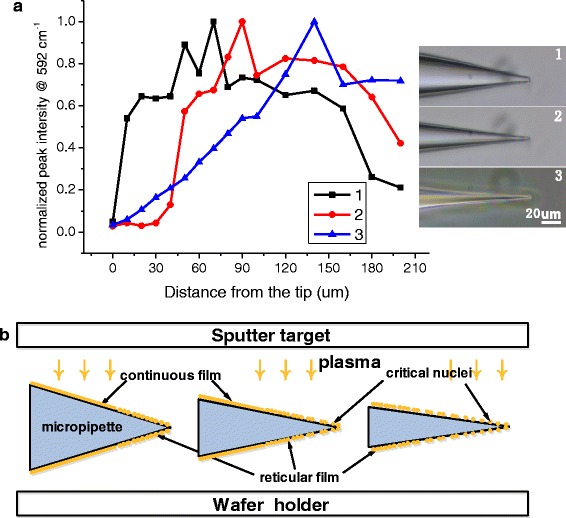
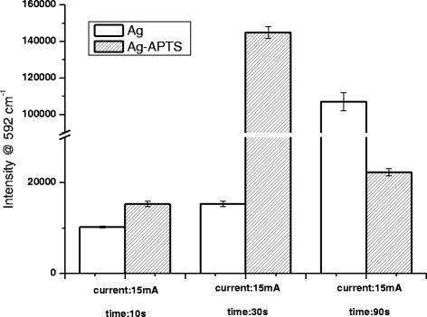
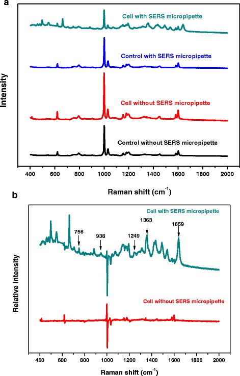
In this work, we established a convenient while reproduceable method for stabilizer-free silver nanoparticle (AgNP)-coated micropipettes by the combination of magnetron sputtering and surface coupling agent. The clear surfaces of the AgNPs are beneficial for absorbing biological or functional molecules on their surfaces. By optimizing the operating parameters, such as sputtering current and sputtering time, the tip of micropipettes coated with AgNPs exhibits excellent surface-enhanced Raman scattering (SERS) performance. Finally, the Raman spectra of a single A549 lung adenocarcinoma cell are successfully acquired by these advanced SERS-active micropipettes.
Keywords: Magnetron sputtering; Micropipette; SERS; Silver nanoparticles; Single cell detection.
Figures
Similar articles
-
An investigation of the surface enhanced Raman scattering (SERS) from a new substrate of silver-modified silver electrode by magnetron sputtering.Spectrochim Acta A Mol Biomol Spectrosc. 2007 Apr;66(4-5):994-1000. doi: 10.1016/j.saa.2006.05.012. Epub 2006 May 22. Spectrochim Acta A Mol Biomol Spectrosc. 2007. PMID: 16875867
-
Highly Sensitive Microarray Immunoassay for Multiple Mycotoxins on Engineered 3D Porous Silicon SERS Substrate with Silver Nanoparticle Magnetron Sputtering.Anal Chem. 2024 Feb 13;96(6):2425-2434. doi: 10.1021/acs.analchem.3c04359. Epub 2024 Jan 30. Anal Chem. 2024. PMID: 38291775
-
The role of adatoms in chloride-activated colloidal silver nanoparticles for surface-enhanced Raman scattering enhancement.Beilstein J Nanotechnol. 2018 Aug 22;9:2236-2247. doi: 10.3762/bjnano.9.208. eCollection 2018. Beilstein J Nanotechnol. 2018. PMID: 30202692 Free PMC article.
-
Synthesis of MBA-Encoded Silver/Silica Core-Shell Nanoparticles as Novel SERS Tags for Biosensing Gibberellin A3 Based on Au@Fe3O4 as Substrate.Sensors (Basel). 2019 Nov 25;19(23):5152. doi: 10.3390/s19235152. Sensors (Basel). 2019. PMID: 31775290 Free PMC article.
-
Fabrication of surface-enhanced Raman spectroscopy substrates using silver nanoparticles produced by laser ablation in liquids.Spectrochim Acta A Mol Biomol Spectrosc. 2023 Aug 5;296:122694. doi: 10.1016/j.saa.2023.122694. Epub 2023 Apr 5. Spectrochim Acta A Mol Biomol Spectrosc. 2023. PMID: 37030254 Review.
Cited by
-
A Novel SERS Substrate Platform: Spatially Stacking Plasmonic Hotspots Films.Nanoscale Res Lett. 2019 Mar 13;14(1):94. doi: 10.1186/s11671-019-2928-8. Nanoscale Res Lett. 2019. PMID: 30868395 Free PMC article.
-
Common Aspects Influencing the Translocation of SERS to Biomedicine.Curr Med Chem. 2018;25(35):4638-4652. doi: 10.2174/0929867325666180105101841. Curr Med Chem. 2018. PMID: 29303073 Free PMC article. Review.
References
-
- Lu X, Huang W, Wang Z, Cheng J. Recent developments in single-cell analysis. Anal Chim Acta. 2004;510(2):127–38. doi: 10.1016/j.aca.2004.01.014. - DOI
LinkOut - more resources
Full Text Sources
Other Literature Sources
Miscellaneous
