. 2015 Nov 10;6(35):37737-49.
doi: 10.18632/oncotarget.5648.

Host JDP2 expression in the bone marrow contributes to metastatic spread

Affiliations

Host JDP2 expression in the bone marrow contributes to metastatic spread

Yelena Barbarov et al. Oncotarget. .

Abstract

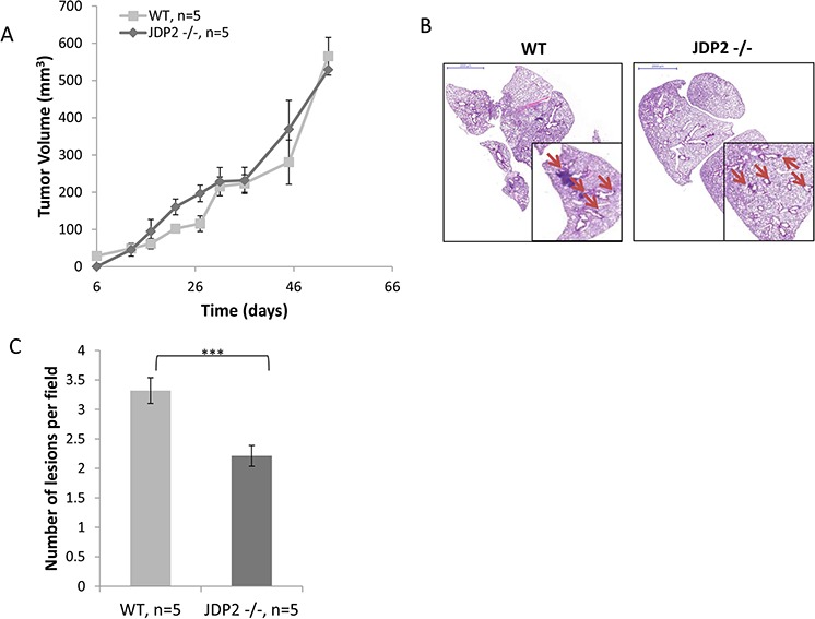
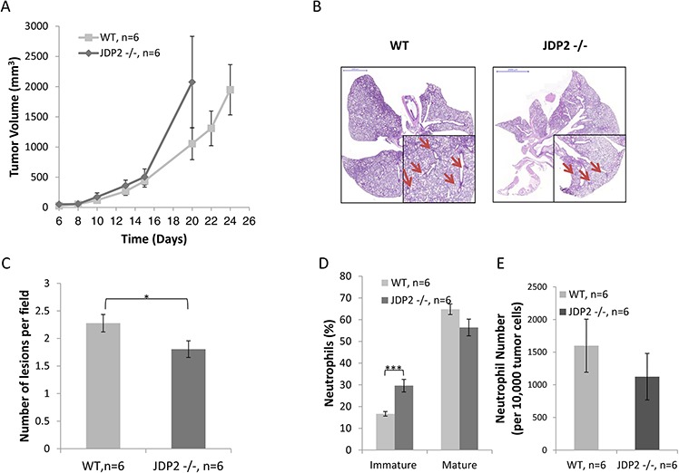
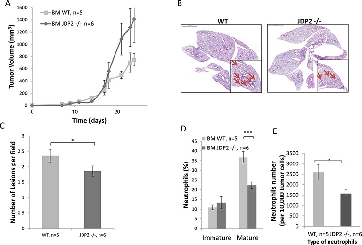
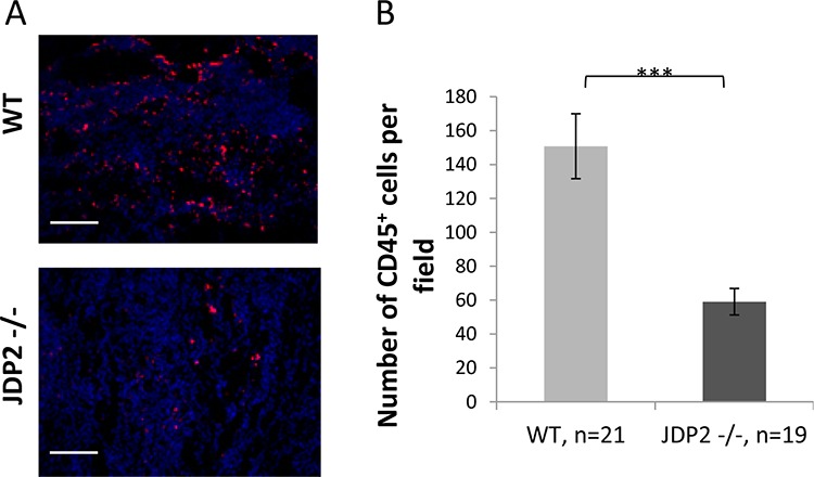
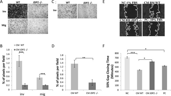
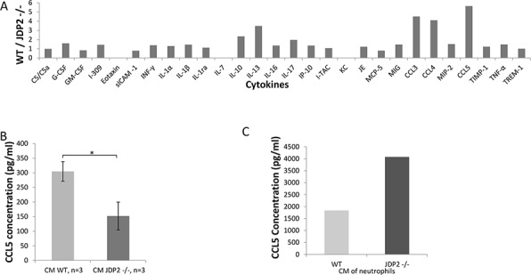
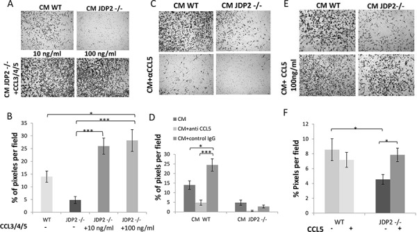
The c-Jun Dimerization Protein 2, JDP2, is a basic leucine zipper protein member of the activator protein-1 (AP-1) family of transcription factors. JDP2 typically suppresses gene transcription through multiple mechanisms and plays a dual role in multiple cellular processes, including cell differentiation and proliferation which is dependent on AP-1 function. Whereas the role of JDP2 expression within cancer cells has been studied, its role in stromal cells at the tumor microenvironment is largely unknown. Here we show that mice lacking JDP2 (JDP2-/-) display a reduced rate of metastasis in Lewis lung carcinoma (LLC) and polyoma middle T-antigen (PyMT) breast carcinoma mouse models. The replacement of wild-type bone marrow derived cells (BMDCs) with JDP2-deficient BMDCs recapitulates the metastatic phenotype of JDP2-/- tumor-bearing mice. In vitro, conditioned medium of wild-type BMDCs significantly potentiates the migration and invasion capacity of LLC cells as compared to that of JDP2-/- BMDCs. Furthermore, wild-type BMDCs secrete CCL5, a chemokine known to contribute to metastasis, to a greater extent than JDP2-/- BMDCs. The supplementation of CCL5 in JDP2-/- BMDC conditioned medium was sufficient to potentiate the invasion capacity of LLC. Overall, this study suggests that JDP2-expressing BMDCs within the tumor microenvironment contribute to metastatic spread.

Keywords: CCL5; JDP2; Lewis lung carcinoma; bone marrow derived cells; metastasis.


Conflict of interest statement

CONFLICTS OF INTEREST

The authors declare no conflicts of interest.

Figures

Figure 1
Figure 1. Host derived JDP2 expression promotes metastasis of mammary tumors
A. Six-to-eight week old female WT and JDP2 −/− mice were orthotopically implanted to the mammary fat pad with 2 × 10⁶ PyMT cells mixed with Matrigel, and tumor volume was monitored over time. B-C. When tumors reached an average volume of 600 mm3, mice were sacrificed and lungs were harvested. Lungs were embedded in paraffin, sectioned, and subsequently stained with H&E. Arrows indicate metastatic lesions. Scale bars = 2000 μm. Small micrographs are 2X magnification. B. The number of pulmonary metastatic lesions per field was quantified (n > 6/group) C.***, p < 0.001 of a two-tailed t-test.
Figure 2
Figure 2. Host derived JDP2 expression inhibits tumor growth but promotes metastasis
A. Six-to-eight week old wild-type (WT) and JDP2 −/− mice were subcutaneously implanted with 0.5 × 10⁶ LLC cells, and tumor volume was monitored over time. B–E. When tumors reached an average volume of 2000 mm3, mice were sacrificed and tumors and lungs were harvested. Lungs were embedded in paraffin, sectioned, and subsequently stained with H&E. Arrows indicate metastatic lesions Scale bars = 2000 μm. Small micrographs are 2X magnification. (B) The number of pulmonary metastatic lesions per field was quantified (C) Tumors were prepared as single cell suspensions and the percentage of mature (CD11b+Ly6C(low)Ly6G+) and immature (CD11b+Ly6C(high)Ly6G+) neutrophils (D), as well as the number of total neutrophils count per 10,000 cells (E), were determined by flow cytometry. *, p < 0.05; ***p < 0.001 of a two-tailed t test.
Figure 3
Figure 3. LLC-bearing mice harboring JDP2-deficient bone marrow exhibit reduced metastasis
A. Wild-type C57B1/6 mice were transplanted with BMDCs from either wild-type (WT) or JDP2−/− mice. The chimeric mice were then subcutaneously implanted with 0.5 × 10⁶ LLC cells, and tumor volume was monitored over time. B–E. At end point, tumors and lungs were excised. Lungs were embedded in paraffin, sectioned, and subsequently stained with H&E. Arrows indicate metastatic lesions. Scale bars = 2000 μm. Small micrographs are 2X magnification (B) The number of pulmonary metastatic lesions per field was quantified (C) Tumors were prepared as single cell suspensions and the percentage of mature (CD11b+Ly6C(low)Ly6G+) and immature (CD11b+Ly6C(high)Ly6G+) neutrophils (D), as well as the total number of neutrophils per 10,000 cells (E), were determined by flow cytometry. *, p < 0.05; ***p < 0.001 of a two-tailed t-test.
Figure 4
Figure 4. Wild-type BMDCs infiltrate into LLC tumors at a higher rate as compared with JDP2 −/− BMDCs
A. OCT fixed LLC tumor slices were stained for αCD45, a pan hematopoietic marker (red). Nuclei were stained by DAPI (blue). Scale bar = 200 μm B. The number of CD45+BMDCs per filed was counted. ***p < 0.001 of a two-tailed t-test.
Figure 5
Figure 5. Conditioned medium of JDP2-expressing BMDCs promotes invasion and migration
A–B. BMDCs of wild-type and JDP2−/− mice were cultured for 24 h to generate conditioned medium (CM). The migration and invasion properties of LLC cells were assessed in the presence of CM using the Boyden chamber assay. Cells invading (Inv) or migrating (Mig) through the membrane were stained with crystal violet and images were captured (A). The percentage of cell coverage per field was quantified (n > 8 fields/group) (B). C–D. The invasion properties of PyMT cells were assessed in the presence of CM derived from wild-type and JDP2−/− BMDC cultures by the modified Boyden chamber assay, as described in (A–B) E–F. PyMT cell motility was assessed by migration closure assay in the presence of CM derived from wild-type and JDP2−/− BMDC cultures. Serum-free and serum-rich medium were used as negative and positive controls (NC and PC), respectively. Gap closure was monitored over time. Representative images are shown when 50% of the gap was closed (E). The time necessary to achieve 50% gap closure is shown (n > 11/group) (F). *p < 0.05; **, p < 0.01; ***p < 0.001 of a one way ANOVA followed by Tukey post-hoc test when a comparison between more than two groups was performed, and unpaired student t-test when comparison between two groups was performed.
Figure 6
Figure 6. CCL5 is secreted by the JDP2-expressing BMDCs and is functionally important
A. A mini cytokine array was probed with conditioned medium derived from wild-type and JDP2 −/− BMDCs. The level of each cytokine was assessed by densitometry. Shown is the ratio of wild-type to JDP2−/− groups. B-C. The level of CCL5 in conditioned medium derived from wild-type and JDP2−/− BMDCs (n = 3 mice/group) B. or conditioned medium derived from isolated neutrophils (pooled sample from 6 mice/group) C. was determined by ELISA. *p < 0.05 assessed by two-tailed t-test.
Figure 7
Figure 7. CCL5 is necessary and sufficient for increased LLC cell invasion
A. The invasion properties of LLC cells were assessed by the Boyden chamber assay in the presence of JDP2 −/− BMDC CM supplemented with a mixture of purified CCL3, 4 and 5 (10 ng/ml and 100 ng/ml). C. CM from wild type or JDP2 −/− BMDCs in the presence of anti-CCL5 neutralizing antibodies or anti-cFos antibodies (as control) or E. CCL5 (100 ng/ml) added to the BMDC CM of either wild type or JDP2 −/− mice. The percentage of cell coverage per field was quantified (n > 8 fields/group) (B, D and F, respectively). *, p < 0.05; ***, p < 0.001 using one way ANOVA followed by a Tukey post-hoc test.

References

    1. Hanahan D, Weinberg RA. Hallmarks of cancer: the next generation. Cell. 2011;144:646–674. - PubMed
    1. Hanahan D, Coussens LM. Accessories to the crime: functions of cells recruited to the tumor microenvironment. Cancer cell. 2012;21:309–322. - PubMed
    1. Houghton AM. The paradox of tumor-associated neutrophils: fueling tumor growth with cytotoxic substances. Cell Cycle. 2010;9:1732–1737. - PubMed
    1. Mao Y, Keller ET, Garfield DH, Shen K, Wang J. Stromal cells in tumor microenvironment and breast cancer. Cancer Met. Rev. 2013;32:303–315. - PMC - PubMed
    1. Lewis CE, Pollard JW. Distinct role of macrophages in different tumor microenvironments. Cancer Res. 2006;66:605–612. - PubMed

Publication types

MeSH terms