Review
. 2015 Dec;40(12):765-778.
doi: 10.1016/j.tibs.2015.09.003. Epub 2015 Oct 21.

IFT-Cargo Interactions and Protein Transport in Cilia

Affiliations
Review

IFT-Cargo Interactions and Protein Transport in Cilia

Karl F Lechtreck. Trends Biochem Sci. 2015 Dec.

Abstract

The motile and sensory functions of cilia and flagella are indispensable for human health. Cilia assembly requires a dedicated protein shuttle, intraflagellar transport (IFT), a bidirectional motility of multi-megadalton protein arrays along ciliary microtubules. IFT functions as a protein carrier delivering hundreds of distinct proteins into growing cilia. IFT-based protein import and export continue in fully grown cilia and are required for ciliary maintenance and sensing. Large ciliary building blocks might depend on IFT to move through the transition zone, which functions as a ciliary gate. Smaller, freely diffusing proteins, such as tubulin, depend on IFT to be concentrated or removed from cilia. As I discuss here, recent work provides insights into how IFT interacts with its cargoes and how the transport is regulated.

Keywords: diffusion; dynein; flagella; intraflagellar transport; kinesin-2; microtubule; tubulin.


Figures

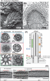
Figure 1
Figure 1. Cilia and intraflagellar transport (IFT)
(A) Scanning electron micrograph and (B) transmission electron micrograph showing multiciliated epithelial cells from mouse airway. (C - E) Cross-sections through cilia of C. reinhardtii (left) and mouse ependymal cilia (right) showing 9+2 axonemes (C), Y-shaped linkers (D), a conserved structural element of the transition zone, and the transitional fibers (E), which link the basal bodies to the plasma membrane and are the putative assembly sites of IFT trains (shown in G). Arrowheads in C mark IFT trains. (F) Schematic presentation of a cilium and IFT trains. The colored arrowheads indicate the level of the cross-sections shown in (C - E). (G) Longitudinal sections through IFT trains in mouse ependymal cilia and C. reinhardtii flagella. Arrowheads mark the periodicities of long and short IFT trains. Bar = 100 nm. The images of C. reinhardtii IFT trains are a curtesy of Dr. Gaia Pagino, MPI Dresden.
Figure 2
Figure 2. The IFT building blocks
IFT-A and IFT-B proteins will assemble into IFT-A and IFT-B subcomplexes; the proteins are believed to be represented in equimolar amounts in each complex. IFT complexes assemble into IFT particles and IFT particles associate with the IFT motors into IFT trains. The BBSome, consisting of at least 7 BBS proteins and BBIP10, appears to be a substoichiometric component of the IFT trains. Peptide coverage in the proteome of whole C. reinhardtii cilia suggests a 10:5:1 ratio for IFT-B/IFT motors, IFT-A, and BBSomes, respectively [2, 22]. Anterograde IFT trains are moved by kinesin-2, which is thought to be associated to IFT-B. IFT dynein is moved as a cargo on anterograde IFT trains to the ciliary tip. Retrograde IFT trains are powered by IFT dynein. How IFT dynein, here depicted as being associated with IFT-A, binds to IFT trains is currently unknown.
Figure 3
Figure 3. Cargo transport by IFT
A) Kymogram (time-distance plot) showing the movement of the axonemal protein DRC4 in a Chlamydomonas cilium as recorded by total internal reflection fluorescence (TIRF) microscopy. The position of the ciliary tip and base are indicated. A trajectory running from the bottom left to the top right indicates transport to the ciliary tip; the slope represents transport velocity. The kymogram depicts the phases of cargo delivery into cilia: DRC4-GFP first moves by IFT(open arrow) to the ciliary tip (arrowhead with * marks the arrival at the tip); after a ~ 2-second dwell time the protein begins to diffuse (arrowhead ‘unloading’) until it docks to the axoneme (arrowhead ‘docking’). Reprinted in modified form from reference (doi: 10.1016/j.cub.2013.10.044). B) Kymograms from two-color TIRF imaging showing DRC4-GFP (middle and green) and IFT20-mCherry (top and red) of a partially bleached cilium. Anterograde trains are marked by filled arrowheads, open arrowheads mark retrograde trains. The two trains marked by arrows carry DRC4-GFP as a cargo. While one cargo is transported to the ciliary tip, the other one is released prematurely along the ciliary shaft (dashed circle) followed by an extended time of diffusion. C) IFT loading and the regulation of ciliary length. The model suggests that cells are able to measure the length of their cilia. Cilia that fall short of a set length will trigger a two-pronged response of increasing the amount of cargo carried by IFT trains and putatively suppressing ciliary disassembly. Cilia that exceed the set length of cilia will suppress cargo loading onto IFT and activate a ciliary disassembly pathway [102]. Since growing and non-growing cilia can be present on the same cell body, IFT loading and ciliary disassembly appear to be regulated locally within each basal body-cilium entity. Tubulin dimers, as an example of a cargo, are indicated by grey and green dots.
Figure 4
Figure 4. Cargo binding by IFT
(A) Schematic presentation of the IFT-B core complex. The crystal structures of partial and complete IFT proteins are included where available and the putative cargo binding sites are indicated. Note that the exact positions of IFT27/25 and IFT52/46 on the C-terminal part of IFT74/81 are unknown. Reprinted in modified form from reference [50] with permission of the authors and journal. ©1967 Taschner et al. Journal of Cell Biology. 207:269-282. doi:10.1083/jcb.201408002. (B) Tubulin binding site. The N-terminal domain of IFT81 forms a CH domain, which binds tubulin dimers. This interaction is stabilized by the N-terminal domain of IFT74, which is positively charged and might interact with the glutamate-rich C-terminal domain of β-tubulin [53]. (C) ODA binding site. Cells expressing an N-terminally truncated IFT46 fail to assemble ODAs onto the axoneme. Mutants in ODA16p have a similar phenotype and ODA16p interacts with IFT46 suggesting that ODA16p stabilizes the interaction of IFT46 and ODAs allowing them to travel on IFT trains [87, 88]. (D) BBSome binding site. Mutant analysis revealed that IFT25/27 are required for the export of BBSomes from cilia suggesting that these IFT proteins form a BBSome binding site. LZTF1 accumulates in Ift27−/− cilia but not those of Bbs−/− mutants and might mediate BBSome binding to IFT27/27. BBS3/Arl6 binds to BBS1 and recruits BBSomes to membranes [69, 103]. The BBSome might function as a cargo adapter allowing various transmembrane and membrane-associated proteins to attach to IFT for ciliary import or export [23, 49].

References

    1. Gealt MA, Adler JH, Nes WR. The sterol and fatty acids from purified flagella of Chlamydomonas reinhardtii. Lipids. 1981;16:133–136.
    1. Pazour GJ, Agrin N, Leszyk J, Witman GB. Proteomic analysis of a eukaryotic cilium. The Journal of cell biology. 2005;170:103–113. - PMC - PubMed
    1. Ostrowski LE, Blackburn K, Radde KM, Moyer MB, Schlatzer DM, Moseley A, Boucher RC. A proteomic analysis of human cilia: identification of novel components. Molecular & cellular proteomics : MCP. 2002;1:451–465. - PubMed
    1. Johnson KA, Rosenbaum JL. Polarity of flagellar assembly in Chlamydomonas. The Journal of cell biology. 1992;119:1605–1611. - PMC - PubMed
    1. Rosenbaum JL, Child FM. Flagellar regeneration in protozoan flagellates. The Journal of cell biology. 1967;34:345–364. - PMC - PubMed

Publication types

LinkOut - more resources