

Lymphocyte senescence in COPD is associated with decreased histone deacetylase 2 expression by pro-inflammatory lymphocytes
- PMID: 26498345
- PMCID: PMC4619495
- DOI: 10.1186/s12931-015-0287-2
Lymphocyte senescence in COPD is associated with decreased histone deacetylase 2 expression by pro-inflammatory lymphocytes
Abstract
Background: Histone acetyltransferases (HAT) and histone deacetylases (HDAC) are enzymes that upregulate and down-regulate pro-inflammatory gene transcription respectively. HDAC2 is required by corticosteroids to switch off activated inflammatory genes and is reduced in lung macrophages in COPD. We have shown that COPD patients have increased steroid resistant CD28null (senescent) pro-inflammatory T and NKT-like peripheral blood cells (particularly CD8+ subsets) and we hypothesized that these changes would be associated with a loss of HDAC2 from these senescent pro-inflammatory lymphocytes.
Methods: Blood was collected from 10 COPD and 10 aged-matched controls. Intracellular pro-inflammatory cytokines, IFNγ and TNFα, and expression of CD28, HDAC2 and HAT, were determined in lymphocyte subsets in the presence of ± 5 mg/ml theophylline (HDAC2 activator), 10 μM prednisolone and 2.5 ng/ml cyclosporine A (immunosuppressant), using flow cytometry.
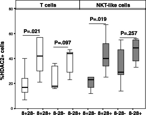
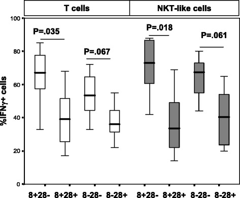
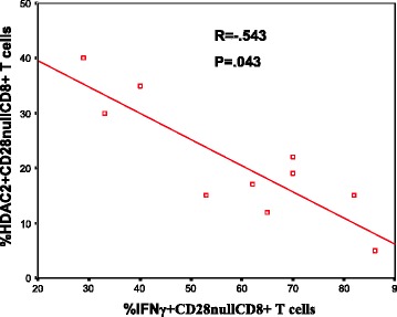
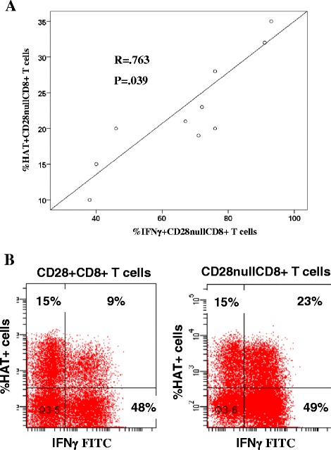
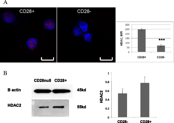
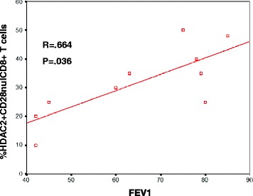
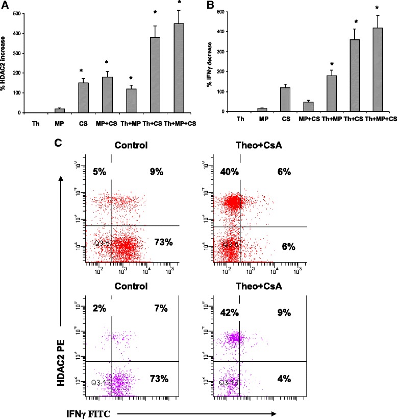
Results: There was a loss of HDAC2 from CD28null CD8+ T and NKT-like cells in COPD. There was a significant negative correlation between HDAC2 expression and the percentage of CD28null CD8+ T and NKT-like cells producing IFNγ or TNFα in all subjects (eg, COPD: R = -.763, p < 0.001 for T-cell IFNγ). There was a synergistic upregulation of HDAC2 and associated decrease in pro-inflammatory cytokine production in CD28nullCD8+ T and NKT-like cells in the presence of 5 mg/L theophylline + 10(-6) M prednisolone or 2.5 ng/mL cyclosporine A (CsA).
Conclusions: Lymphocyte senescence in COPD is associated with loss of HDAC2 in CD28nullCD8+ T and NKT-like cells. Alternative treatment options such as combined theophylline with low-dose CsA, that inhibit these pro-inflammatory cells, may reduce systemic inflammation in COPD.
Figures
Similar articles
-
Lymphocyte senescence in COPD is associated with decreased sirtuin 1 expression in steroid resistant pro-inflammatory lymphocytes.Ther Adv Respir Dis. 2020 Jan-Dec;14:1753466620905280. doi: 10.1177/1753466620905280. Ther Adv Respir Dis. 2020. PMID: 32270742 Free PMC article.
-
Lymphocyte senescence in COPD is associated with loss of glucocorticoid receptor expression by pro-inflammatory/cytotoxic lymphocytes.Respir Res. 2015 Jan 9;16(1):2. doi: 10.1186/s12931-014-0161-7. Respir Res. 2015. PMID: 25573300 Free PMC article.
-
Steroid resistance in COPD is associated with impaired molecular chaperone Hsp90 expression by pro-inflammatory lymphocytes.Respir Res. 2016 Oct 21;17(1):135. doi: 10.1186/s12931-016-0450-4. Respir Res. 2016. PMID: 27769261 Free PMC article.
-
Steroid Resistant CD8+CD28null NKT-Like Pro-inflammatory Cytotoxic Cells in Chronic Obstructive Pulmonary Disease.Front Immunol. 2016 Dec 19;7:617. doi: 10.3389/fimmu.2016.00617. eCollection 2016. Front Immunol. 2016. PMID: 28066427 Free PMC article. Review.
-
Role of HDAC2 in the pathophysiology of COPD.Annu Rev Physiol. 2009;71:451-64. doi: 10.1146/annurev.physiol.010908.163257. Annu Rev Physiol. 2009. PMID: 18817512 Review.
Cited by
-
Senescence: Pathogenic Driver in Chronic Obstructive Pulmonary Disease.Medicina (Kaunas). 2022 Jun 17;58(6):817. doi: 10.3390/medicina58060817. Medicina (Kaunas). 2022. PMID: 35744080 Free PMC article. Review.
-
Age-associated phenotypic imbalance in TCD4 and TCD8 cell subsets: comparison between healthy aged, smokers, COPD patients and young adults.Immun Ageing. 2022 Feb 14;19(1):9. doi: 10.1186/s12979-022-00267-y. Immun Ageing. 2022. PMID: 35164774 Free PMC article.
-
Aging and aging-related diseases: from molecular mechanisms to interventions and treatments.Signal Transduct Target Ther. 2022 Dec 16;7(1):391. doi: 10.1038/s41392-022-01251-0. Signal Transduct Target Ther. 2022. PMID: 36522308 Free PMC article. Review.
-
Mechanisms, Pathophysiology and Currently Proposed Treatments of Chronic Obstructive Pulmonary Disease.Pharmaceuticals (Basel). 2021 Sep 26;14(10):979. doi: 10.3390/ph14100979. Pharmaceuticals (Basel). 2021. PMID: 34681202 Free PMC article. Review.
-
HDAC4 is expressed on multiple T cell lineages but dispensable for their development and function.Oncotarget. 2017 Mar 14;8(11):17562-17572. doi: 10.18632/oncotarget.15077. Oncotarget. 2017. PMID: 28177888 Free PMC article.
References
-
- Hodge G, Nairn J, Holmes M, Reynolds P, Hodge S. Increased intracellular Th1 pro-inflammatory cytokine production in peripheral blood, bronchoalveolar lavage and intraepithelieal T cells of COPD subjects. Clin and Exp Immunol. 2007;150:22–29. doi: 10.1111/j.1365-2249.2007.03451.x. - DOI - PMC - PubMed
Publication types
MeSH terms
Substances
LinkOut - more resources
Full Text Sources
Other Literature Sources
Medical
Research Materials
