. 2015 Dec;20(8):1239-51.
doi: 10.1007/s00775-015-1303-1. Epub 2015 Oct 26.

Organic and inorganic mercurials have distinct effects on cellular thiols, metal homeostasis, and Fe-binding proteins in Escherichia coli

Affiliations

Organic and inorganic mercurials have distinct effects on cellular thiols, metal homeostasis, and Fe-binding proteins in Escherichia coli

Stephen P LaVoie et al. J Biol Inorg Chem. 2015 Dec.

Abstract

The protean chemical properties of the toxic metal mercury (Hg) have made it attractive in diverse applications since antiquity. However, growing public concern has led to an international agreement to decrease its impact on health and the environment. During a recent proteomics study of acute Hg exposure in E. coli, we also examined the effects of inorganic and organic Hg compounds on thiol and metal homeostases. On brief exposure, lower concentrations of divalent inorganic mercury Hg(II) blocked bulk cellular thiols and protein-associated thiols more completely than higher concentrations of monovalent organomercurials, phenylmercuric acetate (PMA) and merthiolate (MT). Cells bound Hg(II) and PMA in excess of their available thiol ligands; X-ray absorption spectroscopy indicated nitrogens as likely additional ligands. The mercurials released protein-bound iron (Fe) more effectively than common organic oxidants and all disturbed the Na(+)/K(+) electrolyte balance, but none provoked efflux of six essential transition metals including Fe. PMA and MT made stable cysteine monothiol adducts in many Fe-binding proteins, but stable Hg(II) adducts were only seen in CysXxx(n)Cys peptides. We conclude that on acute exposure: (a) the distinct effects of mercurials on thiol and Fe homeostases reflected their different uptake and valences; (b) their similar effects on essential metal and electrolyte homeostases reflected the energy dependence of these processes; and (c) peptide phenylmercury-adducts were more stable or detectable in mass spectrometry than Hg(II)-adducts. These first in vivo observations in a well-defined model organism reveal differences upon acute exposure to inorganic and organic mercurials that may underlie their distinct toxicology.

Keywords: EPR; EXAFS; Electrolyte balance; Metal toxicity; Proteomics.


Figures
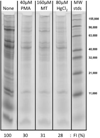
Figure 1
Figure 1. Differential effects of Hg(II), PMA, or merthiolate (MT) on total detectable protein thiols
Lysates of cells exposed (or not) for 30 minutes during growth to mercurials as indicated were reacted with BODIPY-I. Their proteins were separated by SDS-PAGE and imaged by fluorescence (GE Typhoon: Ex: 633 nm, Em: 670BP30, PMT: 750 V, 200 µm resolution, normal sensitivity). Lanes presented are from a single gel that was trimmed for this figure; lanes not shown were identical to these, confirming that the additional 15 minutes at 37° for DF and DTPA treatment had no effect on BODIPY detection of protein thiols. Fluorescence Intensities (FI) for each condition are normalized to the corresponding unexposed cells (lane 1). See Materials and Methods
Figure 2
Figure 2. Effects of Hg(II), PMA, or merthiolate (MT) on total detectable protein thiols
Lysates of cells exposed (or not) to mercurials as indicated for 15 minutes during growth were reacted (or not) with 10 M urea and then with BODIPY-I. Their proteins were separated by SDS-PAGE and imaged by fluorescence (GE Typhoon: Ex: 633 nm, Em: 670BP30, PMT: 750 V, 200 µm resolution, normal sensitivity). Images from several gels prepared under the same conditions were compiled for this figure. Total densitometric lane intensity of unexposed cultures varied by 17% s.d. without urea (e.g. lane 1) and by 13% s.d. with urea (e.g. lane 2). Total densitometric lane intensity for each condition was normalized to the corresponding lane intensity of unexposed cells. (see Materials and Methods)
Figure 3
Figure 3. Increases in free intracellular iron levels in cells exposed to mercurials and common organic oxidants
The increase in intracellular free iron is represented as the average X-fold increase in the Fe(III):DF complex EPR signal at g = 4.3 for each stress condition relative to the unexposed control whose average free iron concentration was 24.4 µM (+/− 4.9 µM). Error bars are standard deviation of biological replicates. Replicates for each condition were: Unexposed (6): H2O2 (2); t-BuOOH (2); MT (3); PMA (3); 16µM HgCl2 (3); and 80 µM HgCl2 (5)
Figure 4
Figure 4. Effect of mercurials on intracellular levels of essential metal ions
Exponential phase MG1655 cells were unexposed (black) or exposed to 40 µM PMA (dark grey), 160 µM MT (medium grey), 16 µM HgCl2 (light grey) or 80 µM HgCl2 (off-white) for 30 minutes, harvested, washed, and assayed for nine essential elements by ICP-MS. Inset: K+ and Na+ content on smaller scale. Error bars represent standard deviation of biological replicates: Unexposed (7); PMA (4); MT (4); 16 µM HgCl2 (3); 80 µM HgCl2 (6). Significance (*) was determined by t-test comparison of the means at the 95% confidence level with two-tailed p-values equal to <0.05.
Figure 5
Figure 5. Changes in cellular ligands as Hg concentration is increased
HgCl2 (10 mM, gray) was added to a known amount of growing cells at 10 µM (blue), 20 µM (green), 40 µM (red), or 80 µM (pink). Panels are the near edge (A) and EXAFS (B) spectra of the concentrated washed cell suspensions and the HgCl2 standard. Panel C is the corresponding Fourier transforms of the EXAFS data in (B) and panel D contains the EXAFS fitting results. Best Fit subscripts denote the number of scatterers per metal atom. Ras is the observed metal-scatterer distance. σas2 is the Debye-Waller or temperature factor.

References

    1. Barkay T, Miller SM, Summers AO. FEMS Microbiol Rev. 2003;27:355–384. - PubMed
    1. Mason RP, Fitzgerald WF, Morel FMM. Geochimica Et Cosmochimica Acta. 1994;58:3191–3198.
    1. Norn S, Permin H, Kruse E, Kruse PR. Dan Medicinhist Arbog. 2008;36:21–40. - PubMed
    1. Crinnion WJ. Altern Med Rev. 2000;5:209–223. - PubMed
    1. Richardson GM, Wilson R, Allard D, Purtill C, Douma S, Graviere J. Sci Total Environ. 2011;409:4257–4268. - PubMed

Publication types

MeSH terms