. 2015 Nov 18;137(45):14358-66.
doi: 10.1021/jacs.5b08729. Epub 2015 Nov 9.

Long-Term Passivation of Strongly Interacting Metals with Single-Layer Graphene

Affiliations

Long-Term Passivation of Strongly Interacting Metals with Single-Layer Graphene

Robert S Weatherup et al. J Am Chem Soc. .

Abstract

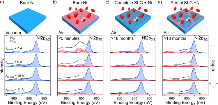
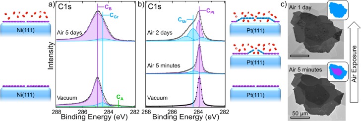
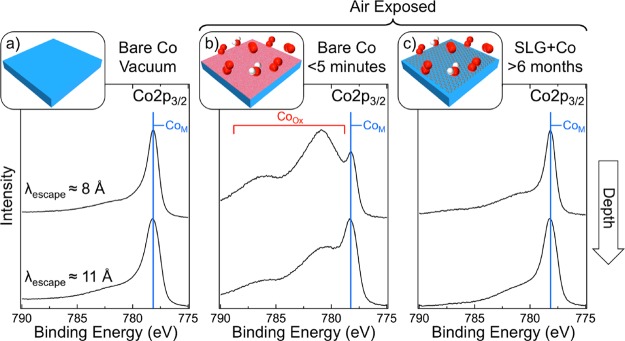
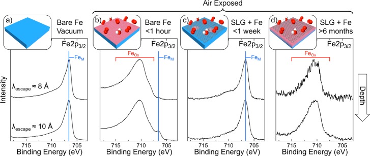
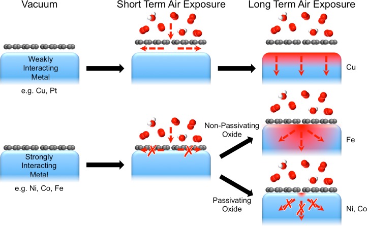
The long-term (>18 months) protection of Ni surfaces against oxidation under atmospheric conditions is demonstrated by coverage with single-layer graphene, formed by chemical vapor deposition. In situ, depth-resolved X-ray photoelectron spectroscopy of various graphene-coated transition metals reveals that a strong graphene-metal interaction is of key importance in achieving this long-term protection. This strong interaction prevents the rapid intercalation of oxidizing species at the graphene-metal interface and thus suppresses oxidation of the substrate surface. Furthermore, the ability of the substrate to locally form a passivating oxide close to defects or damaged regions in the graphene overlayer is critical in plugging these defects and preventing oxidation from proceeding through the bulk of the substrate. We thus provide a clear rationale for understanding the extent to which two-dimensional materials can protect different substrates and highlight the key implications for applications of these materials as barrier layers to prevent oxidation.


Figures

Figure 1
Figure 1
Depth-resolved XP Ni 2p3/2 core level spectra for polycrystalline Ni (25 μm) in situ immediately following annealing [600 °C, H2 (10–1 mbar) for 15 min] (a) and after subsequent exposure to atmosphere for <5 min (b), as well as for Ni (25 μm) covered with a complete SLG layer (c), and Ni (250 μm) covered with noncontinuous SLG islands (d) following exposure to atmosphere for >18 months. The SLG was grown by CVD [600 °C, C6H6 (10–5 mbar) for 15 min]. Spectra are collected at photon energies, Ephoton, of 1010, 1150, 1300, and 1450 eV [from upper to lower spectra, respectively, λescape ≈ 7, 9, 10, and 11 Å] and are normalized to have the same maximum intensity. Fitted components for metallic Ni (NiM) and Ni oxide/hydroxide (NiOx) are shaded blue and red, respectively.
Figure 2
Figure 2
(a) XP C 1s core level lines of Ni(111) covered with SLG grown by CVD [400 °C, C2H4 (10–6 mbar) for 2 h] in situ immediately following growth (lower) and after exposure to atmosphere for 5 days (upper). (b) XP C 1s core level lines of Pt(111) covered with SLG grown by vacuum annealing [10–8 mbar, 1000 °C, for 2 h] in situ immediately following growth (lower) and after exposure to atmosphere for 5 min (middle) and 2 days (upper). All spectra are collected at photon energy, Ephoton, of 425 eV (λescape ≈ 7 Å) and are normalized to have the same maximum intensity. (c) SEM micrographs of SLG island on polycrystalline Pt (25 μm) after exposure to atmosphere for 5 min (lower) and 1 day (upper), with inset schematic indicating the coupled (purple) and decoupled (blue) regions.
Figure 3
Figure 3
Depth-resolved XP Co 2p3/2 core level spectra for polycrystalline Co (25 μm) in situ immediately following annealing [600 °C, H2 (10–1 mbar) for 15 min] (a) and after subsequent exposure to atmosphere for <5 min (b); and for Co (25 μm) covered with a complete SLG layer grown by CVD [700 °C, C2H2 (∼10–6 mbar) for 15 min followed by C2H2 (∼10–5 mbar) for 5 min] following exposure to atmosphere for >6 months. Spectra are collected at photon energies, Ephoton, of 1020 (upper) and 1400 eV (lower) [respectively, λescape ≈ 8 and 11 Å] and are normalized to have the same maximum intensity.
Figure 4
Figure 4
Depth-resolved XP Fe 2p3/2 core level spectra for polycrystalline Fe (100 μm) in situ immediately following annealing [1000 °C, H2 (10–1 mbar) for 15 min] (a) and after subsequent exposure to atmosphere for 1 h (b); and for Fe (100 μm) covered with a complete SLG layer grown by CVD [650 °C, C2H2 (∼10–4 mbar) for 30 min] following exposure to atmosphere for 1 week (c) and >6 months (d). Spectra are collected at photon energies, Ephoton, of 920 (upper) and 1150 eV (lower) [respectively, λescape ≈ 8 and 10 Å] and are normalized to have the same maximum intensity.
Figure 5
Figure 5
Schematic illustrating the passivation behavior of different graphene-covered metals. Graphene is easily decoupled from the surface of weakly interacting metals (e.g., Cu, Pt) on air exposure, providing a pathway for the intercalation of oxidizing species at the graphene–catalyst interface and ready access for these oxidizing species to the whole metal surface. For strongly interacting metals (e.g., Ni, Co, Fe), graphene is not decoupled on air exposure, and the oxidizing species are thus only able to access the metal surface close to graphene defects. For metals that form a passivating oxide (e.g., Ni, Co), these exposed regions near to defects are quickly “plugged” by oxide formation, protecting the substrate from oxidation over the long term. For metals whose oxide is not passivating (e.g., Fe), oxidation is initially slowed by the already formed oxide, and thus the graphene coverage provides short-term passivation. However, oxidation can proceed through the already formed oxide, eventually allowing the metal to become oxidized throughout for long-term air exposures.

References

    1. Bunch J. S.; Verbridge S. S.; Alden J. S.; van der Zande A. M.; Parpia J. M.; Craighead H. G.; McEuen P. L. Nano Lett. 2008, 8, 2458–246210.1021/nl801457b. - DOI - PubMed
    1. Bohm S. Nat. Nanotechnol. 2014, 9, 741–74210.1038/nnano.2014.220. - DOI - PubMed
    1. Weatherup R. S.; Dlubak B.; Hofmann S. ACS Nano 2012, 6, 9996–1000310.1021/nn303674g. - DOI - PubMed
    1. Weatherup R. S.; Baehtz C.; Dlubak B.; Bayer B. C.; Kidambi P. R.; Blume R.; Schloegl R.; Hofmann S. Nano Lett. 2013, 13, 4624–463110.1021/nl401601x. - DOI - PMC - PubMed
    1. Caneva S.; Weatherup R. S.; Bayer B.; Brennan B.; Spencer S. J.; Mingard K.; Cabrero-Vilatela A.; Baehtz C.; Pollard A. J.; Hofmann S. Nano Lett. 2015, 15, 1867–187510.1021/nl5046632. - DOI - PMC - PubMed

Publication types