

Characterization of a novel metastatic prostate cancer cell line of LNCaP origin
- PMID: 26499105
- PMCID: PMC4729204
- DOI: 10.1002/pros.23115
Characterization of a novel metastatic prostate cancer cell line of LNCaP origin
Abstract
Background: The LNCaP cell line was originally isolated from the lymph node of a patient with metastatic prostate cancer. Many cell lines have been derived from LNCaP by selective pressures to study different aspects of prostate cancer progression. When injected subcutaneously into male athymic nude mice, LNCaP and its derivatives rarely metastasize.
Methods: Here, we describe the characteristics of a new LNCaP derivative, JHU-LNCaP-SM, which was generated by long term passage in normal cell culture conditions.
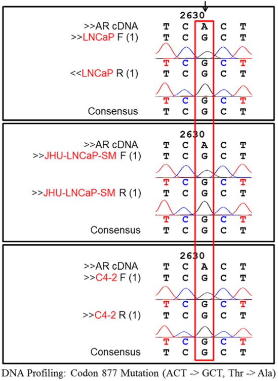
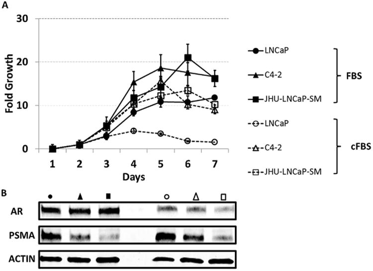
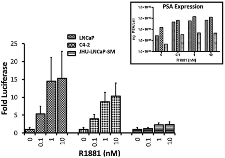
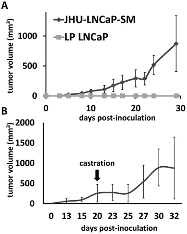
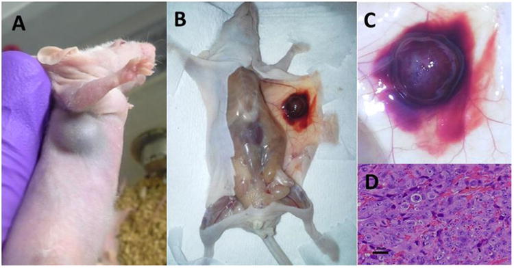
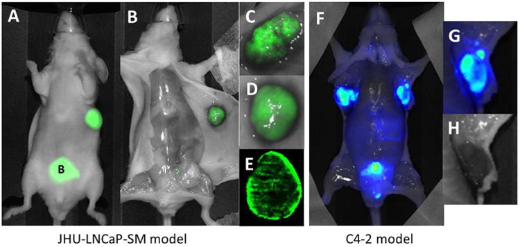
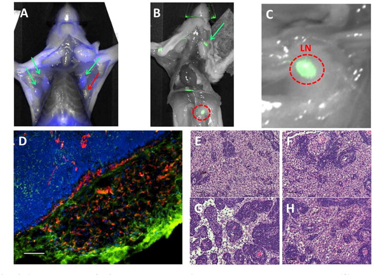
Results: Short tandem repeat (STR) analysis and genomic sequencing verified JHU-LNCaP-SM derivation from parental LNCaP cells. JHU-LNCaP-SM cells express the same mutated androgen receptor (AR) but unlike LNCaP, are no longer androgen dependent for growth. The cells demonstrate an attenuated androgen responsiveness in transcriptional assays and retain androgen sensitive expression of PSA, AR, and PSMA. Unlike parental LNCaP, JHU-LNCaP-SM cells quickly form subcutaneous tumors in male athymic nude mice, reliably metastasize to the lymph nodes and display a striking intra-tumoral and spreading hemorrhagic phenotype as tumor xenografts.
Conclusions: The JHU-LNCaP-SM cell line is a new isolate of LNCaP, which facilitates practical, preclinical studies of spontaneous metastasis of prostate cancer through lymphatic tissues.
Keywords: JHU-LNCaP-SM; PSMA; androgen; lymph node; metastasis.
© 2015 Wiley Periodicals, Inc.
Conflict of interest statement
Disclosures: There are no affiliations or conflicts to disclose.
Figures

Similar articles
-
Efficacy of PACE4 pharmacotherapy in JHU-LNCaP-SM preclinical model of androgen independent prostate cancer.Sci Rep. 2022 Oct 19;12(1):17489. doi: 10.1038/s41598-022-21593-7. Sci Rep. 2022. PMID: 36261691 Free PMC article.
-
LNCaP progression model of human prostate cancer: androgen-independence and osseous metastasis.Prostate. 2000 Jul 1;44(2):91-103 Jul 1;44(2). doi: 10.1002/1097-0045(20000701)44:2<91::aid-pros1>3.0.co;2-l. Prostate. 2000. PMID: 10881018
-
Monomethylated selenium inhibits growth of LNCaP human prostate cancer xenograft accompanied by a decrease in the expression of androgen receptor and prostate-specific antigen (PSA).Prostate. 2006 Jul 1;66(10):1070-5. doi: 10.1002/pros.20329. Prostate. 2006. PMID: 16637076
-
Antiandrogenic effects of novel androgen synthesis inhibitors on hormone-dependent prostate cancer.Cancer Res. 2000 Dec 1;60(23):6630-40. Cancer Res. 2000. PMID: 11118046
-
Application of Prostate Cancer Models for Preclinical Study: Advantages and Limitations of Cell Lines, Patient-Derived Xenografts, and Three-Dimensional Culture of Patient-Derived Cells.Cells. 2019 Jan 20;8(1):74. doi: 10.3390/cells8010074. Cells. 2019. PMID: 30669516 Free PMC article. Review.
Cited by
-
Tuning Pharmacokinetics to Improve Tumor Accumulation of a Prostate-Specific Membrane Antigen-Targeted Phototheranostic Agent.Bioconjug Chem. 2018 Nov 21;29(11):3746-3756. doi: 10.1021/acs.bioconjchem.8b00636. Epub 2018 Oct 29. Bioconjug Chem. 2018. PMID: 30350576 Free PMC article.
-
Establishment and characterization of patient-derived xenografts for hormone-naïve and castrate-resistant prostate cancers to improve treatment modality evaluation.Aging (Albany NY). 2020 Feb 24;12(4):3848-3861. doi: 10.18632/aging.102854. Epub 2020 Feb 24. Aging (Albany NY). 2020. PMID: 32092044 Free PMC article.
-
In situ Generated 212Pb-PSMA Ligand in a 224Ra-Solution for Dual Targeting of Prostate Cancer Sclerotic Stroma and PSMA-positive Cells.Curr Radiopharm. 2020;13(2):130-141. doi: 10.2174/1874471013666200511000532. Curr Radiopharm. 2020. PMID: 32389119 Free PMC article.
-
Quantifying the invasion and migration ability of cancer cells with a 3D Matrigel drop invasion assay.Biol Methods Protoc. 2021 Jul 21;6(1):bpab014. doi: 10.1093/biomethods/bpab014. eCollection 2021. Biol Methods Protoc. 2021. PMID: 34377838 Free PMC article.
-
Evaluation of micro-RNA in extracellular vesicles from blood of patients with prostate cancer.PLoS One. 2021 Dec 31;16(12):e0262017. doi: 10.1371/journal.pone.0262017. eCollection 2021. PLoS One. 2021. PMID: 34972164 Free PMC article.
References
-
- Horoszewicz JS, Leong SS, Kawinski E, Karr JP, Rosenthal H, Chu TM, Mirand EA, Murphy GP. LNCaP model of human prostatic carcinoma. Cancer Res. 1983;43(4):1809–1818. - PubMed
-
- Lim DJ, Liu XL, Sutkowski DM, Braun EJ, Lee C, Kozlowski JM. Growth of an androgen-sensitive human prostate cancer cell line, LNCaP, in nude mice. Prostate. 1993;22(22):109–118. - PubMed
-
- Culig Z, Hoffmann J, Erdel M, Eder EI, Hobisch A, Hittmair A, Bartsch G, Utermann G, Schneider MR, Parczyk K, Klocker H. Switch from antagonist to agonist of the androgen receptor bicalutamide is associated with prostate tumour progression in a new model system. Br J Cancer. 1999;81(2):242–251. - PMC - PubMed
-
- Gao M, Ossowski L, Ferrari AC. Activation of Rb and decline in androgen receptor protein precede retinoic acid-induced apoptosis in androgen-dependent LNCaP cells and their androgen-independent derivative. J Cell Physiol. 1999;179(3):336–346. - PubMed
-
- Kokontis JM, Hay N, Liao S. Progression of LNCaP prostate tumor cells during androgen deprivation: hormone-independent growth, repression of proliferation by androgen, and role for p27Kip1 in androgen-induced cell cycle arrest. Mol Endocrinol. 1998;12(7):941–953. - PubMed
Publication types
MeSH terms
Grants and funding
LinkOut - more resources
Full Text Sources
Other Literature Sources
Medical
Research Materials
Miscellaneous
