. 2015 Dec 18;290(51):30540-50.
doi: 10.1074/jbc.M115.683342. Epub 2015 Oct 23.

D-Amino Acid Probes for Penicillin Binding Protein-based Bacterial Surface Labeling

Affiliations

D-Amino Acid Probes for Penicillin Binding Protein-based Bacterial Surface Labeling

Jonathan M Fura et al. J Biol Chem. .

Abstract

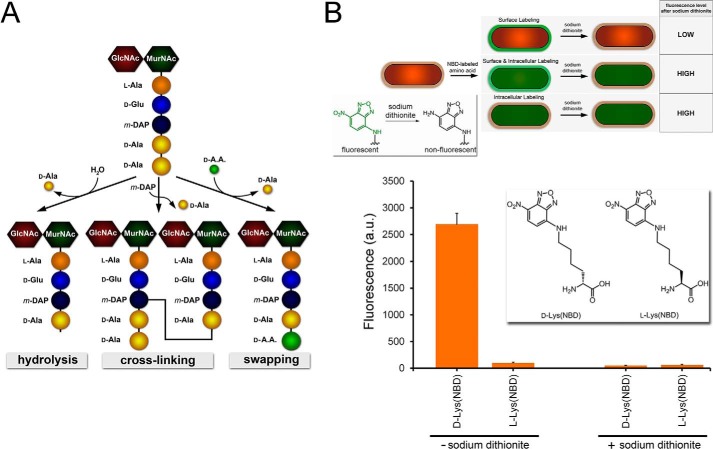
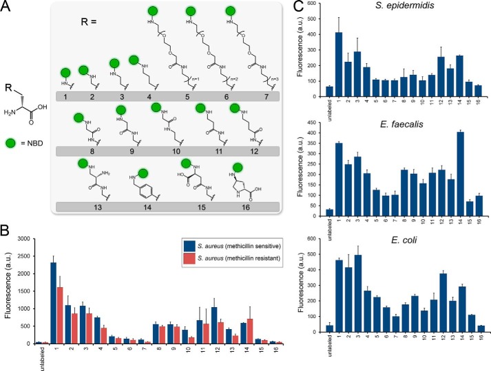
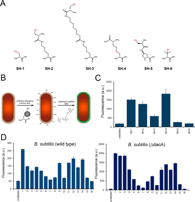
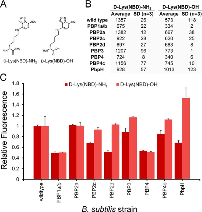
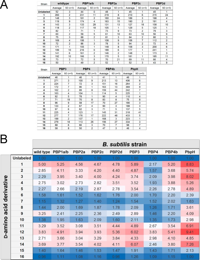
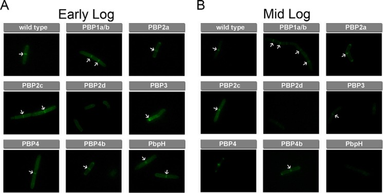
Peptidoglycan is an essential and highly conserved mesh structure that surrounds bacterial cells. It plays a critical role in retaining a defined cell shape, and, in the case of pathogenic Gram-positive bacteria, it lies at the interface between bacterial cells and the host organism. Intriguingly, bacteria can metabolically incorporate unnatural D-amino acids into the peptidoglycan stem peptide directly from the surrounding medium, a process mediated by penicillin binding proteins (PBPs). Metabolic peptidoglycan remodeling via unnatural D-amino acids has provided unique insights into peptidoglycan biosynthesis of live bacteria and has also served as the basis of a synthetic immunology strategy with potential therapeutic implications. A striking feature of this process is the vast promiscuity displayed by PBPs in tolerating entirely unnatural side chains. However, the chemical space and physical features of this side chain promiscuity have not been determined systematically. In this report, we designed and synthesized a library of variants displaying diverse side chains to comprehensively establish the tolerability of unnatural D-amino acids by PBPs in both Gram-positive and Gram-negative organisms. In addition, nine Bacillus subtilis PBP-null mutants were evaluated with the goal of identifying a potential primary PBP responsible for unnatural D-amino acid incorporation and gaining insights into the temporal control of PBP activity. We empirically established the scope of physical parameters that govern the metabolic incorporation of unnatural D-amino acids into bacterial peptidoglycan.

Keywords: Bacillus; amino acid; bacteria; bacterial conjugation; cell surface; cell wall; peptide biosynthesis; peptide chemical synthesis; peptides; peptidoglycan.


Figures

FIGURE 1.
FIGURE 1.
A, schematic showing the monomeric unit of peptidoglycan. The pentapeptide is processed by PBPs CP and TP to generate three possible products, all resulting from the acyl intermediate following the release of the terminal d-Ala. B, schematic demonstrating the three possible labeling locations within the bacterial cell. Only extracellular labeling would lead to complete reduction of the signal in the presence of the reducing agent sodium dithionite. Inset, reduction of the nitro group on NBD by sodium dithionite quenches the green fluorescence. B. subtilis cells were labeled with either d-Lys(NBD) or l-Lys(NBD) overnight. Cells were analyzed by flow cytometry. Data are represented as mean ± S.D. (n = 3).
FIGURE 2.
FIGURE 2.
A, metabolic labeling of bacterial surfaces with d-lysine conjugated to the small fluorophore NBD. B, labeling of S. aureus cells (methicillin-sensitive and -resistant) was performed with all the members of the d-amino acid NBD conjugates. Cells were incubated overnight at 100 μm and analyzed by flow cytometry. Data are represented as mean ± S.D. (n = 3). C, S. epidermidis and E. faecalis cells were labeled with the panel of NBD-conjugated d-amino acids for 4 h at stationary phase at 100 μm. E. coli cells were labeled with the panel of NBD-conjugated d-amino acids for 4 h at stationary phase at 500 μm. Cells were analyzed by flow cytometry, and data are represented as mean ± S.D. (n = 3).
FIGURE 3.
FIGURE 3.
A, chemical structures of B. subtilis cells were labeled with the panel of sulfhydryl containing d-amino acids. Cells were incubated for 4 h at stationary phase at 500 μm, incubated with NBD-l-Lys(maleimide), and analyzed by flow cytometry. Data are represented as mean ± S.D. (n = 3). D, wild-type and ΔdacA mutant B. subtilis cells were labeled with the panel of NBD-conjugated d-amino acids. Cells were incubated for 4 h at stationary phase at 100 μm and analyzed by flow cytometry. Data are represented as mean ± S.D. (n = 3).
FIGURE 4.
FIGURE 4.
A, chemical structures of d-Lys(NBD)-OH and d-Lys(NBD)-NH2. B, B. subtilis cells were labeled with the panel of NBD-conjugated d-amino acids. Cells were incubated overnight at 100 μm and analyzed by flow cytometry. Data are represented as mean ± S.D. (n = 3). C, data are represented as mean relative to the wild type + S.D. (n = 3).
FIGURE 5.
FIGURE 5.
A, mutant strains of B. subtilis lacking specified PBPs were labeled with NBD-conjugated d-amino acids. Cells were incubated for 4 h at stationary phase at 100 μm and analyzed by flow cytometry. Data are represented as mean ± S.D. (n = 3). B, heat map of labeling a series of B. subtilis mutants with the NBD-conjugated d-amino acids. (Data were derived from A). The heat map of incorporation shows relative fluorescence levels to unlabeled cells. Red cells indicate elevated incorporation, and blue cells indicate lower incorporation.
FIGURE 6.
FIGURE 6.
A, chemical structures of d-Lys(FITC)-OH. B, B. subtilis cells were labeled with the panel of NBD-conjugated d-amino acids. Cells were incubated for 4 h at stationary phase at 100 μm and analyzed by flow cytometry. Data are represented as mean ± S.D. (n = 3).
FIGURE 7.
FIGURE 7.
Pulse labeling of mutant strains of B. subtilis lacking specified PBPs with d-Lys(FITC)-OH. Cells were incubated for 15 min at specified phases at 1 mm and analyzed by flow cytometry. Data are represented as mean ± S.D. (n = 3).
FIGURE 8.
FIGURE 8.
Pulse labeling of mutant strains of B. subtilis lacking specified PBPs with d-Lys(FITC)-OH. Cells were incubated for 15 min at specified phases at 1 mm and analyzed by flow cytometry. Data are represented as mean relative to wild type + S.D. (n = 3).
FIGURE 9.
FIGURE 9.
A and B, pulse labeling of mutant strains of B. subtilis lacking specified PBPs with d-Lys(FITC)-OH. Cells were incubated for 15 min at early log (A) and mid-log phase (B) at 1 mm and analyzed by fluorescence microscopy. Arrows are indicative of increased septal labeling.

References

    1. Vollmer W., Blanot D., and de Pedro M. A. (2008) Peptidoglycan structure and architecture. FEMS Microbiol. Rev. 32, 149–167 - PubMed
    1. Frère J. M., and Joris B. (1985) Penicillin-sensitive enzymes in peptidoglycan biosynthesis. Crit. Rev. Microbiol. 11, 299–396 - PubMed
    1. van Heijenoort J. (2001) Recent advances in the formation of the bacterial peptidoglycan monomer unit. Nat. Prod. Rep. 18, 503–519 - PubMed
    1. Bugg T. D., and Walsh C. T. (1992) Intracellular steps of bacterial cell wall peptidoglycan biosynthesis: enzymology, antibiotics, and antibiotic resistance. Nat. Prod. Rep. 9, 199–215 - PubMed
    1. Scheffers D. J., and Pinho M. G. (2005) Bacterial cell wall synthesis: new insights from localization studies. Microbiol. Mol. Biol. Rev. 69, 585–607 - PMC - PubMed

Publication types

MeSH terms

LinkOut - more resources