. 2015 Oct 6:9:130.
doi: 10.3389/fnana.2015.00130. eCollection 2015.

Extra-neurohypophyseal axonal projections from individual vasopressin-containing magnocellular neurons in rat hypothalamus

Affiliations

Extra-neurohypophyseal axonal projections from individual vasopressin-containing magnocellular neurons in rat hypothalamus

Vito S Hernández et al. Front Neuroanat. .

Abstract

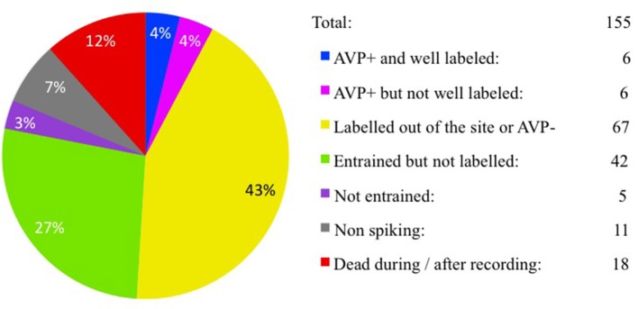
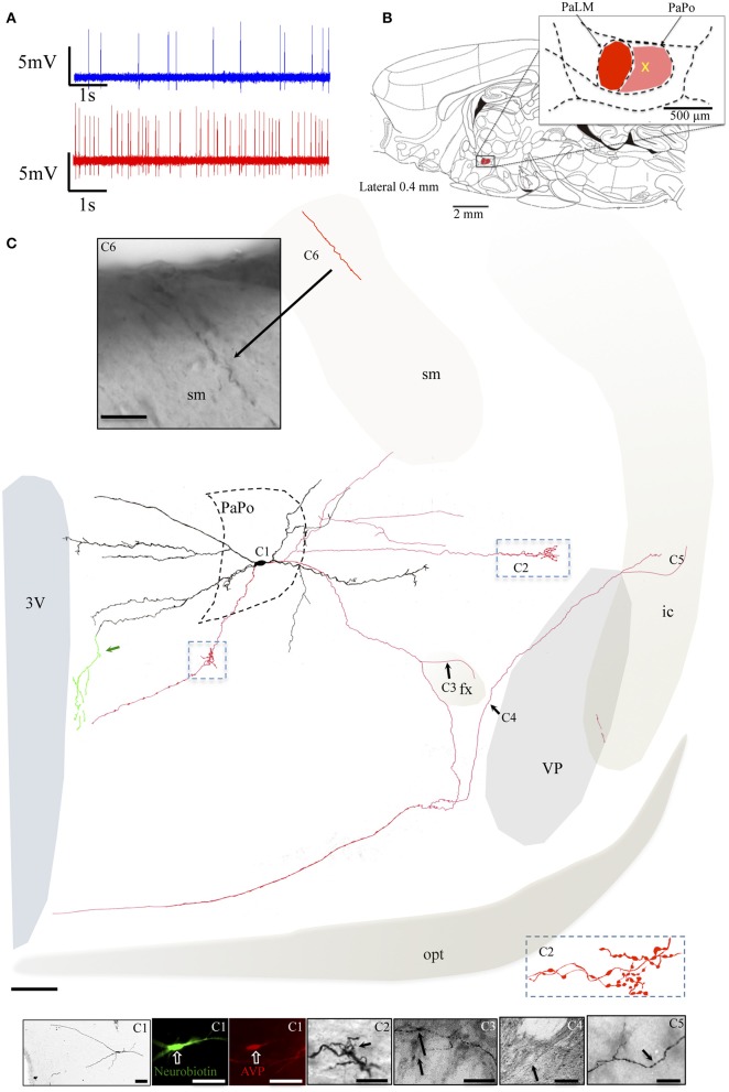
Conventional neuroanatomical, immunohistochemical techniques, and electrophysiological recording, as well as in vitro labeling methods may fail to detect long range extra-neurohypophyseal-projecting axons from vasopressin (AVP)-containing magnocellular neurons (magnocells) in the hypothalamic paraventricular nucleus (PVN). Here, we used in vivo extracellular recording, juxtacellular labeling, post-hoc anatomo-immunohistochemical analysis and camera lucida reconstruction to address this question. We demonstrate that all well-labeled AVP immunopositive neurons inside the PVN possess main axons joining the tract of Greving and multi-axon-like processes, as well as axonal collaterals branching very near to the somata, which project to extra-neurohypophyseal regions. The detected regions in this study include the medial and lateral preoptical area, suprachiasmatic nucleus (SCN), lateral habenula (LHb), medial and central amygdala and the conducting systems, such as stria medullaris, the fornix and the internal capsule. Expression of vesicular glutamate transporter 2 was observed in axon-collaterals. These results, in congruency with several previous reports in the literature, provided unequivocal evidence that AVP magnocells have an uncommon feature of possessing multiple axon-like processes emanating from somata or proximal dendrites. Furthermore, the long-range non-neurohypophyseal projections are more common than an "occasional" phenomenon as previously thought.

Keywords: axon collaterals; extra-hypothalamic projections; juxtacellular labeling; magnocellular neurosecretory system; paraventricular nucleus (PVN); vasopressin.


Figures

Figure 1
Figure 1
Juxtacellular recording/labeling census (cell per rat). Pie-chart showing recording/labeling census of this study. One hundred fifty-five rats were used for in vivo extracellular recording and juxtacellular labeling of hypothalamic paraventricular vasopressin-containing magnocellular neurons (AVP-magnocells). The fraction of cases that satisfied the selection criteria is labeled in blue.
Figure 2
Figure 2
Neuron EV16. (A) Spike trains under basal (blue trace) and hypertonic (red trace) conditions. (B) Sagittal plate of lateral 0.40 mm from Rat Brain Atlas (Paxinos and Watson, 2006) and the amplification of the paraventricular nucleus (inset): red and pink shadows indicate the lateral magnocellular division of paraventricular nucleus (PaLM) and the posterior division (PaPo) respectively. The yellow cross indicates the rostro-caudal position of EV16. (C) Camera lucida reconstruction of the neuron's soma, dendrites and axons. Drawings from serial sections were superimposed manually in a 2-dimension (2D) projection drawing on a coronal view of the rat hypothalamus. The soma and dendrites were represented in black and axonal segments were represented in red. (C1) AVP-containing nature was ascertained by AVP immunoreaction. The soma gave rise initially to two short thick primary dendrites, which branched proximally. The bottom dendrite branched extensively until the fifth order of branches; all directed medially reaching the wall of the third ventricle (3V). The top dendrite gave rise to two secondary branches, the medial and the lateral ones. The medial branch was similar to the bottom group. The lateral portion curled up proximally near the soma but gave rise to the main axon. Photomicrograph (C1a, from neurobiotin HRP/DAB reaction); (C1b,C1c, from neurobiotin-streptavidin-Alexa 488 fluorescence in two consecutive sections) showing the sites where three axons were originated (red arrows). The main axon coursed laterally passing the fornix (fx), turned ventro-caudally toward the posterior pituitary gland (PPit) (D). Two main collaterals emanated from this axon (C2,C3). The first collateral (C2) coursed dorsomedially joining the stria medularis (sm). Neurobiotin labeled processes were found inside the lateral habenula (C4). The second collateral originated passing the fx and coursed medially (C3). The labeling of one of the ventrally directed axons coursed further ventrally along the periventricular region, reaching the suprachiasmatic nucleus (SCN). (D) Computer aided schematic reconstruction based on observations of serial coronal sections to facilitate the perspective of axonal projections from a sagittal view. Note that the size of cell vs. brain is not in strict proportion. LHb and MHb, lateral and medial habenula; PPit, posterior pituitary gland; sm, stria medularis; fx, fornix; 3V, third ventricle; opt, optical tract. Scale bars: 50 μm for (C1), 20 μm for the rest of panels.
Figure 3
Figure 3
Neuron MM22. (A) Spike trains under basal (blue trace) and hypertonic (red trace) conditions. (B) Sagittal plate of lateral 0.40 mm from Rat Brain Atlas (Paxinos and Watson, 2006) and the amplification of the paraventricular nucleus (inset): red and pink shadows indicate the lateral magnocellular division of paraventricular nucleus (PaLM) and the posterior division (PaPo) respectively. The yellow cross indicates the rostro-caudal position of MM22. (C) Camera lucida reconstruction of the neuron's soma, dendrites and axons. Drawings from serial sections were superimposed manually in a 2-dimension (2D) projection drawing from a coronal view of the rat hypothalamus. The soma and dendrites were represented in black and axonal segments were represented in red. AVP containing nature of MM22 was ascertained by AVP immunoreaction (C1). Photomicrographs from neurobiotin HRP/DAB reaction in (C2,C3) show the sites where two main axons were originated (red arrows) and where the axon turned medially (green arrowhead) and coursed ventrally along the periventricular zone emitting numerous branches toward the suprachiasmatic nucleus (SCN). opt: optical tract, fx: fornix. The light gray irregular line symbolizes the site where the brain tissue was broken. The optical tract, symbolized by dashed line was drawn only for reference. Scale bars: 100 μm in main drawing, 50 μm in (C1), and 30 μm in (C2,C3).
Figure 4
Figure 4
Neuron VH25. (A) Spike trains under basal (blue trace) and hypertonic (red trace) conditions. (B) Sagittal plate of lateral 0.40 mm from Rat Brain Atlas (Paxinos and Watson, 2006) and the amplification of the paraventricular nucleus (inset): red and pink shadows indicate the lateral magnocellular division of paraventricular nucleus (PaLM) and the posterior division (PaPo) respectively. The yellow cross indicates the rostro-caudal position of VH25. (C) Camera lucida reconstruction of the neuron's soma, dendrites and axons. Drawings from serial sections were superimposed manually in a 2-dimension (2D) projection drawing on a coronal view of the rat hypothalamus. The soma and dendrites were represented in black and axonal segments were represented in red. The green processes indicate the beaded processes originated from dendritic processes. This kind of processes were described in an early study and were considered as axonal processes (Sofroniew and Glasmann, 1981). Numbered regions/segments in (C) correspond to the numbered photomicrographs. (C1) Vasopressin (AVP)-containing nature of neuron VH25 was ascertained by AVP immunoreaction; (C2) photomicrograph from neurobiotin HRP/DAB reaction showing the sites where the two main axons were originated (red arrows) opt, optical tract; fx, fornix. Scale bars: 100 μm for main panel, 25 μm for (C1,C2).
Figure 5
Figure 5
Neuron EV40. (A) Spike trains under basal (blue trace) and hypertonic (red trace) conditions. (B) Sagittal plate of lateral 0.40 mm from Rat Brain Atlas (Paxinos and Watson, 2006) and the amplification of the paraventricular nucleus (inset): red and pink shadows indicate the lateral magnocellular division of paraventricular nucleus (PaLM) and the posterior division (PaPo) respectively. The yellow cross indicates the rostro-caudal position of EV40. (C) Camera lucida reconstruction of the coronal projection and corresponding photomicrographs (C1–C4) showing dendritic and axonal patterns. Red, blue and pink arrows indicate the origins of three parent-axons. Red and blue arrowheads indicate the subsequent axonal branches. The green arrowheads indicate the beaded processes originated from the dendritic processes, some of them entering the fornix (green arrowheads). This kind of processes were described in an early study and were considered as axonal processes (Sofroniew and Glasmann, 1981). (C1a) AVP-containing nature was ascertained by AVP immunoreaction; (C2) photomicrograph of the section contiguous caudally to the soma (depicted in C1b) showing the branching of the axon labeled with a blue arrow. (C3,C4) Show axonal processes found in amygdala. Scale bars: 100 μm for (C); 50 μm for all photomicrographs. MeA, medial amygdala; CeA, central amygdala; opt, optical tract; fx, fornix.
Figure 6
Figure 6
Neuron VH52. (A) Spike trains under basal (blue trace) and hypertonic (red trace) conditions. (B) Sagittal plate of lateral 0.40 mm from Rat Brain Atlas (Paxinos and Watson, 2006) and the amplification of the paraventricular nucleus (inset): red and pink shadows indicate the lateral magnocellular division of paraventricular nucleus (PaLM) and the posterior division (PaPo), respectively. The yellow cross indicates the rostro-caudal position of VH52. (C) Coronal projection of camera lucida two-dimensional reconstruction and corresponding photomicrographs (C1–C6) showing dendritic and axonal patterns. (C1) Shows that the AVP-containing nature was ascertained by AVP immunoreaction; (C2) shows an axon with several small branches and classical image of strings of pearls in a small region in the lateral preoptical area; (C3–C6) show the presence of labeled axon segments in or near the conducting systems, fornix (fx), internal capsule (ic), and stria medullaris (sm). Green traces indicated by the green arrow, showing the beaded processes originated from the dendritic processes branching extensively in the periventricular region. This kind of processes were described in an early study and were considered as axonal processes (Sofroniew and Glasmann, 1981). Scale bars: 100 μm for (C); 50 μm for all photomicrographs. VP, Ventral pallidum (symbolized with gray shadow); conducting systems (i.e., optical tract, opt, stria medullaris st, fornix, fx, internal capsule, ic) were symbolized with light beige shadows.
Figure 7
Figure 7
Neuron MM15. (A) Spike trains under basal (blue trace) and hypertonic (red trace) conditions. (B) Sagittal plate of lateral 0.40 mm from Rat Brain Atlas (Paxinos and Watson, 2006) and the amplification of the paraventricular nucleus (inset): red and pink shadows indicate the lateral magnocellular division of paraventricular nucleus (PaLM) and the posterior division (PaPo) respectively. The yellow cross indicates the rostro-caudal position of MM15. (C) Camera lucida reconstruction of coronal projection and corresponding photomicrographs (C1–C3) showing dendritic and axonal patterns. The soma was located in the PaPo anterolateral border with PaLM. It emitted three primary dendrites. In contrast to the previous cells, this cell's main dendritic arborizations were directed laterally and ventrally. Four axonal processes (colored asterisks) arose from the proximal dendritic loci belonging to the same primary dendrite. Two of them projected ventrally joining the tract of Greving. The two other axon-like processes bended dorsally. (C1a) Shows a projection of several confocal photomicrographs of the juxtacellular labeled cell. Note that there was a process labeled from an unlabeled soma, indicated by a pink arrow. It seemed that the tracer passed from MM15 to this segment through the contact indicated by a pink arrowhead, also see the Supplementary Video. This phenomenon has been reported in the literature and the presence of gap-junctions between the two structures had been suggested (Andrew et al., 1981), (C1b) and insets show the AVP-containing nature of the labeled cell ascertained by AVP immunoreaction. (C2) Shows a thin axon-like process positive to vGluT2. (C3a) Is a digital rotation of the 3D reconstruction (Supplementary information) aiming to show the double axons (labeled with red and pink asterisk) emitting from the same point of the proximal dendrite (C3b is a coronal view of the same feature). (C3c,d) Show the emerging sites of the two other axons. Scale bars: for (C,C1a,b) 50 μm; rest: 10 μm.

References

    1. Andrew R. D., Macvicar B. A., Dudek F. E., Hatton G. I. (1981). Dye transfer through gap junctions between neuroendocrine cells of rat hypothalamus. Science 211, 1187–1189. 10.1126/science.7466393 - DOI - PubMed
    1. Armstrong W. (2004). Hypothalamic supraoptic and paraventricular nuclei, in The Rat Nervous System, 3rd Edn., Chapter 15, ed Paxinos G. (Amsterdam: Elsevier; ), 369–388.
    1. Armstrong W. E., Warach S., Hatton G. I., McNeill T. H. (1980). Subnuclei in the rat hypothalamic paraventricular nucleus: a cytoarchitectural, horseradish peroxidase and immunocytochemical analysis. Neuroscience 5, 1931–1958. 10.1016/0306-4522(80)90040-8 - DOI - PubMed
    1. Bargmann W., Scharrer E. (1951). The site of origin of the hormones of the posterior pituitary. Am. Sci. 39, 255–259. - PubMed
    1. Bisset G. W., Hilton S. M., Poisner A. M. (1967). Hypothalamic pathways for independent release of vasopressin and oxytocin. Proc. R. Soc. Lond. B Biol. Sci. 166, 422–442. 10.1098/rspb.1967.0005 - DOI - PubMed

LinkOut - more resources