. 2015 Oct 6:9:266.
doi: 10.3389/fnbeh.2015.00266. eCollection 2015.

Neural correlates of rules and conflict in medial prefrontal cortex during decision and feedback epochs

Affiliations

Neural correlates of rules and conflict in medial prefrontal cortex during decision and feedback epochs

Gregory B Bissonette et al. Front Behav Neurosci. .

Abstract

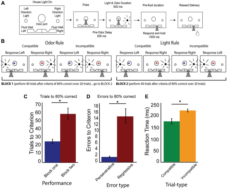
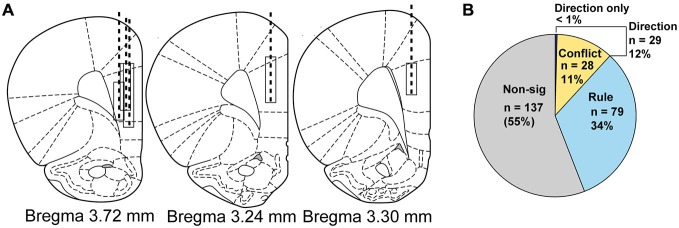
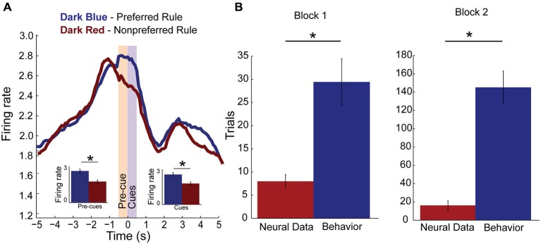
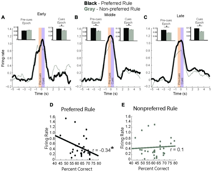
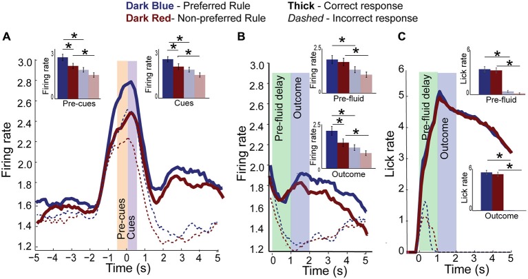
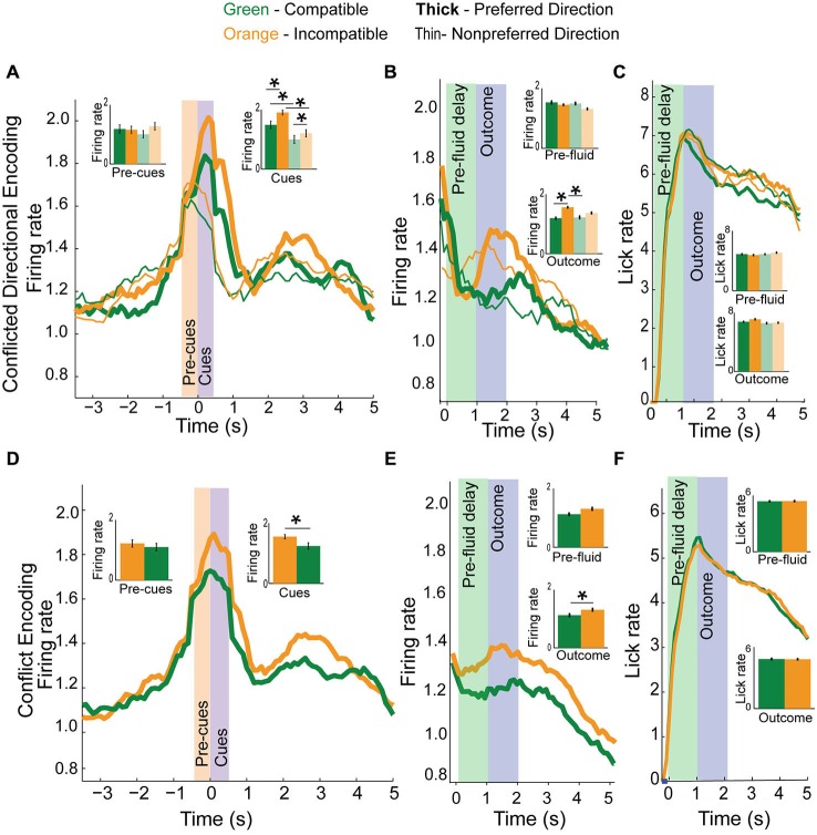
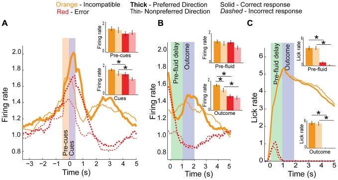
The ability to properly adjust behavioral responses to cues in a changing environment is crucial for survival. Activity in the medial Prefrontal Cortex (mPFC) is thought to both represent rules to guide behavior as well as detect and resolve conflicts between rules in changing contingencies. However, while lesion and pharmacological studies have supported a crucial role for mPFC in this type of set-shifting, an understanding of how mPFC represents current rules or detects and resolves conflict between different rules is unclear. Here, we directly address the role of rat mPFC in shifting rule based behavioral strategies using a novel behavioral task designed to tease apart neural signatures of rules, conflict and direction. We demonstrate that activity of single neurons in rat mPFC represent distinct rules. Further, we show increased firing on high conflict trials in a separate population of mPFC neurons. Reduced firing in both populations of neurons was associated with poor performance. Moreover, activity in both populations increased and decreased firing during the outcome epoch when reward was and was not delivered on correct and incorrect trials, respectively. In addition, outcome firing was modulated by the current rule and the degree of conflict associated with the previous decision. These results promote a greater understanding of the role that mPFC plays in switching between rules, signaling both rule and conflict to promote improved behavioral performance.

Keywords: conflict; in vivo electrophysiology; mPFC; rule encoding; set-shifting.


Figures

Figure 1
Figure 1
Rule shifting behavior schematic and measures. (A) Schematic of odor port, fluid wells and simultaneous directional cues as well as flow chart of successful trial completion denoting specific time delays. (B) Example correct responses within different rule blocks, demonstrating compatible and incompatible trials with simultaneous directional information presented by both cues. Blue arrow indicates the “left” direction odor, Red arrow indicates the “right” direction odor, while black radiating circle represents the side of the light. Rat head represents the correct choice made by a rat which would have previously received the simultaneous presentation of both odor and light cues before making his choice. (C) Data showing increased number of trials required to reach criterion after a switched rule, with trials to criterion in block one (blue bar) and block two (red bar). (D) Data demonstrating a significant number of errors are regressive (red bar), rather than perseverative (blue bar). (E) Reaction time data (cue offset to port exit), demonstrating significant slowing on incompatible (orange bars) compared to compatible trial-types (green bars). Significance of at least p < 0.05 denoted with asterisk *.
Figure 2
Figure 2
Electrode implant beginning and final positions, drive paths and classification of neural activity by regression. (A) Atlas schematics showing electrode wire bundle implant locations (top of box) and location at completion of study (bottom of box) and general drive path (dashed line) for five rats. (B) Pie chart showing by number and percentage the breakdown of neural activity types from regression analysis on neural firing, with non-modulated neuron data shown in gray, rule neurons in light blue, conflict neurons in yellow and direction neurons in purple.
Figure 3
Figure 3
Rule encoding in the medial Prefrontal Cortex (mPFC). (A) Population activity of rule neurons, plotted by preferred (dark blue) and non-preferred (dark red) rule blocks centered on cue presentation (time 0). Activity significantly divides during pre-cue epoch (yellow bar, −500–0 ms, inset) and remains higher for one rule over another during the cue epoch (purple bar, 0–500 ms, inset). (B) Bar plots comparing when rule neurons first significantly modulate activity in preferred rule block (red bar) and when rats first reached behavioral criterion (blue bar). Change in rule neuron activity precedes behavioral criterion regardless of whether the neuron’s preferred rule was in block one or two. Significance of at least p < 0.05 denoted with asterisk *.
Figure 4
Figure 4
Rule modulated neurons signal rule change. (A–C) baseline subtracted neural activity for preferred (black) and non-preferred (gray) rule blocks with inset bar plots comparing neural firing during pre-cue and cue epochs between rule representations. (A) In first two trials of a rule block, neuronal activity is significantly higher during cue epoch for preferred rule. (B) In the next eight trials, neural activity for preferred rule increases compared to the early trials, and is significantly higher for preferred rule during both the pre-cue epoch and cue epoch. (C) In the last ten trials, neural activity for rules has significantly decreased compared to the middle eight, though it remains significantly higher for pre-cue and cue epochs compared to non-preferred rule blocks. (D) Correlation of firing rate during the cue epoch with percent correct, showing a significant negative correlation between neural activity and percent correct for the preferred rule block. (E) Unlike during preferred rule blocks, firing rate and percent correct for the non-preferred rule block demonstrates no significant correlation. Significance denoted of at least p < 0.05 with asterisk *.
Figure 5
Figure 5
Rule activity is diminished on error trials. (A) Population histogram of all rule-modulated neurons by preferred (dark blue) and non-preferred (dark red) rule blocks for correct (solid) and error (dashed) trials with inset bar graphs showing data from pre-cue and cue epochs for correct trials and error trials (faded blue and red bars). Though neural activity is the same as correct trials at baseline, it is significantly reduced during pre-cue and cue epochs. (B) Aligning the data from (A) to fluid-well entry and plotting pre-fluid delay and reward delivery epochs, we see that neural activity of rule-modulated neurons also reflects trial outcomes, being significantly reduced on error trials and highest during preferred rule correct trials. (C) Licking aligned to fluid well entry. Reward is delivered 1 s later on correct trials. Licking did not significantly differ between rules. Significance denoted of at least p < 0.05 with asterisk *.
Figure 6
Figure 6
Conflict and direction activity in the mPFC. (A) Top panel plots direction neurons based on preferred direction (thick) and non-preferred direction (thin) and by trial-type, compatible (green) and incompatible (orange) with inset bar graphs showing data during both pre-cue and cue epochs for correct and error (faded green and yellow) trials. While direction neurons were more active for one direction over another, activity in these neurons was more active when directional cues were in conflict than when they were compatible with each other. (B) Direction and conflict data demonstrates elevated activity for preferred direction incompatible trials during the outcome epoch, but not pre-fluid epoch. (C) Plots licking data demonstrating no difference in lick rate between trial-types. (D) Collapsing the data from (A) into compatible or incompatible trials, we see that directional neurons fire more for incompatible trial-types than for compatible trial-types when presented with cue information. (E) Activity plotted by compatibility averaged across direction during the pre-fluid and outcome epochs demonstrates a significant elevation of activity on correct incompatible trial-types compared to compatible trial-types but not during the pre-fluid epoch. (F) Plots licking rate by compatibility, and demonstrates no difference in lick rate between compatible or incompatible trial-types in either the pre-fluid or outcome epochs. Significance denoted of at least p < 0.05 with asterisk *.
Figure 7
Figure 7
Conflict activity on error trials. (A) Population histogram of conflict neurons on incompatible trial-types (orange) plotted by direction (full color are preferred direction, faded color are non-preferred directions in bar graph insets) showing decreased neural activity on error trials (dashed red or faded red for inset bar graphs) during the cue epoch. (B) Aligning data to fluid-well entry demonstrates a significant reduction in activity during pre-fluid delay and after reward should have been delivered, suggesting these neurons also represent the outcome of the trials. (C) Licking did not significantly differ between directions. Significance denoted of at least p < 0.05 with asterisk *.

References

    1. Alexander W. H., Brown J. W. (2011). Medial prefrontal cortex as an action-outcome predictor. Nat. Neurosci. 14, 1338–1344. 10.1038/nn.2921 - DOI - PMC - PubMed
    1. Antzoulatos E. G., Miller E. K. (2011). Differences between neural activity in prefrontal cortex and striatum during learning of novel abstract categories. Neuron 71, 243–249. 10.1016/j.neuron.2011.05.040 - DOI - PMC - PubMed
    1. Baker P. M., Ragozzino M. E. (2014). Contralateral disconnection of the rat prelimbic cortex and dorsomedial striatum impairs cue-guided behavioral switching. Learn. Mem. 21, 368–379. 10.1101/lm.034819.114 - DOI - PMC - PubMed
    1. Balleine B. W., O’Doherty J. P. (2010). Human and rodent homologies in action control: corticostriatal determinants of goal-directed and habitual action. Neuropsychopharmacology 35, 48–69. 10.1038/npp.2009.131 - DOI - PMC - PubMed
    1. Birrell J. M., Brown V. J. (2000). Medial frontal cortex mediates perceptual attentional set shifting in the rat. J. Neurosci. 20, 4320–4324. - PMC - PubMed

LinkOut - more resources