

A muscle stem cell for every muscle: variability of satellite cell biology among different muscle groups
- PMID: 26500547
- PMCID: PMC4595652
- DOI: 10.3389/fnagi.2015.00190
A muscle stem cell for every muscle: variability of satellite cell biology among different muscle groups
Abstract
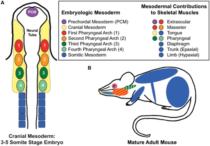
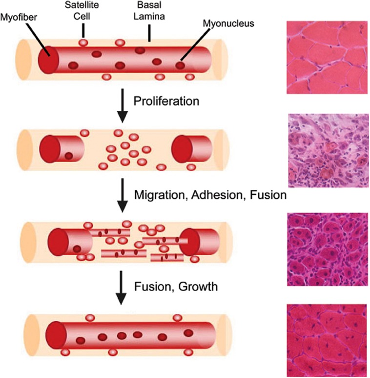
The human body contains approximately 640 individual skeletal muscles. Despite the fact that all of these muscles are composed of striated muscle tissue, the biology of these muscles and their associated muscle stem cell populations are quite diverse. Skeletal muscles are affected differentially by various muscular dystrophies (MDs), such that certain genetic mutations specifically alter muscle function in only a subset of muscles. Additionally, defective muscle stem cells have been implicated in the pathology of some MDs. The biology of muscle stem cells varies depending on the muscles with which they are associated. Here we review the biology of skeletal muscle stem cell populations of eight different muscle groups. Understanding the biological variation of skeletal muscles and their resident stem cells could provide valuable insight into mechanisms underlying the susceptibility of certain muscles to myopathic disease.
Keywords: craniofacial; diaphragm; epaxial; hypaxial; muscle; muscle stem cell; muscular dystrophy; satellite cell.
Figures
Similar articles
-
Human skeletal muscle organoids model fetal myogenesis and sustain uncommitted PAX7 myogenic progenitors.Elife. 2023 Nov 14;12:RP87081. doi: 10.7554/eLife.87081. Elife. 2023. PMID: 37963071 Free PMC article.
-
Enhanced Diaphragm Muscle Function upon Satellite Cell Transplantation in Dystrophic Mice.Int J Mol Sci. 2024 Feb 21;25(5):2503. doi: 10.3390/ijms25052503. Int J Mol Sci. 2024. PMID: 38473751 Free PMC article.
-
Distinct Embryonic Origin and Injury Response of Resident Stem Cells in Craniofacial Muscles.Front Physiol. 2021 Jul 1;12:690248. doi: 10.3389/fphys.2021.690248. eCollection 2021. Front Physiol. 2021. PMID: 34276411 Free PMC article. Review.
-
Novel concept for the epaxial/hypaxial boundary based on neuronal development.J Anat. 2020 Sep;237(3):427-438. doi: 10.1111/joa.13219. Epub 2020 Aug 12. J Anat. 2020. PMID: 32786168 Free PMC article.
-
Direct effects of the pathogenic mutation on satellite cell function in muscular dystrophy.Exp Cell Res. 2010 Nov 1;316(18):3100-8. doi: 10.1016/j.yexcr.2010.05.014. Epub 2010 May 28. Exp Cell Res. 2010. PMID: 20546725 Review.
Cited by
-
The body region specificity in murine models of muscle regeneration and atrophy.Acta Physiol (Oxf). 2021 Jan;231(1):e13553. doi: 10.1111/apha.13553. Epub 2020 Sep 17. Acta Physiol (Oxf). 2021. PMID: 32875719 Free PMC article.
-
Myogenic marker expression as a function of age and exercise-based therapy in the tongue.Exp Gerontol. 2020 Dec;142:111104. doi: 10.1016/j.exger.2020.111104. Epub 2020 Oct 2. Exp Gerontol. 2020. PMID: 33017670 Free PMC article.
-
Dosage effect of multiple genes accounts for multisystem disorder of myotonic dystrophy type 1.Cell Res. 2020 Feb;30(2):133-145. doi: 10.1038/s41422-019-0264-2. Epub 2019 Dec 18. Cell Res. 2020. PMID: 31853004 Free PMC article.
-
One-Month-Old Rabbits Exhibit a Longer Postoperative Remodeling in Extraocular Muscles Compared to 3-Month-Old Rabbits.Invest Ophthalmol Vis Sci. 2025 Mar 3;66(3):12. doi: 10.1167/iovs.66.3.12. Invest Ophthalmol Vis Sci. 2025. PMID: 40042875 Free PMC article.
-
Wnt7a promotes muscle regeneration in branchiomeric orbicularis oris muscle.Int J Clin Exp Pathol. 2021 Jun 15;14(6):693-704. eCollection 2021. Int J Clin Exp Pathol. 2021. PMID: 34239670 Free PMC article.
References
Publication types
Grants and funding
LinkOut - more resources
Full Text Sources
Other Literature Sources
Research Materials
Miscellaneous