Review
. 2015 Oct 6:6:781.
doi: 10.3389/fpls.2015.00781. eCollection 2015.

Chloroplast signaling within, between and beyond cells

Affiliations
Review

Chloroplast signaling within, between and beyond cells

Krzysztof Bobik et al. Front Plant Sci. .

Abstract

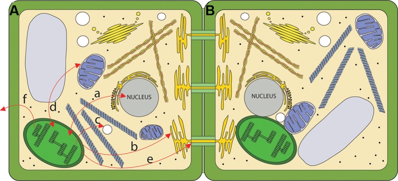
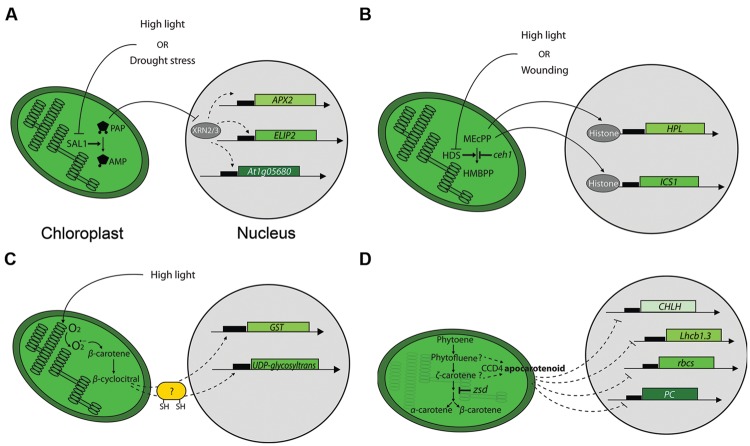
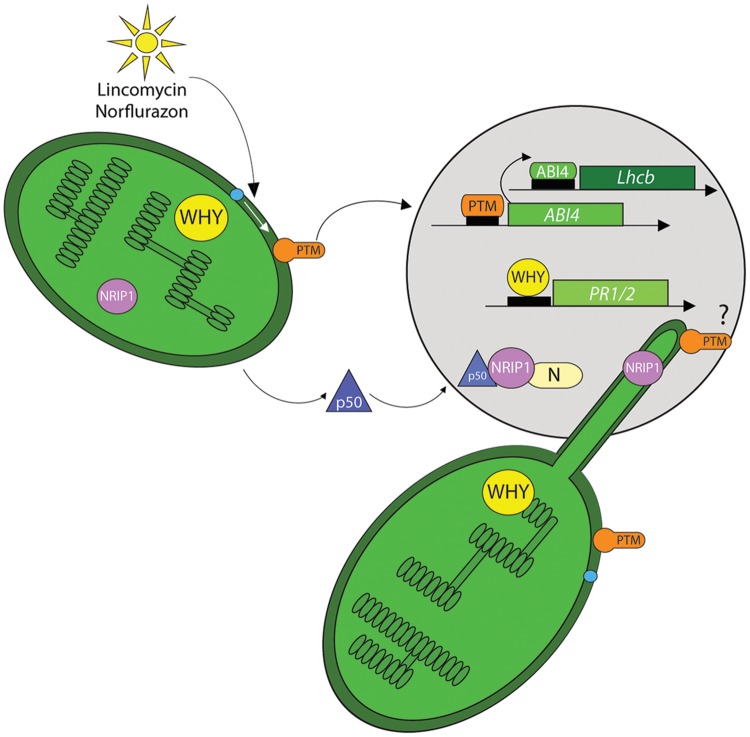
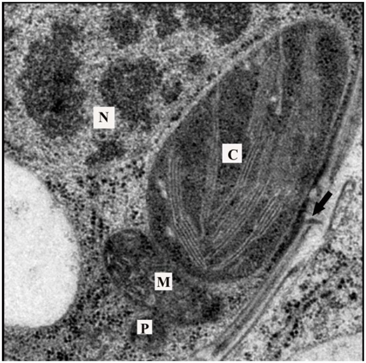
The most conspicuous function of plastids is the oxygenic photosynthesis of chloroplasts, yet plastids are super-factories that produce a plethora of compounds that are indispensable for proper plant physiology and development. Given their origins as free-living prokaryotes, it is not surprising that plastids possess their own genomes whose expression is essential to plastid function. This semi-autonomous character of plastids requires the existence of sophisticated regulatory mechanisms that provide reliable communication between them and other cellular compartments. Such intracellular signaling is necessary for coordinating whole-cell responses to constantly varying environmental cues and cellular metabolic needs. This is achieved by plastids acting as receivers and transmitters of specific signals that coordinate expression of the nuclear and plastid genomes according to particular needs. In this review we will consider the so-called retrograde signaling occurring between plastids and nuclei, and between plastids and other organelles. Another important role of the plastid we will discuss is the involvement of plastid signaling in biotic and abiotic stress that, in addition to influencing retrograde signaling, has direct effects on several cellular compartments including the cell wall. We will also review recent evidence pointing to an intriguing function of chloroplasts in regulating intercellular symplasmic transport. Finally, we consider an intriguing yet less widely known aspect of plant biology, chloroplast signaling from the perspective of the entire plant. Thus, accumulating evidence highlights that chloroplasts, with their complex signaling pathways, provide a mechanism for exquisite regulation of plant development, metabolism and responses to the environment. As chloroplast processes are targeted for engineering for improved productivity the effect of such modifications on chloroplast signaling will have to be carefully considered in order to avoid unintended consequences on plant growth and development.

Keywords: cell wall; phytohormones; plasmodesmata; plastid signaling; redox; retrograde signaling; stress responses; stromules.


Figures

FIGURE 1
FIGURE 1
Routes for chloroplast signaling. (A) Chloroplasts generate signals that target multiple intercellular targets. (a) The majority of chloroplast proteins are encoded by the nucleus, and the import of those proteins into the chloroplast is anterograde signaling. In turn, several chloroplast products act as retrograde signals to regulate expression of nucleus-encoded genes. (b) Chloroplasts are metabolically coupled to the ER and it is likely that signals may move from the chloroplast to the ER. (c) Chloroplasts and peroxisomes are also closely associated, and numerous chloroplast products are substrates for peroxisomal pathways. (d) Mitochondria and chloroplasts are known to signal to each other. (e) Chloroplast signals regulate intercellular trafficking via plasmodesmata. It is not clear if this signaling is direct or involves retrograde signaling to the nucleus. (f) Chloroplasts produce volatile compounds that can signal to neighboring plants during pathogen attack. (B) The physical interaction between chloroplasts and various organelles may serve as a direct route for signaling.
FIGURE 2
FIGURE 2
Mechanisms of chloroplast-to-nucleus signaling. (A) Retrograde signaling by PAP. High light or drought stress inhibits SAL1 phosphatase and leads to the accumulation of PAP. PAP likely inhibits specific exoribonucleases (XRNs) to modify nuclear genes expression. APX2 and ELIP2 stand for ASCORBATE PEROXIDASE 2 and EARLY LIGHT INDUCIBLE PROTEIN 2 genes, respectively. (B) Retrograde signaling by MEcPP. High light or wounding inhibits 1-hydroxy-2-methyl-2-(E)-butenyl-4-diphosphate synthase (HDS), leading to the subsequent accumulation of MEcPP. MEcPP affects nuclear gene expression via a mechanism proposed to involve chromatin remodeling by destabilizing DNA-histone interactions. HPL and ICS1 stand for HYDROPEROXIDE LYASE and ISOCHORISMATE SYNTHASE 1genes, respectively. (C) Carotenoid-derivative β-cyclocitral mediates retrograde signaling. The ROS singlet oxygen induces formation of β-cyclocitral during high light treatment. β-cyclocitral’s action on selected nuclear genes is proposed to involve proteins containing sulphydryl groups. The genes depicted are GLUTATHIONE-S-TRANSPHERASE (GST) and UDP-glycosyltransferase. (D) An unidentified apocarotenoid affects expression of nuclear genes. It is proposed that the putative signaling apocarotenoid accumulates in chloroplasts due to compromised ζ-carotene desaturase activity that results in accumulation of phytofluene and ζ-carotene, putative substrates for the carotenoid cleavage deoxygenase 4 (CCD4) enzyme that is prerequisite for the putative apocarotenoid synthesis. CHLH, Lhcb1.3, rbcs and PC stand for genes encoding the subunit H of the Mg-chelatase complex, light-harvesting complex 1.3 isoform, the Rubisco small subunit and plastocyanin, respectively.
FIGURE 3
FIGURE 3
Chloroplast proteins as retrograde signals. A few chloroplast proteins have been implicated in directly modulating nuclear gene expression by their nuclear localization. These proteins may transit the cytoplasm by an unknown mechanism. Alternatively, it has also been proposed that they may move through stromules to enter the nucleus. High light, lincomycin or norflurazon treatments induce a serine protease-dependent (blue dot) proteolytic-cleavage of the PTM, a chloroplast envelope-bound plant homeodomain (PHD) transcription factor. The cleavage product is found in the nucleus where it binds to promoter region of the ABI4 transcription factor. ABI4 in turn associates with the regulatory sequences of the Lhcb genes and prevents their transcription. The chloroplast protein Whirly1 also localizes to the nucleus and this is correlated with increased expression of PATHOGENESIS RELATED GENE 1 and 2 (PR1/2). Upon TMV infection the chloroplast-localized NRIP1 is also detected in the nucleus where it interacts with the helicase domain of the TMV replicase (p50). Finally a trimeric complex of p50, NRIP1 and the N protein is localized to nucleus to provide resistance against the virus. It is suggested that NRIP1 may use stromules to translocate from the chloroplast to the nucleus.
FIGURE 4
FIGURE 4
Arrangement of organelles in a leaf cell. Transmission electron microscopy images often reveal chloroplasts in close proximity with peroxisomes and mitochondria. Chloroplasts can also be observed near the nucleus and cell wall. Note the presence of a plasmodesma in the cell wall (arrow). Such arrangements of organelles would minimize distances that signals must traverse to arrive at their target. C, chloroplast; M, mitochondria; P, peroxisome; N, nucleus.
FIGURE 5
FIGURE 5
Chloroplast behavior during defense. (A) Stromules (arrows) are observed intermittently from chloroplasts in the epidermis of Nicotiana benthamiana leaves. (B) Upon infection with Tobacco mosaic virus, chloroplasts cluster around the nucleus (asterisk), and stromule formation is induced (arrow). The chloroplasts shown are expressing NRIP1-CFP (Caplan et al., 2008, 2015). Note the NRIP1-CFP signal detected in the nucleus in TMV-infected chloroplasts, indicative of the translocation of NRIP1 from the chloroplasts to the nucleus. Images were collected on a Zeiss LSM 710 confocal laser scanning microscope and single focal plane images are shown. Scale bar is 10 μm.

References

    1. Abbink T. E., Peart J. R., Mos T. N., Baulcombe D. C., Bol J. F., Linthorst H. J. (2002). Silencing of a gene encoding a protein component of the oxygen-evolving complex of photosystem II enhances virus replication in plants. Virology 295 307–319. 10.1006/viro.2002.1332 - DOI - PubMed
    1. Adam Z. (2015). Plastid intramembrane proteolysis. Biochim. Biophys. Acta 1847 910–914. 10.1016/j.bbabio.2014.12.006 - DOI - PubMed
    1. Alazem M., Lin N. S. (2015). Roles of plant hormones in the regulation of host-virus interactions. Mol. Plant Pathol. 16 529–540. 10.1111/mpp.12204 - DOI - PMC - PubMed
    1. Albrecht V., Simkova K., Carrie C., Delannoy E., Giraud E., Whelan J., et al. (2010). The cytoskeleton and the peroxisomal-targeted snowy cotyledon3 protein are required for chloroplast development in Arabidopsis. Plant Cell 22 3423–3438. 10.1105/tpc.110.074781 - DOI - PMC - PubMed
    1. Ali M., Sugimoto K., Ramadan A., Arimura G. (2013). Memory of plant communications for priming anti-herbivore responses. Sci. Rep. 3 1872 10.1038/srep01872 - DOI - PMC - PubMed