. 2015 Oct 20:3:11.
doi: 10.1186/s40170-015-0138-0. eCollection 2015.

Targeting mitochondrial complex I using BAY 87-2243 reduces melanoma tumor growth

Affiliations

Targeting mitochondrial complex I using BAY 87-2243 reduces melanoma tumor growth

Laura Schöckel et al. Cancer Metab. .

Abstract

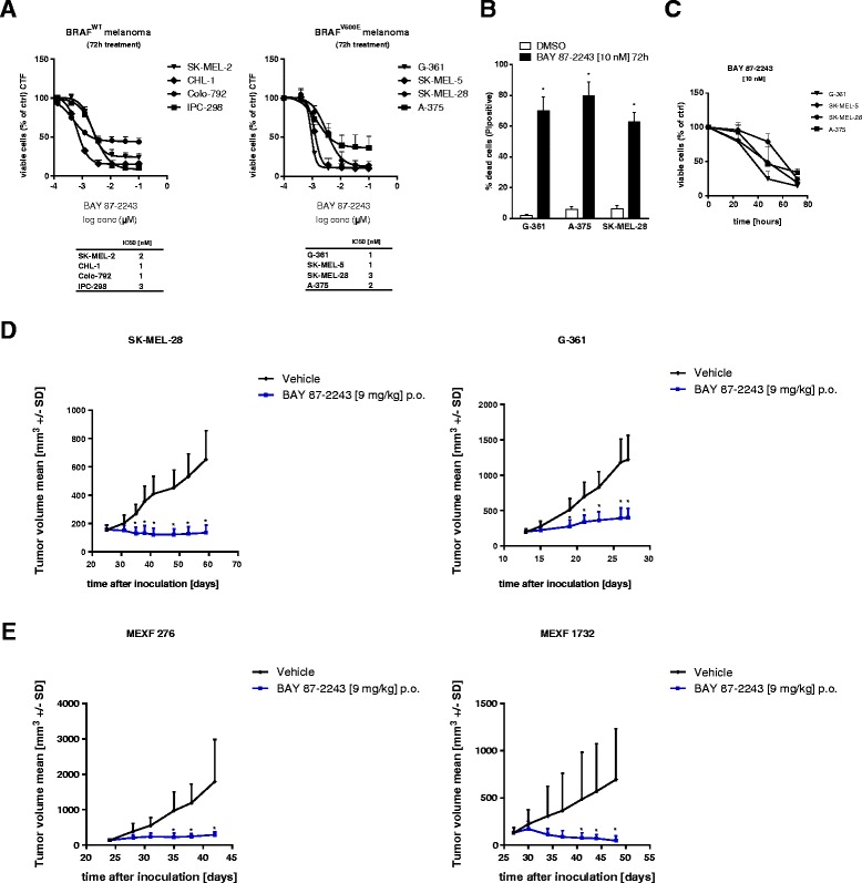
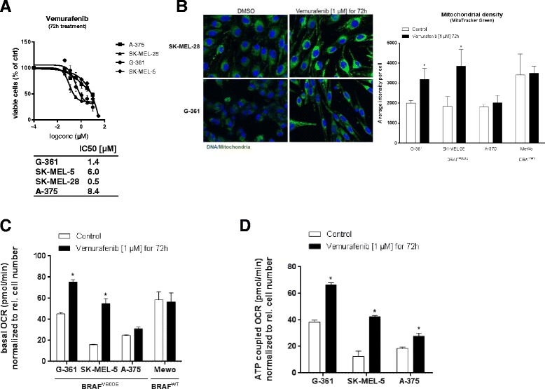
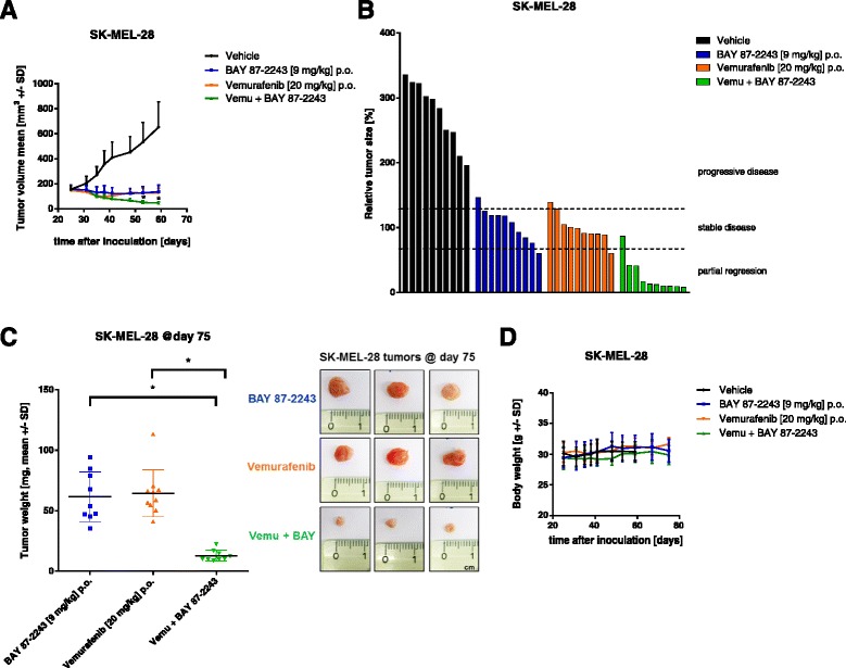
Background: Numerous studies have demonstrated that functional mitochondria are required for tumorigenesis, suggesting that mitochondrial oxidative phosphorylation (OXPHOS) might be a potential target for cancer therapy. In this study, we investigated the effects of BAY 87-2243, a small molecule that inhibits the first OXPHOS enzyme (complex I), in melanoma in vitro and in vivo.

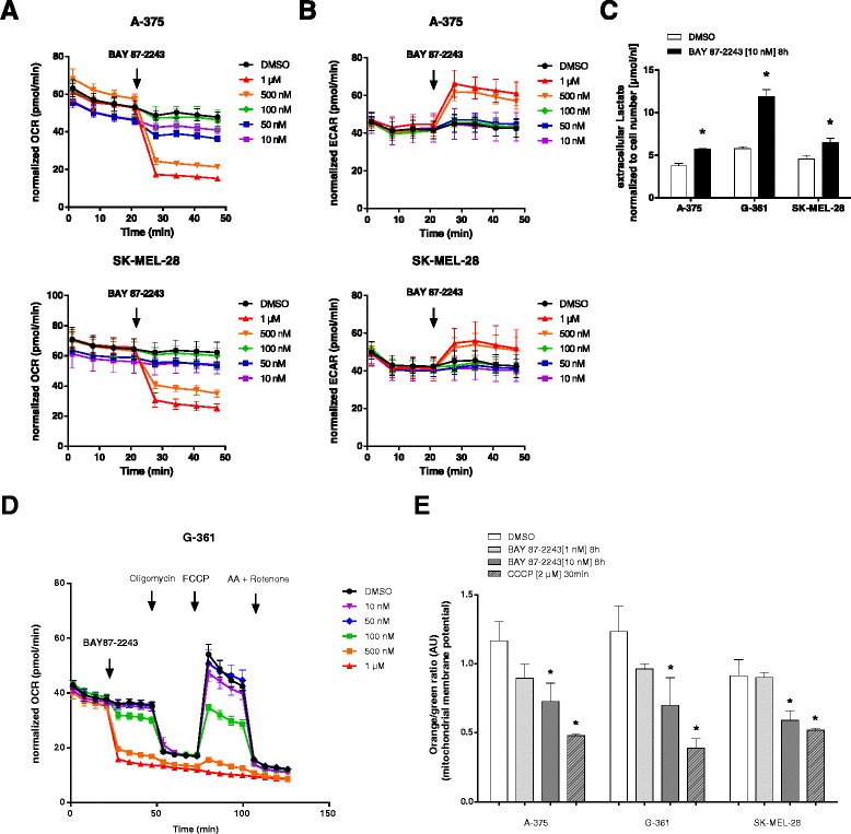
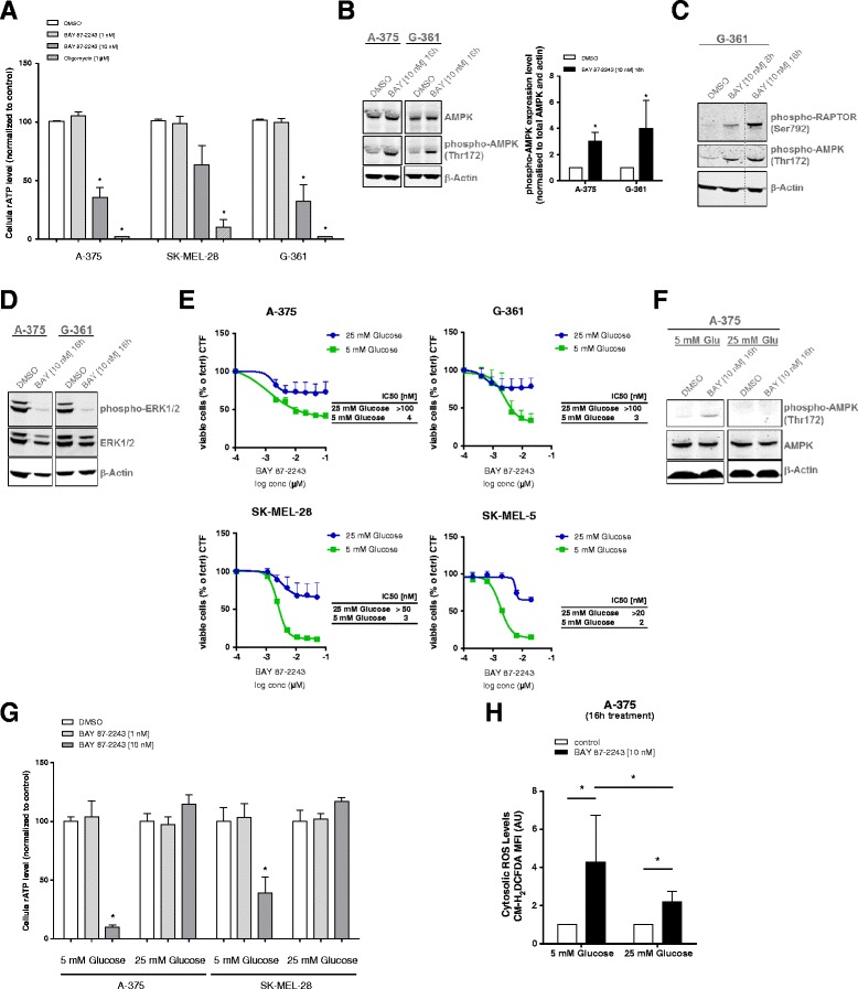
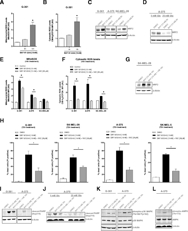
Results: BAY 87-2243 decreased mitochondrial oxygen consumption and induced partial depolarization of the mitochondrial membrane potential. This was associated with increased reactive oxygen species (ROS) levels, lowering of total cellular ATP levels, activation of AMP-activated protein kinase (AMPK), and reduced cell viability. The latter was rescued by the antioxidant vitamin E and high extracellular glucose levels (25 mM), indicating the involvement of ROS-induced cell death and a dependence on glycolysis for cell survival upon BAY 87-2243 treatment. BAY 87-2243 significantly reduced tumor growth in various BRAF mutant melanoma mouse xenografts and patient-derived melanoma mouse models. Furthermore, we provide evidence that inhibition of mutated BRAF using the specific small molecule inhibitor vemurafenib increased the OXPHOS dependency of BRAF mutant melanoma cells. As a consequence, the combination of both inhibitors augmented the anti-tumor effect of BAY 87-2243 in a BRAF mutant melanoma mouse xenograft model.

Conclusions: Taken together, our results suggest that complex I inhibition has potential clinical applications as a single agent in melanoma and also might be efficacious in combination with BRAF inhibitors in the treatment of patients with BRAF mutant melanoma.

Keywords: Anti-tumor efficacy; BRAF mutant melanoma; Cancer metabolism; Mitochondrial complex I; Oxidative phosphorylation (OXPHOS); Reactive oxygen species (ROS); Small molecule inhibitor.


Figures

Fig. 1
Fig. 1
Inhibition of mitochondrial complex I with BAY 87-2243 in melanoma cells induces cell death in vitro and reduces tumor growth in vivo. a BRAF wild type and mutant melanoma cells were treated with various concentrations of BAY 87-2243. Cell viability was measured after 72 h. IC50 values were calculated using GraphPad prism (n = 4). b Melanoma cells were treated with BAY 87-2243 (10 nM). Cell death was measured after 72 h using propidium iodine (n = 4). c Melanoma cells were treated with BAY 87-2243 (10 nM). Cell viability was measured after 24, 48, and 72 h (n = 3). d Nude mice were xenotransplanted with either 3 × 106 SK-MEL-28 (50 % matrigel) or 5 × 106 G-361 (100 % matrigel) human melanoma cells (n = 10 per group). When tumor area reached around 50 mm2, mice were treated once a day with vehicle or 9 mg/kg BAY 87-2243 per oral gavage (p.o.). e Nude mice bearing patient-derived (MEXF 276, MEXF 1732, n = 8 per group) melanoma xenograft tumors were treated orally (p.o.), once a day with vehicle or 9 mg/kg BAY 87-2243. Data are represented as the mean ± SD. *p < 0.05
Fig. 2
Fig. 2
Inhibition of mitochondrial complex I with BAY 87-2243 blocks OXPHOS and triggers glycolysis in melanoma cells. a OCR was measured using Seahorse analyzer in BRAF mutant melanoma cells. BAY 87-2243 was injected (black arrow) in different concentrations (n = 6). b ECAR was measured using Seahorse analyzer in BRAF mutant melanoma cells. BAY 87-2243 was injected (black arrow) in different concentrations (n = 6). c Melanoma cells were treated with BAY 87-2243 (10 nM). Extracellular lactate was measured in cell culture supernatant after 8 h (n = 4). d OCR was measured using Seahorse analyzer in G-361 cells. BAY 87-2243 was injected (black arrow) in different concentrations followed by consecutive injections of oligomycin (1 μM), FCCP (0.5 μM), and antimycin A (1 μM)/rotenone (1 μM) (n = 6). e Melanoma cells were treated with different concentrations of BAY 87-2243. After 8 h, mitochondrial membrane potential was measured using Mito-ID assay (n = 3). CCCP (2 μM) was used as positive control. Data are represented as the mean ± SD. *p < 0.05
Fig. 3
Fig. 3
Complex I inhibition using BAY 87-2243 reduces ATP levels and induces an energy crisis in melanoma cells. a Melanoma cells were treated with different concentrations of BAY 87-2243 and ATP was measured after 8 h. Oligomycin (1 μM) served as positive control (n = 3). b, c Melanoma cells were treated with BAY 87-2243 (10 nM). Cell lysates were collected from treated cells (16 h) and probed with antibodies recognizing phospho-AMPK (Thr172), total AMPK, c phospho-RAPTOR (Ser792), d anti-phospho-ERK1/2 (Thr202/Tyr204), and anti-ERK1/2. Actin was used as a loading control. e Melanoma cells were treated with various concentrations of BAY 87-2243. Cell viability was measured in high (25 mM) and low (5 mM) glucose medium after 72 h (n = 3). IC50 values were calculated using GraphPad Prism. f A-375 cells were treated with BAY 87-2243 (10 nM) under low- (5 mM) and high (25 mM) glucose conditions. Cell lysates were collected from treated cells (16 h) and probed with antibodies recognizing phospho-AMPK (Thr172) and total AMPK. Actin was used as a loading control. g Melanoma cells were treated with BAY 87-2243 (10 nM) under low- (5 mM) and high (25 mM) glucose conditions and ATP was measured after 16 h (n = 3). h A-375 cells were treated with BAY 87-2243 (10 nM) under low- (5 mM) and high (25 mM) glucose conditions. Cytosolic ROS levels were measured using the ROS-reactive dye CM-H2DCFDA after 16 h (n = 3). Data are represented as the mean ± SD. *p < 0.05
Fig. 4
Fig. 4
BAY 87-2243-mediated complex I inhibition increases mitochondrial and cytosolic ROS levels resulting in ROS-mediated cell death. a G-361 cells were treated with BAY 87-2243 (10 nM). Mitochondrial ROS levels were measured using the MitoSOX dye after 8 and 16 h (n = 3). b G-361 cells were treated with BAY 87-2243 (10 nM). Cytosolic ROS levels were measured using the ROS-reactive dye CM-H2DCFDA after 8 and 16 h (n = 3). cd Melanoma cells were treated with BAY 87-2243 (10 nM) under (c and d) low- (5 mM) and d high (25 mM) glucose conditions. Cell lysates were collected from treated melanoma cell lines (8 h) and probed with antibodies recognizing NRF2. Actin was used as a loading control. ef Melanoma cells were treated for 16 h with BAY 87-2243 (10 nM) alone or in combination with the antioxidant vitamin E (25 μM). e Mitochondrial and f cytosolic ROS levels were analyzed using flow cytometry (n = 3). g SK-MEL-28 cells were treated with BAY 87-2243 (10 nM) alone or in combination with vitamin E (25 μM). Cell lysates collected from treated SK-MEL-28 cells (16 h) were probed with antibodies recognizing NRF2. Actin was used as a loading control. h BRAF mutant melanoma cells were treated with BAY 87-2243 (10 nM) alone or in combination with vitamin E (25 μM). Cell death was measured after 72 h using propidium iodine (n = 3). ij Melanoma cells were treated with BAY 87-2243 (10 nM) alone or in combination with vitamin E (25 μM) under (i and j) low- and j high glucose conditions. Cell lysates collected from treated A-375 and G-361 cells (16 h) were probed with antibodies recognizing cleaved PARP. Actin was used as a loading control. kl BRAF mutant melanoma cells were treated with BAY 87-2243 (10 nM) alone or in combination with vitamin E (25 μM). Cell lysates collected from treated cells (8 and 16 h) were probed with antibodies recognizing k phospho-p38 MAPK and total p38 MAPK and l phospho-AMPK (Thr172) and total AMPK. Actin was used as a loading control. Data are represented as the mean ± SD. *p < 0.05
Fig. 5
Fig. 5
Vemurafenib increases mitochondrial density and respiration in BRAF mutant melanoma cells. a BRAF mutant melanoma cells were treated with various concentrations of vemurafenib. Cell proliferation was measured after 72 h. IC50 values were calculated using GraphPad prism (n = 4). b MitoTracker Green fluorescence (left) and quantification (right) (average intensity per cell) of BRAF mutant and BRAF wild type melanoma cells treated with vemurafenib (1 μM) for 72 h (n = 3). c OCR was measured using Seahorse analyzer in BRAF mutant and BRAF wild type melanoma cells after treatment with vemurafenib (1 μM) for 72 h (n = 3). d ATP-coupled OCR (after injection of oligomycin (1 μM)) was analyzed using Seahorse in BRAF mutant melanoma cells after treatment with vemurafenib (1 μM) for 72 h (n = 3). Data are represented as the mean ± SD. *p < 0.05
Fig. 6
Fig. 6
Inhibition of complex I using BAY 87-2243 in combination with vemurafenib attenuated melanoma tumor growth in vivo. a Nude mice were xenotransplanted with 3 × 106 SK-MEL-28 (50 % matrigel) human melanoma cells (n = 10 per group). Mice were treated orally (p.o.) with vemurafenib (20 mg/kg, twice a day) or for combination studies with vemurafenib (20 mg/kg, twice a day) and BAY 87-2243 (9 mg/kg, once a day). Tumor volume was measured. b Relative tumor size (%) of individual SK-MEL-28 xenograft tumors (tumor area at study end × 100 / tumor area at study begin). Partial regression, <70 % (tumor size at study end > 30 % smaller than original tumor size at study start); stable disease, 70–120 % (tumor size between 30 % smaller and 20 % bigger of original size); progressive disease, >120 % (tumor size at study end > 20 % bigger than original size). c Tumor weights of individual SK-MEL-28 xenograft tumors. d Body weights of mice (n = 10) bearing human SK-MEL-28 xenografts treated orally (p.o.) once daily with vehicle (ethanol/solutol/water = 10:40:50), or with vemurafenib (20 mg/kg, twice a day), or for combination studies with vemurafenib (20 mg/kg, twice a day) and BAY 87-2243 (9 mg/kg, once a day). Data are represented as the mean ± SD. *p < 0.05

Similar articles

Cited by

References

    1. Cantor JR, Sabatini DM. Cancer cell metabolism: one hallmark, many faces. Cancer Discov. 2012;2(10):881–98. doi: 10.1158/2159-8290.CD-12-0345. - DOI - PMC - PubMed
    1. Warburg O. On the origin of cancer cells. Science. 1956;123(3191):309–14. doi: 10.1126/science.123.3191.309. - DOI - PubMed
    1. Warburg O, Wind F, Negelein E. The metabolism of tumors in the body. J Gen Physiol. 1927;8(6):519–30. doi: 10.1085/jgp.8.6.519. - DOI - PMC - PubMed
    1. Hensley CT, Wasti AT, DeBerardinis RJ. Glutamine and cancer: cell biology, physiology, and clinical opportunities. J Clin Invest. 2013;123(9):3678–84. doi: 10.1172/JCI69600. - DOI - PMC - PubMed
    1. Zaugg K, Yao Y, Reilly PT, Kannan K, Kiarash R, Mason J, et al. Carnitine palmitoyltransferase 1C promotes cell survival and tumor growth under conditions of metabolic stress. Genes Dev. 2011;25(10):1041–51. doi: 10.1101/gad.1987211. - DOI - PMC - PubMed