

Imaging the spontaneous obliteration of a cerebral arteriovenous malformation using c-arm cone beam computed tomography: A case report
- PMID: 26500802
- PMCID: PMC4596051
- DOI: 10.4103/2152-7806.166174
Imaging the spontaneous obliteration of a cerebral arteriovenous malformation using c-arm cone beam computed tomography: A case report
Abstract
Background: Spontaneous occlusion of a cerebral arteriovenous malformation (AVM) without treatment is a rare occurrence.
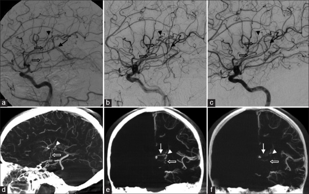
Case description: We report the case of a 56-year-old male who presented with aphasia and right hemiparesis secondary to intracerebral and intraventricular hemorrhage. Diagnostic digital subtraction angiography (DSA) and c-arm cone beam computed tomography (CBCT) demonstrated a 5 mm Spetzler-Martin Grade III left thalamic AVM drained by the internal cerebral vein. Subsequent DSA and CBCT studies confirmed the spontaneous obliteration of the AVM.
Conclusions: In this case, CBCT provided high resolution imaging of the AVM. Future clinical use of CBCT as an adjunct to DSA may enhance the diagnostic and therapeutic imaging of vascular lesions.
Keywords: Arteriovenous malformation; c-arm cone beam computed tomography; digital subtraction angiography.
Figures

Similar articles
-
Cerebral arteriovenous malformation: Spetzler-Martin classification at subsecond-temporal-resolution four-dimensional MR angiography compared with that at DSA.Radiology. 2008 Jan;246(1):205-13. doi: 10.1148/radiol.2453061684. Epub 2007 Oct 19. Radiology. 2008. PMID: 17951352
-
Microsurgery for cerebral arteriovenous malformations: subgroup outcomes in a consecutive series of 288 cases.J Neurosurg. 2017 Apr;126(4):1056-1063. doi: 10.3171/2016.4.JNS153017. Epub 2016 Jun 10. J Neurosurg. 2017. PMID: 27285541 Clinical Trial.
-
Radiosurgery for low-grade intracranial arteriovenous malformations.J Neurosurg. 2014 Aug;121(2):457-67. doi: 10.3171/2014.1.JNS131713. Epub 2014 Mar 7. J Neurosurg. 2014. PMID: 24605839
-
Venous malformation serving as the draining vein of an adjoining arteriovenous malformation. Case report and review of the literature.Surg Neurol. 2001 Sep;56(3):170-4. doi: 10.1016/s0090-3019(01)00457-8. Surg Neurol. 2001. PMID: 11597644 Review.
-
Development of De Novo Arteriovenous Malformation Following Ischemic Stroke: Case Report and Review of Current Literature.World Neurosurg. 2016 Dec;96:608.e5-608.e12. doi: 10.1016/j.wneu.2016.09.062. Epub 2016 Sep 23. World Neurosurg. 2016. PMID: 27671884 Review.
Cited by
-
Usefulness of preoperative cone beam computed tomography and intraoperative digital subtraction angiography for dural arteriovenous fistula at craniocervical junction: Technical case report.Surg Neurol Int. 2019 Jan 18;10:5. doi: 10.4103/sni.sni_439_17. eCollection 2019. Surg Neurol Int. 2019. PMID: 30775059 Free PMC article.
-
Thalamic Arteriovenous Malformations: A Systematic Review of Presentation, Diagnostic Modalities, and Treatment Approaches.Cureus. 2025 Jan 6;17(1):e76995. doi: 10.7759/cureus.76995. eCollection 2025 Jan. Cureus. 2025. PMID: 39912036 Free PMC article. Review.
References
-
- Abdulrauf SI, Malik GM, Awad IA. Spontaneous angiographic obliteration of cerebral arteriovenous malformations. Neurosurgery. 1999;44:280–7. - PubMed
-
- Lee SK, Vilela P, Willinsky R, TerBrugge KG. Spontaneous regression of cerebral arteriovenous malformations: Clinical and angiographic analysis with review of the literature. Neuroradiology. 2002;44:11–6. - PubMed
-
- Mizutani T, Tanaka H, Aruga T. Total recanalization of a spontaneously thrombosed arteriovenous malformation. Case report. J Neurosurg. 1995;82:506–8. - PubMed
-
- Panciani PP, Fontanella M, Carlino C, Bergui M, Ducati A. Progressive spontaneous occlusion of a cerebellar AVM: Pathogenetic hypothesis and review of literature. Clin Neurol Neurosurg. 2008;110:502–10. - PubMed
Publication types
LinkOut - more resources
Full Text Sources
Other Literature Sources
