Review
. 2015 Oct 9:2:57.
doi: 10.3389/fmolb.2015.00057. eCollection 2015.

Metazoan Hsp70-based protein disaggregases: emergence and mechanisms

Affiliations
Review

Metazoan Hsp70-based protein disaggregases: emergence and mechanisms

Nadinath B Nillegoda et al. Front Mol Biosci. .

Abstract

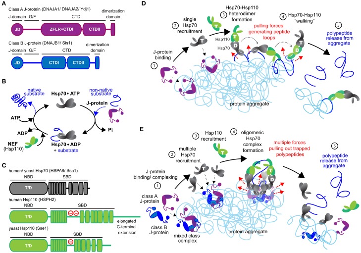
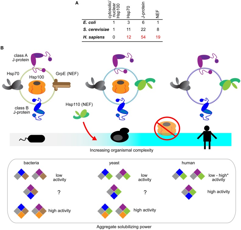
Proteotoxic stresses and aging cause breakdown of cellular protein homeostasis, allowing misfolded proteins to form aggregates, which dedicated molecular machines have evolved to solubilize. In bacteria, fungi, protozoa and plants protein disaggregation involves an Hsp70•J-protein chaperone system, which loads and activates a powerful AAA+ ATPase (Hsp100) disaggregase onto protein aggregate substrates. Metazoans lack cytosolic and nuclear Hsp100 disaggregases but still eliminate protein aggregates. This longstanding puzzle of protein quality control is now resolved. Robust protein disaggregation activity recently shown for the metazoan Hsp70-based disaggregases relies instead on a crucial cooperation between two J-protein classes and interaction with the Hsp110 co-chaperone. An expanding multiplicity of Hsp70 and J-protein family members in metazoan cells facilitates different configurations of this Hsp70-based disaggregase allowing unprecedented versatility and specificity in protein disaggregation. Here we review the architecture, operation, and adaptability of the emerging metazoan disaggregation system and discuss how this evolved.

Keywords: Hsp110; Hsp70; J-protein; metazoan; protein disaggregation.


Figures

Figure 1
Figure 1
Mechanistic models for Hsp70-based metazoan protein disaggregation. (A) Domain organization of class A and B J-proteins (as protomers). JD designates the conserved N-terminal J-domain. G/F denotes the glycine/phenylalanine rich flexible region; ZFLR, Zinc finger-like region; CTDI and CTDII, two homologous C-terminal β-sandwich substrate-binding domains. CTDs together with ZFLR provide substrate specificity. The dimerization domain forms functional J-protein homodimers. (B) Hsp70•J-protein•Hsp110 functional cycle. Concomitant interaction of Hsp70 with a J-protein and substrate results in allosteric stimulation of ATP hydrolysis trapping the substrate in Hsp70. Subsequent Hsp110 mediated ADP release from Hsp70 allows ATP rebinding, which triggers substrate release to complete Hsp70 cycle. (C) Schematic representation of the domain organization of yeast and human Hsp110 and Hsp70. (−) in red indicates the acidic region inserted between the terminal strands of the predicted β-sheet structure. The acidic loop determines the nuclear/cytoplasmic localization of human HSPH1 (Saito et al., 2009). The extended C-terminal domain is noted in HSPH2. ATP and ADP nucleotides bound to the NBD of Hsp70 and Hsp110s are denoted as “T” and “D,” respectively. (D) “Clamp and walk” model for Hsp70 and Hsp110 mediated protein disaggregation. Hsp70, J-protein and Hsp110 indicated in gray, purple and green, respectively. Nucleotide state at the NBDs of Hsp70 and Hsp110 indicated by T and D. Sequential reaction steps (encircled numbers): 1, J-protein targets aggregate; 2, J-protein recruits Hsp70; 3, Hsp110 recruitment and formation of Hsp70•Hsp110 heterodimer; 4, Hsp70•Hsp110 heterodimer “walking” on aggregate by alternating scanning (ATP state) and clamping (ADP state) substrate-interaction modes that generate pulling forces (dashed red arrows) on trapped polypeptides. Pulling forces result in forming peptide loops (dark blue) that fold to native-like conformations; 5, Releasing of polypeptides from aggregate due to accumulation of native-like folding events in trapped substrates. (E) Metazoan “nucleation” model for efficient Hsp70-based protein disaggregation. 1, J-protein targets and nucleates on aggregate; 2, Localized, multiple Hsp70 recruitment by J-protein assemblies on aggregates; 3, Hsp110 recruitment; 4, formation of oligomeric chaperone complex containing J-protein, Hsp110 and multiple Hsp70 molecules and buildup of entropic pulling forces (dashed red arrows) leading to extraction of trapped polypeptides (dark blue). 5, Hsp110 NEF activity triggered releasing of polypeptides from aggregate.
Figure 2
Figure 2
Evolution of the Hsp70-based protein disaggregases. (A) Tabulation of Hsp100, Hsp70, J-protein and nucleotide exchange factors in prokaryotes (E. coli), non-metazoan eukaryotes (S. cerevisiae), and metazoa (H. sapiens). The listings include experimentally established isoforms of the respective chaperones and co-chaperones (Genevaux et al., ; Lu et al., ; Saito et al., ; Kampinga and Craig, 2010). (B) Schematic diagram depicting the emergence of Hsp70-based protein disaggregases and loss of Hsp100 and Hsp70 bi-chaperone machines during evolution. Hsp100, GrpE (bacterial NEF), Hsp70, J-proteins and Hsp110 indicated in orange, brown, gray, purple (class A J-protein), blue (class B J-protein) and green, respectively. Hsp110 NEFs only appear in eukaryotes. Hsp100 is absent in metazoan cytosol and nucleus. Aggregate solubilization power of bacterial, yeast and human disaggregation systems indicated bellow. Color-coding depicts different components of the disaggregation systems. *Denotes the high protein disaggregation activity by Hsp70•JB1•Hsp110 configuration that rapidly disassemble α-synuclein fibrils (Gao et al., 2015).

References

    1. Andréasson C., Fiaux J., Rampelt H., Druffel-Augustin S., Bukau B. (2008). Insights into the structural dynamics of the Hsp110-Hsp70 interaction reveal the mechanism for nucleotide exchange activity. Proc. Natl. Acad. Sci. U.S.A. 105, 16519–16524. 10.1073/pnas.0804187105 - DOI - PMC - PubMed
    1. Arrasate M., Mitra S., Schweitzer E. S., Segal M. R., Finkbeiner S. (2004). Inclusion body formation reduces levels of mutant huntingtin and the risk of neuronal death. Nature 431, 805–810. 10.1038/nature02998 - DOI - PubMed
    1. Ben-Zvi A., De Los Rios P., Dietler G., Goloubinoff P. (2004). Active solubilization and refolding of stable protein aggregates by cooperative unfolding action of individual hsp70 chaperones. J. Biol. Chem. 279, 37298–37303. 10.1074/jbc.M405627200 - DOI - PubMed
    1. Bukau B., Horwich A. L. (1998). The Hsp70 and Hsp60 chaperone machines. Cell 92, 351–366. 10.1016/S0092-8674(00)80928-9 - DOI - PubMed
    1. Carroni M., Kummer E., Oguchi Y., Wendler P., Clare D. K., Sinning I., et al. . (2014). Head-to-tail interactions of the coiled-coil domains regulate ClpB activity and cooperation with Hsp70 in protein disaggregation. Elife 3:e02481. 10.7554/eLife.02481 - DOI - PMC - PubMed