Review
. 2015 Nov-Dec;7(6):518-26.
doi: 10.1177/1941738115587270. Epub 2015 May 18.

Current Concepts in Hip Preservation Surgery: Part I

Affiliations
Review

Current Concepts in Hip Preservation Surgery: Part I

Kelly L Adler et al. Sports Health. 2015 Nov-Dec.

Abstract

Context: An evolution in conceptual understanding, coupled with technical innovations, has enabled hip preservation surgeons to address complex pathomorphologies about the hip joint to reduce pain, optimize function, and potentially increase the longevity of the native hip joint. Technical aspects of hip preservation surgeries are diverse and range from isolated arthroscopic or open procedures to hybrid procedures that combine the advantages of arthroscopy with open surgical dislocation, pelvic and/or proximal femoral osteotomy, and biologic treatments for cartilage restoration.

Evidence acquisition: PubMed and CINAHL databases were searched to identify relevant scientific and review articles from January 1920 to January 2015 using the search terms hip preservation, labrum, surgical dislocation, femoroacetabular impingement, peri-acetabular osteotomy, and rotational osteotomy. Reference lists of included articles were reviewed to locate additional references of interest.

Study design: Clinical review.

Level of evidence: Level 4.

Results: Thoughtful individualized surgical procedures are available to optimize the femoroacetabular joint in the presence of hip dysfunction.

Conclusion: A comprehensive understanding of the relationship between femoral and pelvic orientation, morphology, and the development of intra-articular abnormalities is necessary to formulate a patient-specific approach to treatment with potential for a successful long-term result.

Keywords: arthroscopy; hip preservation; labrum; peri-acetabular osteotomy; surgical dislocation.


Conflict of interest statement

The following authors declared potential conflicts of interest: P. Christopher Cook, MD, FRCS(C), is a paid consultant for Arthrex. Yi-Meng Yen, MD, PhD, is a paid consultant for Orthopediatrics, Smith & Nephew, and Arthrex. Brian D. Giordano, MD, is a paid consultant for Arthrex.

Figures

Figure 1.
Figure 1.
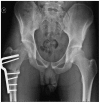
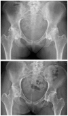
A 25-year-old woman with left lateral hip pain and right anterior groin pain. Plain radiographs demonstrate relative acetabular overcoverage on the right and undercoverage on the left due to leg length inequality and pelvic tilt. Acetabular undercoverage of the left hip placed increased demand on the peri-articular myofascial envelope (TFL, GMax, and ITB) and resulted in fatigue overload of these structures. Overcoverage on the right hip resulted in symptomatic FAI with labral and chondral injuries. Left proximal femoral shortening osteotomy corrected pelvic obliquity, normalized coverage, and rebalanced peri-articular myofascial stabilizers. FAI, femoroacetabular impingement; GMax, gluteus maximus; ITB, iliotibial band/complex; TFL, tensor fascia lata.
Figure 2.
Figure 2.
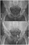
A 16-year-old swimmer with symptomatic right hip acetabualar dysplasia.
Figure 3.
Figure 3.
A 17-year-old soccer player with excessive proximal femoral valgus and concomitant symptomatic femoroacetabular impingement.
Figure 4.
Figure 4.
A 28-year-old recreational athlete with symptomatic left hip femoroacetabular impingement (FAI) and reduced femoral head-neck demonstrated on computed tomography with 3-dimensional reconstruction.
Figure 5.
Figure 5.
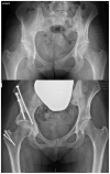
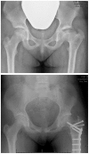
Pre- and postoperative radiographic images of a 32-year-old man with a low-volume acetabulum and concomitant femoroacetabular impingement (FAI) treated with arthroscopic FAI decompression and peri-acetabular osteotomy to correct structural instability, abnormal bony morphology, and intra-articular labral-chondral injury.
Figure 6.
Figure 6.
Pre- and postoperative images of a 26-year-old woman with acetabular dysplasia and AVN of the femoral head treated with peri-acetabular osteotomy and femoral head OATS using a vessel-sparing SD. AVN, avascular necrosis; OATS, osteoarticular allograft transplantation system; SD, surgical dislocation.
Figure 7.
Figure 7.
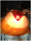
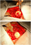
Intraoperative images depicting the exposed femoral head after osteoarticular allograft transplantation system (OATS) with excellent graft contour and smooth press-fit.
Figure 8.
Figure 8.
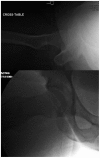
Preoperative lateral and intraoperative image demonstrating reduced femoral head-neck offset and subsequent correction using arthroscopic decompression.
Figure 9.
Figure 9.
Patient from Figure 2 after varus proximal femoral osteotomy. This patient also underwent arthroscopic femoroacetabular impingement decompression and labral repair.
Figure 10.
Figure 10.
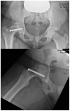
A 16-year-old soccer player with large osteochondral defect of the femoral head. The lesion was noted to have a stable articular surface and viable subchondral bone. Therefore, drilling and bioabsorbable compression screw fixation was used to stabilize the lesion.
Figure 11.
Figure 11.
A 14-year-old girl demonstrating sequelae of treatment for unstable slipped capital femoral epiphysis (SCFE) with subsequent severe rotational and structural abnormalities of the proximal femur.
Figure 12.
Figure 12.
A 15-year-old girl demonstrating sequelae of Legg-Calve-Perthes with severe femoral head deformity, coxa breva, and rotational abnormality of the proximal femur. Treatment consisted of osteochondroplasty of the femoral head via surgical dislocation, relative femoral neck lengthening, and proximal femoral osteotomy.
Figure 13.
Figure 13.
A 26-year-old woman with a history of graft-versus-host disease who developed widespread heterotopic bone formation as a result of her treatment. She underwent bilateral open femoracetabular impingement decompression via surgical dislocation without the use of trochanteric osteotomy.
Figure 14.
Figure 14.
Intraoperative image demonstrating labral-chondral injury treated arthroscopically during combined arthroscopic and open hip preservation surgery.

Similar articles

Cited by

References

    1. Albers C, Steppacher S, Ganz R, Siebenrock K, Tannast M. Optimal acetabular reorientation and offset correction improve the long term results after periacetabular osteotomy. J Bone Joint Surg Br. 2012;94(suppl 37):585.
    1. Albers CE, Steppacher SD, Ganz R, Siebenrock KA, Tannast M. Joint-preserving surgery improves pain, range of motion, and abductor strength after Legg-Calvé-Perthes disease. Clin Orthop Relat Res. 2012;470:2450-2461. - PMC - PubMed
    1. Anderson K, Strickland SM, Warren R. Hip and groin injuries in athletes. Am J Sports Med. 2001;29:521-533. - PubMed
    1. Anderson LA, Erickson JA, Severson E, Peters CL. Sequelae of Perthes disease: treatment with surgical hip dislocation and relative femoral neck lengthening. J Pediatr Orthop. 2010;30:758-766. - PMC - PubMed
    1. Arokoski JP, Jurvelin JS, Vaatainen U, Helminen HJ. Normal and pathological adaptations of articular cartilage to joint loading. Scand J Med Sci Sports. 2000;10:186-198. - PubMed