. 2016 Feb;34(2):405-17.
doi: 10.1002/stem.2235. Epub 2015 Nov 17.

Ataxia Telangiectasia Mutated Dysregulation Results in Diabetic Retinopathy

Affiliations

Ataxia Telangiectasia Mutated Dysregulation Results in Diabetic Retinopathy

Ashay D Bhatwadekar et al. Stem Cells. 2016 Feb.

Abstract

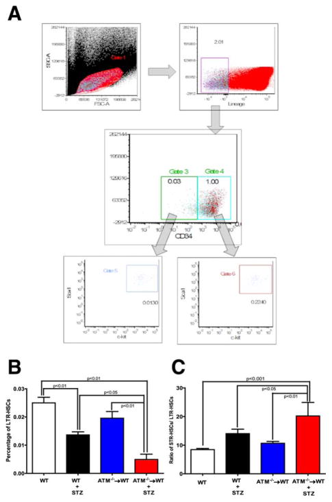
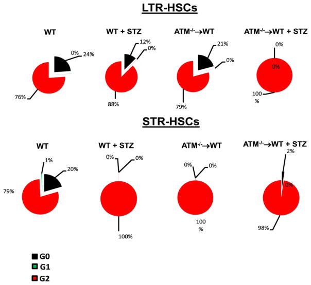
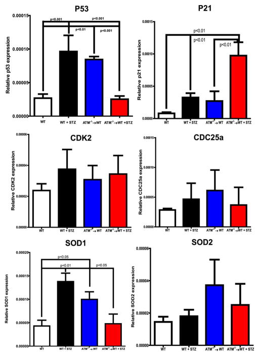
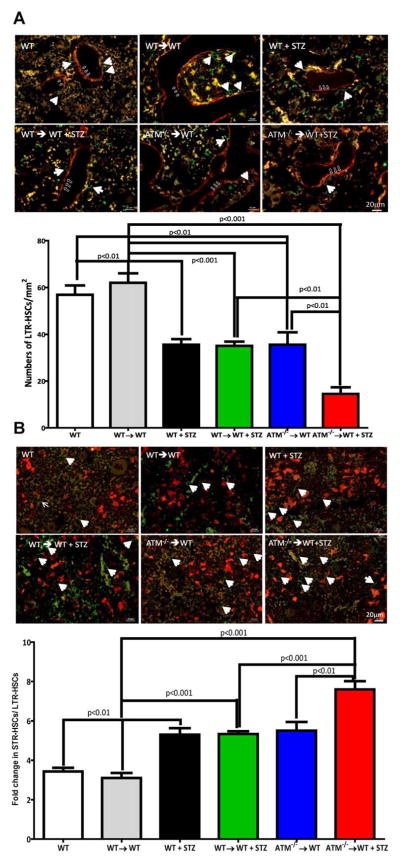
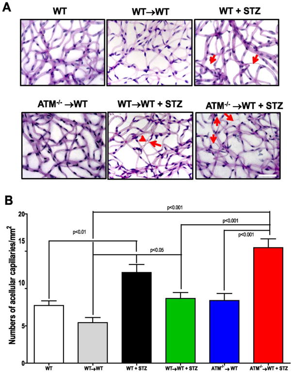
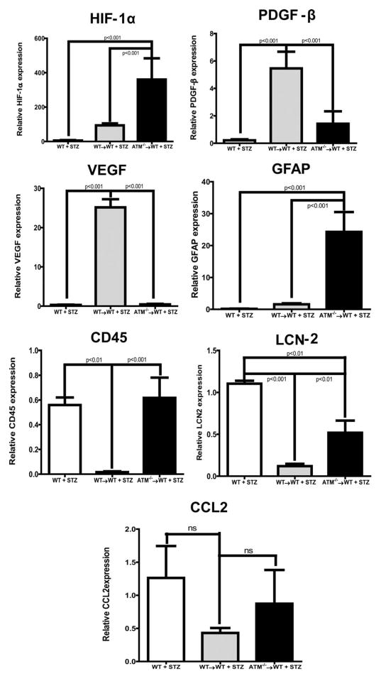
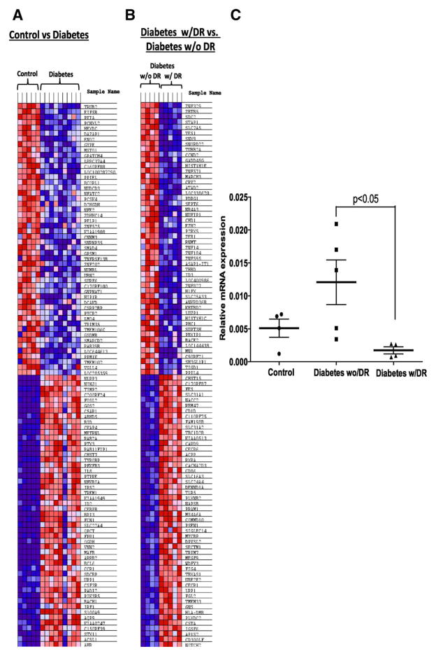
Ataxia telangiectasia mutated (ATM) acts as a defense against a variety of bone marrow (BM) stressors. We hypothesized that ATM loss in BM-hematopoietic stem cells (HSCs) would be detrimental to both HSC function and microvascular repair while sustained ATM would be beneficial in disease models of diabetes. Chronic diabetes represents a condition associated with HSC depletion and inadequate vascular repair. Gender mismatched chimeras of ATM(-/-) on wild type background were generated and a cohort were made diabetic using streptozotocin (STZ). HSCs from the STZ-ATM(-/-) chimeras showed (a) reduced self-renewal; (b) decreased long-term repopulation; (c) depletion from the primitive endosteal niche; (d) myeloid bias; and (e) accelerated diabetic retinopathy (DR). To further test the significance of ATM in hematopoiesis and diabetes, we performed microarrays on circulating angiogenic cells, CD34(+) cells, obtained from a unique cohort of human subjects with long-standing (>40 years duration) poorly controlled diabetes that were free of DR. Pathway analysis of microarrays in these individuals revealed DNA repair and cell-cycle regulation as the top networks with marked upregulation of ATM mRNA compared with CD34(+) cells from diabetics with DR. In conclusion, our study highlights using rodent models and human subjects, the critical role of ATM in microvascular repair in DR.

Keywords: Ataxia telangiectasia mutated; Diabetic retinopathy; Hematopoietic stem cells.


Conflict of interest statement

of Potential Conflicts of Interest The authors indicate no potential conflicts of interest.

Figures

Figure 1
Figure 1
Hematopoietic stem cell (HSC) imbalance in diabetic ataxia telangiectasia mutated (ATM)−/−→ wild type (WT) chimeras. (A): Representative scheme for flow cytomtery dot plot used to differentiate and quantify long-term repopulating (LTR) and short-term repopulating (STR)-HSCs. LinScal1+ c-kit+ CD34 and LinScal1+ c-kit+ CD34+ were identified as LTR-HSCs and STR-HSCs, respectively. Bar chart showing a quantification of (B) LTR-HSCs and (C) STR-HSCs. WT (n = 12), WT+ STZ (n = 5), ATM−/−→WT (n = 5), ATM−/−→WT (n = 5). Statistical test one way analysis of variance followed by Student’s Newman Keul Test. Abbreviations: ATM, ataxia telangiectasia mutated; FSCA, forward-scattered light area; HSC, hematopoietic stem cell; LTR, long-term repopulating; SSCA, side-scattered light area; STZ, streptozotocin; WT, wild type.
Figure 2
Figure 2
Decrease in quiescent long-term repopulating (LTR)-hematopoietic stem cells (HSCs) and short-term repopulating (STR)-HSCs in ataxia telangiectasia mutated (ATM)−/−→ wild type (WT) mice. Cell cycle status for LTR and STR-HSCs was determined following staining with Pyronin Y and Hoechst blue. Top panel showing a pie chart for the percentage of LTR-HSCs in different stages of cell cycle; diabetes and ATM−/−→WT caused a significant decrease in quiescent (G0) LTR-HSCs with an increase in the numbers of cells in active cell cycle (G1 and G2). Bottom panel showing a significant lack in quiescent cells with an increase in cell numbers in the phase of the active cycle in all three groups except wild type (WT) animals. WT (n = 12), WT+ STZ (n = 5), ATM−/−→WT (n = 5), ATM−/−→WT (n = 5) Statistical test-one way analysis of variance followed by Student’s Newman Keul Test. Abbreviations: ATM, ataxia telangiectasia mutated; HSC, hematopoietic stem cell; LTR, long-term repopulating; STR, short-term repopulating; STZ, streptozotocin; WT, wild type.
Figure 3
Figure 3
mRNA expression of cell cycle check points and anti-oxidant enzymes in bone marrow cells. mRNA expression on bone marrow cell pellet was determined using q-RT-PCR. Bar chart showing expression of P53, P21, CDK2, CDC25a, SOD1 and SOD2. Wild type (WT) (n = 4), WT+ STZ (n = 4), ataxia telangiectasia mutated (ATM)−/−→WT (n = 5), ATM−/−→WT (n = 5). Statistical Test- One way analysis of variance followed by Student’s Newman Keul Test. Abbreviations: ATM, ataxia telangiectasia mutated; SOD, Superoxide dismutase; STZ, streptozotocin; WT, wild type.
Figure 4
Figure 4
Reduced bone marrow engraftment in diabetic ataxia telangiectasia mutated (ATM)−/−→wild type (WT) chimeras. (A): Demineralized mouse femurs were stained for n-cadherin (red) and c-kit (green) antibodies to identify long-term repopulating (LTR)-hematopoietic stem cells (HSCs). Representative photomicrographs from respective groups showing c-kit positive cells (white arrows) in an endosteal niche (open arrow) while bar chart showing a quantification of LTR-HSCs. (B): Mouse femurs stained with ve-cadherin (red) to define the vascular niche. c-kit+ cells (white arrows) in ve-cadherin positive regions identified as short-term repopulating (STR)-HSC. Bar chart showing ratio of STR-HSCs to LTR-HSCs. Scale bar = 20 μm. WT (n = 6), WT→WT (n = 4), WT+ STZ (n = 5), WT→WT + STZ (n = 7), ATM−/−→WT (n = 5), ATM−/−→WT (n = 5). Statistical Test- One way analysis of variance followed by Student’s Newman Keul Test. Abbreviations: ATM, ataxia telangiectasia mutated; HSC, hematopoietic stem cell; LTR, long-term repopulating; STZ, streptozotocin; WT, wild type.
Figure 5
Figure 5
Accelerated diabetic retinopathy (DR) in ataxia telangiectasia mutated (ATM)−/−→ wild type (WT) chimeras. (A): Isolated mouse retinas were trypsin digested and stained with PAS-hematoxylin stain, top panel showing representative pictures of retinal trypsin digests from respective groups. Red arrows showing acellular capillaries as a marker of DR. Scale bar = 50 μm (B): Bar chart showing quantification of numbers of acellular capillaries. WT (n = 6), WT→WT (n = 8) WT+ STZ (n = 5), ATM−/−→WT (n = 5) WT→WT + STZ (n = 9), ATM−/−→WT (n = 5) Statistical Test- One way analysis of variance followed by Student’s Newman Keul Test. Abbreviations: ATM, ataxia telangiectasia mutated; STZ, streptozotocin; WT, wild type.
Figure 6
Figure 6
Increase in retinal inflammation of ataxia telangiectasia mutated (ATM)−/−→ wild type (WT) chimeric animals. qRT-PCR was performed on isolated retinas, bar chart showing mRNA expression of a variety of retinal inflammatory markers (HIF-1α, PDGF-β, vascular endothelial growth factor [VEGF], Glial fibrillary acidic protein [GFAP], CD45, Lipocalin-2 [LCN-2], and CCL2). WT (n = 5), WT→WT (n = 8) WT+ STZ (n = 4), WT→WT + STZ (n = 13) ATM−/−→WT (n = 5), ATM−/−→WT (n = 5). Statistical test-one way analysis of variance followed by Student’s Newman Keul Test. Abbreviations: ATM, ataxia telangiectasia mutated; CCL2, The chemokine (C-C motif) ligand 2; GFAP, Glial fibrillary acidic protein; HIF-1α, Hypoxia-inducible factor 1-alpha; LCN-2, Lipocalin-2; PDGF-β, Platelet-Derived Growth Factor Beta; STZ, streptozotocin; VEGF, vascular endothelial growth factor; WT, wild type.
Figure 7
Figure 7
Diabetic patients protected from microvascular complications map unique targets involved in DNA repair and cell cycle in CD34+ cells. (A): Gene set enrichment cluster analysis showing a heat map exhibiting distinct signature in diabetic group as compared with control. (B): a similar comparison between protected patients from diabetic retinopathy (DR) and patients with DR. (C): Dot plot showing mRNA expression of ataxia telangiectasia mutated in CD34+ cells of study participants; Control (n = 5), Diabetes w/o DR (n = 5), Diabetes w/ DR (n = 4). Statistical test-one way analysis of variance followed by Student’s Newman Keul Test. Abbreviation: DR, diabetic retinopathy.

References

    1. Engerman RL. Pathogenesis of diabetic retinopathy. Diabetes. 1989;38:1203–1206. - PubMed
    1. Kowluru RA, Chan PS. Oxidative stress and diabetic retinopathy. Exp Diabetes Res. 2007;2007:43603. - PMC - PubMed
    1. Curtis TM, Gardiner TA, Stitt AW. Microvascular lesions of diabetic retinopathy: Clues towards understanding pathogenesis? Eye (Lond) 2009;23:1496–1508. - PubMed
    1. Joussen AM, Doehmen S, Le ML, et al. TNF-alpha mediated apoptosis plays an important role in the development of early diabetic retinopathy and long-term histopathological alterations. Mol Vis. 2009;15:1418–1428. - PMC - PubMed
    1. Grant MB, May WS, Caballero S, et al. Adult hematopoietic stem cells provide functional hemangioblast activity during retinal neovascularization. Nat Med. 2002;8:607–612. - PubMed

Publication types

Substances