. 2015 Dec;21(12):917-29.
doi: 10.1093/molehr/gav058. Epub 2015 Oct 26.

Induction of interferon-λ contributes to TLR3 and RIG-I activation-mediated inhibition of herpes simplex virus type 2 replication in human cervical epithelial cells

Affiliations

Induction of interferon-λ contributes to TLR3 and RIG-I activation-mediated inhibition of herpes simplex virus type 2 replication in human cervical epithelial cells

Li Zhou et al. Mol Hum Reprod. 2015 Dec.

Abstract

Study hypothesis: Is it possible to immunologically activate human cervical epithelial cells to produce antiviral factors that inhibit herpes simplex virus type 2 (HSV-2) replication?

Study finding: Our results indicate that human cervical epithelial cells possess a functional TLR3/RIG-I signaling system, the activation of which can mount an Interferon-λ (IFN-λ)-mediated anti-HSV-2 response.

What is known already: There is limited information about the role of cervical epithelial cells in genital innate immunity against HSV-2 infection.

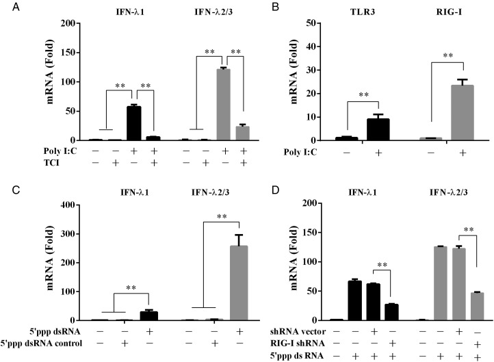
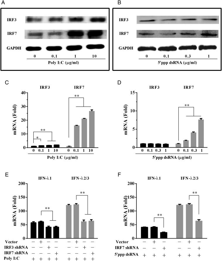
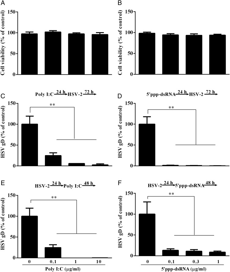
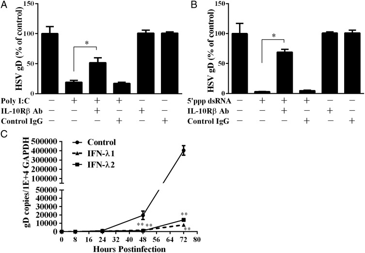
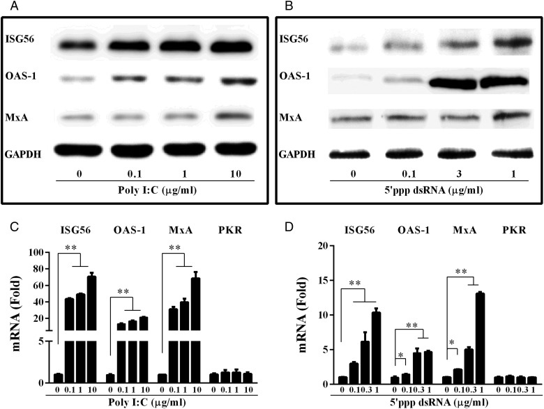
Study design, samples/materials, methods: We examined the expression of toll-like receptors (TLRs) and retinoic acid-inducible I (RIG-I) in End1/E6E7 cells by real-time PCR. The IFN-λ induced by TLR3 and RIG-I activation of End1/E6E7 cells was also examined by real-time PCR and ELISA. HSV-2 infection of End1/E6E7 cells was evaluated by the real-time PCR detection of HSV-2 gD expression. The antibody to IL-10Rβ was used to determine whether IFN-λ contributes to TLR3/RIG-I mediated HSV-2 inhibition. Expression of interferon regulatory factor 3 (IRF3), IRF7, IFN-stimulated gene 56 (ISG56), 2'-5'-oligoadenylate synthetase I (OAS-1) and myxovirus resistance A (MxA) were determined by the real-time PCR and western blot. End1/E6E7 cells were transfected with shRNA to knockdown the IRF3, IRF7 or RIG-I expression. Student's t-test and post Newman-Keuls test were used to analyze stabilized differences in the immunological parameters above between TLR3/RIG-I-activated cells and control cells.

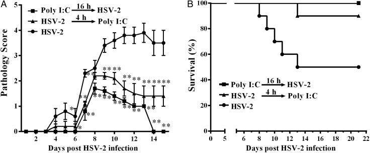
Main results and the role of chance: Human cervical epithelial cells expressed functional TLR3 and RIG-I, which could be activated by poly I:C and 5'ppp double-strand RNAs (5'ppp dsRNA), resulting in the induction of endogenous interferon lambda (IFN-λ). The induced IFN-λ contributed to TLR3/RIG-I-mediated inhibition of HSV-2 replication in human cervical epithelial cells, as an antibody to IL-10Rβ, an IFN-λ receptor subunit, could compromise TLR3/RIG-I-mediated inhibition of HSV-2. Further studies showed that TLR3/RIG-I signaling in the cervical epithelial cells by dsRNA induced the expression of the IFN-stimulated genes (ISGs), ISG56, 2'-5'-oligoadenylate synthetase I (OAS-1) and myxovirus resistance A (MxA), the key antiviral elements in the IFN signaling pathway. In addition, we observed that the topical treatment of genital mucosa with poly I:C could protect mice from genital HSV-2 infection.

Limitations, reasons for caution: Future prospective studies with primary cells and suitable animal models are needed in order to confirm these outcomes.

Wider implications of the findings: The findings provide direct and compelling evidence that there is intracellular expression and regulation of IFN-λ in human cervical epithelial cells, which may have a key role in the innate genital protection against viral infections.

Large scale data: Not applicable.

Study funding and competing interests: This work was supported by the National Natural Science Foundation of China (81301428 to L.Z. and 81271334 to W.-Z.H.), the Fundamental Research Funds for the Central Universities (2042015kf0188 to L.Z.), the China Postdoctoral Science Foundation (2013M531745 to L.Z.), the Development Program of China ('973', 2012CB518900 to W.-Z.H.) from the Ministry of Science and Technology of the People's Republic of China, grants (DA12815 and DA022177 to W.-Z.H.) from the National Institute on Drug Abuse (NIDA) and the open project of Hubei Key Laboratory of Wudang Local Chinese Medicine Research (WDCM005 to M.S.). The authors declare no competing financial interests.

Keywords: Toll-like receptor 3; herpes simplex virus type 2; human cervical epithelial cells; interferon-stimulated genes; interferon-λ; retinoic acid-inducible gene I.


Figures

Figure 1
Figure 1
Effect of TLRs activation on IFN-λ expression. (A) Expression of TLRs in End1/E6E7 cells. Total cellular RNA extracted from End1/E6E7 cells was subjected to the RT–PCR with the primers specific for toll-like receptors (TLRs) 1–10. Amplified PCR products were displayed on 2% agarose gel. (B) End1/E6E7 cells were transfected with poly I:C (10 μg/ml), imiquimod (10 μg/ml), ssRNA40 (10 μg/ml) or ODN2006 (5 μM) for 12 h. (C) End1/E6E7 cells were transfected with or without poly I:C (1 μg/ml) for the indicated times. Total cellular RNA was subjected to the real-time RT–PCR for the mRNA levels of IFN-λ1 and IFN-λ2/3 as indicated. (D and E) End1/E6E7 cells were transfected with or without poly I:C at indicated concentrations (1, 10 μg/ml) for mRNA (D) or protein (E) detection. (F and G) End1/E6E7 cells infected with or without HSV-2 (MOI = 0.001) for 24 h were then transfected with or without poly I:C (1 μg/ml) for mRNA (F) or for protein (G) detection. The results are the mean ± SD of triplicate cultures, representative of three experiments (*P < 0.05, **P < 0.01, N.S. as no significance difference).
Figure 2
Figure 2
Roles of TLR3 and RIG-I in poly I:C-mediated IFN-λ induction. (A) Effect of TLR3/dsRNA complex inhibitor (TCI) on the induction of IFN-λ by poly I:C. End1/E6E7 cells were pretreated with TCI (100 nM) for 1 h prior to poly I:C (1 μg/ml) transfection for 12 h. (B) End1/E6E7 cells were stimulated with 1 μg/ml poly I:C for 12 h. (C) End1/E6E7 cells were stimulated with 1 μg/ml 5′ppp dsRNA or 5′ppp dsRNA control for 12 h. (D) End1/E6E7 cells were pretransfected with RIG-I shRNA or control shRNA for 48 h prior to stimulation with 5′ppp dsRNA (1 μg/ml). Total cellular RNA was subjected to the real-time RT–PCR for the mRNA levels of IFN-λ1 and IFN-λ2/3 (A, C and D), TLR3 and RIG-I (B). The data are expressed as mRNA levels relative (-fold) to control (without poly I:C or 5′ppp dsRNA defined as 1). The results are mean ± SD of triplicate cultures, representative of three experiments (*P < 0.05, **P < 0.01).
Figure 3
Figure 3
Effect of TLR3/RIG-I activation on IFN regulatory factors (IRFs). (A and B) End1/E6E7 cells, transfected with or without poly I:C (A) or 5′ppp dsRNA (B) at indicated concentrations for 24 h, were subjected to western blot assay using antibodies to IRF3, IRF7 or GAPDH. GAPDH was used as the loading control. (C and D) End1/E6E7 cells were transfected with or without poly I:C (C) or 5′ppp dsRNA (D) at indicated concentrations for 12 h. (E and F) shRNA-mediated knockdown of IRF3 and/or IRF7 impaired IFN-λ expression. End1/E6E7 cells were transfected with or without control vector, IRF3 shRNA or IRF7 shRNA for 48 h. Cells were then transfected with or without poly I:C (E) or 5′ppp dsRNA (F) for additional 12 h. Total cellular RNA extracted from End1/E6E7 cells was subjected to the real-time RT–PCR detection for the mRNA levels of IRF3, IRF7 and IFN-λ. The data are expressed as mRNA levels relative (-fold) to control (without poly I:C or 5′ppp dsRNA stimulation, which is defined as 1). The results are mean ± SD of triplicate cultures, representative of three experiments (*P < 0.05, **P < 0.01).
Figure 4
Figure 4
Effect of TLR3, RIG-I activation on HSV-2 replication. (A and B) MTT assay of End1/E6E7 cells transfected with poly I:C or 5′ppp dsRNA. End1/E6E7 cells were transfected with poly I:C (A) or 5′ppp dsRNA (B) at the concentrations indicated at the bottom of the figure. Cells viability was assessed by MTT assay 72 h post transfection. The results are mean ± SD of triplicate cultures, representative of three experiments. (C and E) End1/E6E7 cells were transfected with poly I:C at the concentrations indicated at the bottom of the figure for 24 h prior to HSV-2 infection (MOI = 0.001) (C) or transfected with poly I:C at 24 h post-infection (E). (D and F) End1/E6E7 cells were transfected with 5′ppp dsRNA at concentrations indicated at the bottom of the figure for 24 h prior to HSV-2 infection (MOI = 0.001) (D) or transfected with 5′ppp dsRNA at 24 h post-infection (F). Genomic DNA extracted from the cell cultures at 72 h post-infection was subjected to the real-time PCR for HSV-2 quantification. The data are expressed as HSV-2 gD gene levels relative (%) to the control (without poly I:C treatment, which is defined as 100). The results are mean ± SD of triplicate cultures, representing three independent experiments (**P < 0.01).
Figure 5
Figure 5
The role of IFN-λ in TLR3/RIG-I activation-mediated HSV-2 inhibition. (A and B) End1/E6E7 cells were treated with poly I:C (1 μg/ml) (A) or 5′ppp dsRNA (B) and anti-IL-10Rβ antibody or control IgG at the concentration of 5 μg/ml for 24 h prior to HSV-2 infection. HSV-2 replication was quantified 72 h post-infection. (C) End1/E6E7 cells were treated with recombinant IFN-λ1 or IFN-λ2 (100 ng/ml) for 24 h and then infected with HSV-2 (MOI = 0.001). Genomic DNA extracted from the cell cultures at the indicated time points post-infection was subjected to the real-time PCR for HSV-2 quantification by using serial dilutions of HSV-2 gD standards with known copy numbers. The results are mean ± SD of triplicate cultures, representative of three experiments (*P < 0.05, **P < 0.01).
Figure 6
Figure 6
Effect of poly I:C and 5′ppp dsRNA on the expression of IFN-stimulated genes (ISGs). (A and B) Total proteins extracted from End1/E6E7 cells transfected with or without poly I:C (A) or 5′ppp dsRNA (B) at indicated concentrations for 24 h were subjected to western blot assay using antibodies to ISG56, MxA, OAS-1 or GAPDH. (C and D) End1/E6E7 cells were transfected with or without poly I:C (C) or 5′ ppp dsRNA (D) at indicated concentrations for 12 h. Total cellular RNA extracted from End1/E6E7 cells was subjected to the real-time RT–PCR detection for the mRNA levels of ISG56, OAS-1 MxA and PKR. The data are expressed as mRNA levels relative (-fold) to control (without treatment, defined as 1). The results are mean ± SD of triplicate cultures, representative of three experiments (*P < 0.05, **P < 0.01).
Figure 7
Figure 7
Effect of disease progression in HSV-2 infected mice. Poly I:C 25 μg suspended in 30 μl Lyovec transfection reagent was delivered to each mice prophylactically (n = 10) or therapeutically (n = 10), 16 h before or 4 h after, intravaginal inoculation of HSV-2 (3000 pfu/mouse). Mice were then monitored daily for genital pathology (A) and survival rate (B). Groups were compared for statistical significance relative to HSV-2 infection group (n = 10) by analysis of variance (*P < 0.05, **P < 0.01).

Similar articles

Cited by

References

    1. Alexopoulou L, Holt AC, Medzhitov R, Flavell RA. Recognition of double-stranded RNA and activation of NF-kappaB by Toll-like receptor 3. Nature 2001;413:732–738. - PubMed
    1. Ank N, Paludan SR. Type III IFNs: new layers of complexity in innate antiviral immunity. Biofactors 2009;35:82–87. - PubMed
    1. Ank N, West H, Bartholdy C, Eriksson K, Thomsen AR, Paludan SR. Lambda interferon (IFN-lambda), a type III IFN, is induced by viruses and IFNs and displays potent antiviral activity against select virus infections in vivo. J Virol 2006;80:4501–4509. - PMC - PubMed
    1. Ashkar AA, Yao XD, Gill N, Sajic D, Patrick AJ, Rosenthal KL. Toll-like receptor (TLR)-3, but not TLR4, agonist protects against genital herpes infection in the absence of inflammation seen with CpG DNA. J Infect Dis 2004;190:1841–1849. - PubMed
    1. Azulay-Debby H, Edry E, Melamed D. CpG DNA stimulates autoreactive immature B cells in the bone marrow. Eur J Immunol 2007;37:1463–1475. - PubMed

Publication types

MeSH terms