. 2016 May;124(5):563-9.
doi: 10.1289/ehp.1409200. Epub 2015 Oct 27.

Parabens and Human Epidermal Growth Factor Receptor Ligand Cross-Talk in Breast Cancer Cells

Affiliations

Parabens and Human Epidermal Growth Factor Receptor Ligand Cross-Talk in Breast Cancer Cells

Shawn Pan et al. Environ Health Perspect. 2016 May.

Abstract

Background: Xenoestrogens are synthetic compounds that mimic endogenous estrogens by binding to and activating estrogen receptors. Exposure to estrogens and to some xenoestrogens has been associated with cell proliferation and an increased risk of breast cancer. Despite evidence of estrogenicity, parabens are among the most widely used xenoestrogens in cosmetics and personal-care products and are generally considered safe. However, previous cell-based studies with parabens do not take into account the signaling cross-talk between estrogen receptor α (ERα) and the human epidermal growth factor receptor (HER) family.

Objectives: We investigated the hypothesis that the potency of parabens can be increased with HER ligands, such as heregulin (HRG).

Methods: The effects of HER ligands on paraben activation of c-Myc expression and cell proliferation were determined by real-time polymerase chain reaction, Western blots, flow cytometry, and chromatin immunoprecipitation assays in ERα- and HER2-positive human BT-474 breast cancer cells.

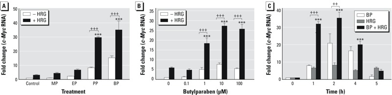
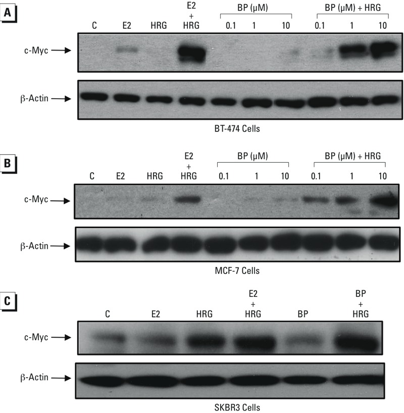
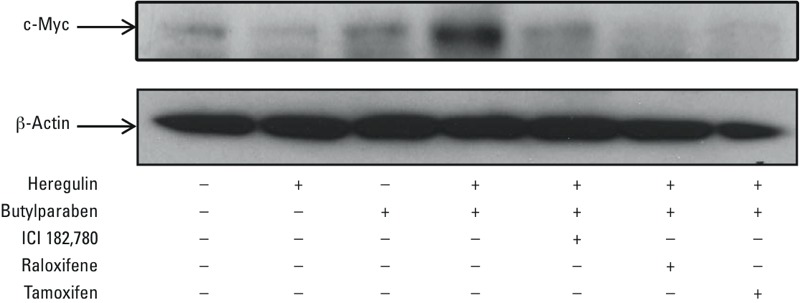
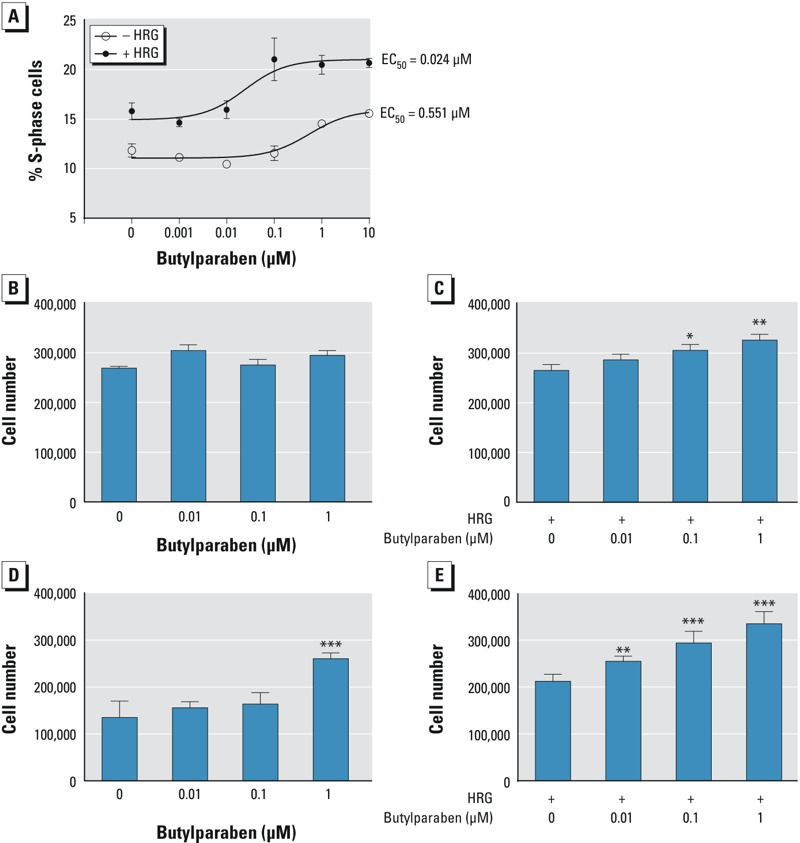
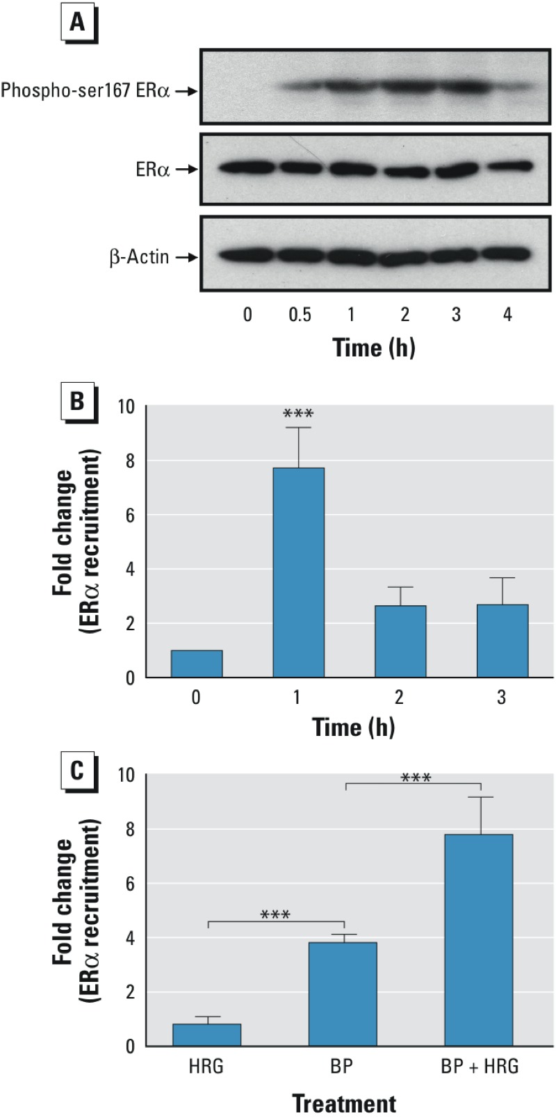
Results: Butylparaben (BP) and HRG produced a synergistic increase in c-Myc mRNA and protein levels in BT-474 cells. Estrogen receptor antagonists blocked the synergistic increase in c-Myc protein levels. The combination of BP and HRG also stimulated proliferation of BT-474 cells compared with the effects of BP alone. HRG decreased the dose required for BP-mediated stimulation of c-Myc mRNA expression and cell proliferation. HRG caused the phosphorylation of serine 167 in ERα. BP and HRG produced a synergistic increase in ERα recruitment to the c-Myc gene.

Conclusion: Our results show that HER ligands enhanced the potency of BP to stimulate oncogene expression and breast cancer cell proliferation in vitro via ERα, suggesting that parabens might be active at exposure levels not previously considered toxicologically relevant from studies testing their effects in isolation.

Citation: Pan S, Yuan C, Tagmount A, Rudel RA, Ackerman JM, Yaswen P, Vulpe CD, Leitman DC. 2016. Parabens and human epidermal growth factor receptor ligand cross-talk in breast cancer cells. Environ Health Perspect 124:563-569; http://dx.doi.org/10.1289/ehp.1409200.


Conflict of interest statement

The authors declare they have no actual or potential competing financial interests.

Figures

Figure 1
Figure 1
Effects of parabens and heregulin (HRG) on c-Myc transcript levels in BT-474 breast cancer cells. (A) Human BT-474 breast cancer cells were treated with 10 μM methylparaben (MP), 10 μM ethylparaben (EP), 10 μM propylparaben (PP), or 10 μM butylparaben (BP) for 2 hr in the absence and presence of HRG (20 ng/mL). (B) BT-474 breast cancer cells were treated with 0.01 μM 17β-estradiol (E2) or increasing concentrations (0.1 μM to 100 μM) of BP in the absence and presence of HRG (20 ng/mL) for 2 hr. (C) BT-474 breast cancer cells were treated with 10 μM BP in the absence or presence of HRG (20 ng/mL) for the indicated time periods. Relative mRNA levels for c-Myc were determined by real-time polymerase chain reaction (PCR) and were normalized to GAPDH using the comparative CT method. The fold changes were obtained by comparing the treated values with the control values. Each data point is the average of triplicate samples ± SEM. The figures are representative of three experiments with similar results. The statistical significance of the means was analyzed by two-way analysis of variance (ANOVA) followed by a post hoc Tukey’s multiple comparisons test to analyze the significance of the differences between the control and various parabens (A), various doses of BP (B), and various time periods (C) in the presence of HRG. Differences in c-Myc expression comparing cells receiving the same paraben treatment (A), the same dose of BP (B), and the same time period (C), but with and without HRG, were tested using a post hoc Bonferroni multiple comparisons test. ***< 0.001. Bonferroni multiple comparisons test: ++ < 0.01 and +++ < 0.001.
Figure 2
Figure 2
Effects of heregulin (HRG) on butylparaben (BP) stimulation of c-Myc protein levels in breast cancer cell lines. (A) BT-474 and (B) MCF-7 breast cancer cells were treated with the indicated concentrations of BP without or with HRG (20 ng/mL) for 2 hr. (C) SKBR3 cells were treated with 10 μM BP in the absence and presence of HRG (20 ng/mL) for 2 hr. The treated cells were then lysed, and the cellular lysates were prepared for Western blots using antibodies against c-Myc as described in “Materials and Methods.” Cells simultaneously treated with 17β-estradiol (E2) (0.01 μM) were also included for comparison. After exposure to an X-ray film, the membranes were washed and reprobed with an antibody against β-actin as a loading control. This figure is representative of two independent experiments with similar results.
Figure 3
Figure 3
Effects of estrogen receptor antagonists on the synergistic increase of c-Myc protein levels. BT-474 cells were treated with 10 μM butylparaben (BP) in the absence or presence of 1 μM ICI 182,780, 1 μM raloxifene, 10 μM tamoxifen, and 20 ng/mL HRG for 2 hr. The treated cells were then lysed, and the cellular lysates were prepared for Western blots using antibodies against c-Myc as described in “Materials and Methods.” After exposure to an X-ray film, the membranes were washed and reprobed by an antibody against β-actin as the loading control. This figure is representative of two independent experiments with similar results.
Figure 4
Figure 4
Effects of heregulin (HRG) on the potency of butylparaben (BP) stimulation of BT-474 cell proliferation. (A) BT-474 cells were treated with the indicated concentrations of BP alone or BP and HRG for 24 hr. Changes in cell cycle distribution were then analyzed by flow cytometry as described in “Materials and Methods.” The percentage of S-phase cells was plotted for different BP concentrations in the absence and presence of HRG. The plotted values are the means (± SD) of biological triplicates, which represent three independent experiments with similar results. (B,C) BT-474 cells were plated in 6-well dishes at 250,000 cells/well. The culture medium was then switched to serum-free medium 24 hr later, and the cells were treated with the indicated concentrations of BP in the absence (B) or presence (C) of HRG (20 ng/mL). The cells were harvested after 24 hr of treatment and then counted with a Coulter counter. The figures are representative of three experiments with similar results. The data are expressed as the means (± SD) obtained from biological triplicates. Statistical significance was analyzed by one-way analysis of variance (ANOVA) followed by a post hoc Tukey’s multiple comparison test to compare the differences between the control and each dose. (C) *< 0.05 and **< 0.01 compared with HRG alone. (D,E) BT-474 cells were plated in 6-well dishes at 50,000 cells/well and treated with indicated amounts of BP in the absence (D) or presence (E) of HRG. The cells were harvested after 5 days of treatment and then counted with a Coulter counter. The data are expressed as means (± SD) obtained from biological triplicates and are representative of three experiments with similar results. The statistical significance was analyzed with one-way ANOVA followed by a post hoc Tukey’s multiple comparison test to compare the differences between the control and each dose of BP with or without HRG. (D) ***< 0.001 compared with the untreated control. (E) **p < 0.01 and ***p < 0.001 compared with cells treated with HRG alone.
Figure 5
Figure 5
Effects of heregulin (HRG) on the phosphorylation of serine 167 (Ser167) in estrogen receptor α (ERα) and the recruitment of ERα to the c-Myc enhancer. (A) BT-474 cells were treated with HRG (20 ng/mL) for 0.5, 1, 2, 3, and 4 hr. The treated cells were lysed and subjected to Western blotting using an anti-ERα Ser167 phosphorylation antibody to probe phosphorylated ERα. Total ERα expression in the treated cells was also determined by Western blotting with anti-ERα monoclonal antibody. (B) BT-474 cells were treated with BP (10 μM) in the presence of HRG (20 ng/mL) for 1, 2, and 3 hr. The ERα recruitment to the c-Myc enhancer was examined by performing ChIP assays with anti-ERα antibody. (C) BT-474 cells were treated with HRG (20 ng/mL), butylparaben (BP) (10 μM), or BP plus HRG for 1 hr and were then subjected to ChIP assays with anti-ERα antibody. The data (B,C) shown are derived from quantitative real-time polymerase chain reaction (PCR) analysis of an ERα binding site in the c-Myc enhancer region. The Ct values of ERα antibody–precipitated DNA were adjusted by nonspecific immunoprecipitated DNA. The fold changes were obtained by comparing the adjusted Ct values of treated samples with those of a nontreated sample (control). The results are expressed as the mean ± SD from triplicate experiments. The statistical significance was analyzed with one-way analysis of variance (ANOVA) followed by a post hoc Tukey’s multiple comparison test to compare the differences between the control at each time point (B) and between the control and HRG, BP, and HRG plus BP. ***< 0.001.

References

    1. Al-Dhaheri MH, Rowan BG. 2006. Application of phosphorylation site-specific antibodies to measure nuclear receptor signaling: characterization of novel phosphoantibodies for estrogen receptor α. Nucl Recept Signal 4 e007, doi:10.1621/nrs.04007 - DOI - PMC - PubMed
    1. Anisimov VN, Popovich IG, Alimova IN, Zabezhinski MA, Semenchenko AV, Yashin AI. Number of pregnancies and ovariectomy modify mammary carcinoma development in transgenic HER-2/neu female mice. Cancer Lett. 2003;193:49–55. - PubMed
    1. Barr L, Metaxas G, Harbach CA, Savoy LA, Darbre PD. Measurement of paraben concentrations in human breast tissue at serial locations across the breast from axilla to sternum. J Appl Toxicol. 2012;32:219–232. - PubMed
    1. Barros FF, Powe DG, Ellis IO, Green AR. Understanding the HER family in breast cancer: interaction with ligands, dimerization and treatments. Histopathology. 2010;56:560–572. - PubMed
    1. Bolger R, Wiese TE, Ervin K, Nestich S, Checovich W. Rapid screening of environmental chemicals for estrogen receptor binding capacity. Environ Health Perspect. 1998;106:551–557. - PMC - PubMed