. 2015 Nov 5;527(7576):59-63.
doi: 10.1038/nature15709. Epub 2015 Oct 21.

Ion channels enable electrical communication in bacterial communities

Affiliations

Ion channels enable electrical communication in bacterial communities

Arthur Prindle et al. Nature. .

Abstract

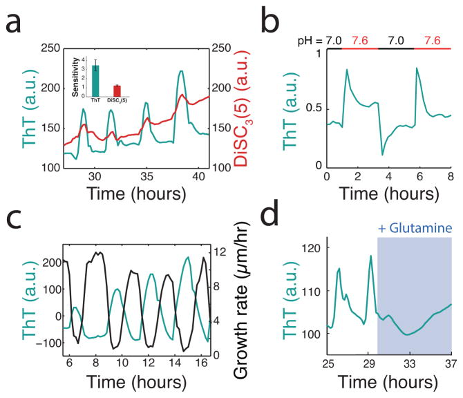
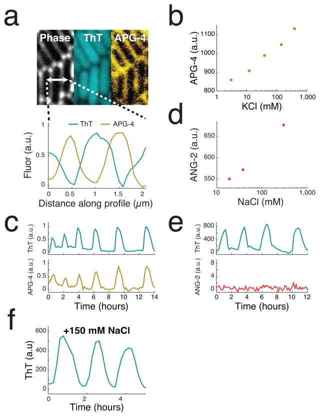
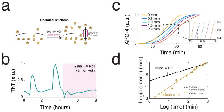
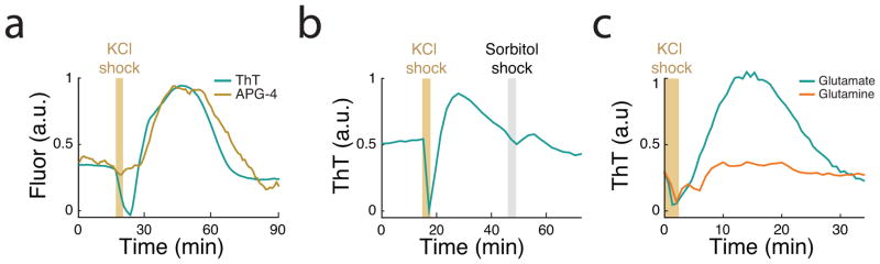
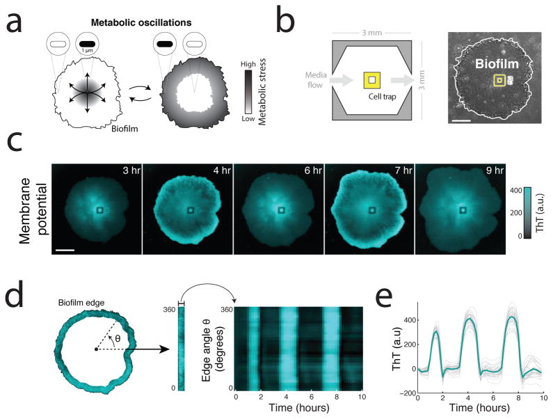
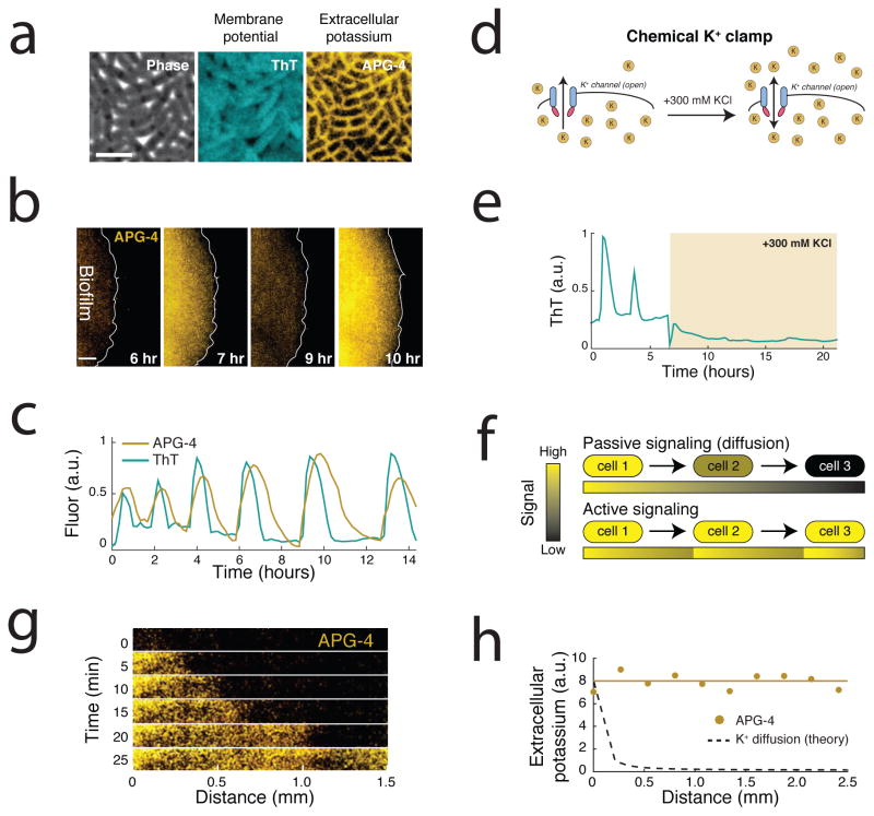
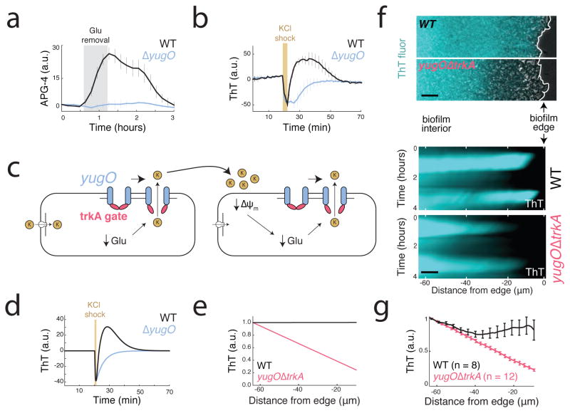
The study of bacterial ion channels has provided fundamental insights into the structural basis of neuronal signalling; however, the native role of ion channels in bacteria has remained elusive. Here we show that ion channels conduct long-range electrical signals within bacterial biofilm communities through spatially propagating waves of potassium. These waves result from a positive feedback loop, in which a metabolic trigger induces release of intracellular potassium, which in turn depolarizes neighbouring cells. Propagating through the biofilm, this wave of depolarization coordinates metabolic states among cells in the interior and periphery of the biofilm. Deletion of the potassium channel abolishes this response. As predicted by a mathematical model, we further show that spatial propagation can be hindered by specific genetic perturbations to potassium channel gating. Together, these results demonstrate a function for ion channels in bacterial biofilms, and provide a prokaryotic paradigm for active, long-range electrical signalling in cellular communities.


Conflict of interest statement

The authors declare no competing financial interest.

Figures

Extended Data Figure 1
Thioflavin T (ThT) is a fluorescent reporter that is inversely related to the membrane potential. (a) ThT and DiSC3(5), an established reporter of membrane potential in bacteria, both oscillate within biofilms. ThT has an approximately three fold higher sensitivity to changes in membrane potential compared to DiSC3(5). Sensitivity is defined as the ratio between peak height and error in peak height. Representative trace is selected from 3 independent biofilms. (b) The cellular ThT fluorescence depends on the external pH, where higher pH results in greater membrane potential as expected. ThT itself is insensitive to these pH changes and the traces are background subtracted to eliminate possible artifacts. Representative trace is selected from 3 independent biofilms. (c) Oscillations in ThT and growth rate are inversely correlated, linking membrane potential oscillations to the metabolic cycle which produces periodic growth pauses. Growth rate is calculated by taking the derivative of biofilm radius over time (Supplementary Information). Representative trace is selected from over 75 independent biofilms. (d) Replacing glutamate with 0.2% glutamine, which eliminates the need to take up glutamate or retain ammonium, quenches ThT oscillations. This further suggests that ThT oscillations are specific to the metabolic cycle involving glutamate and ammonium. Representative trace is selected from 3 independent experiments.
Extended Data Figure 2
A fluorescent reporter of extracellular potassium (APG-4) indicates that potassium plays a role in membrane potential oscillations. (a) High-resolution images showing the intracellular localization of ThT and primarily extracellular localization of APG-4 (left). Quantification of ThT and APG-4 along the 2μm profile indicated in the phase image indicates that APG-4 does not significantly diffuse into the cell (right). Representative images are selected from 6 independent experiments. (b) Induction curve for APG-4 generated using externally supplemented KCl. The experiment was repeated twice. (c) Oscillations in extracellular potassium in the surrounding cell-free region during biofilm oscillations. These oscillations occurred during the experiment shown in Figure 2bc and the pulses are synchronized between the biofilm and the surrounding cell-free region. Representative trace is selected from 6 independent experiments. (d) Induction curve for ANG-2 generated using externally supplemented NaCl. Experiment was repeated twice. (e) Simultaneous measurement of ThT and ANG-2 within the biofilm indicates a lack of oscillations in extracellular sodium. Representative trace selected from 3 independent biofilms. (f) Furthermore, perturbing extracellular sodium concentrations in the media had no detectable effect on membrane potential oscillations. Experiment was repeated twice.
Extended Data Figure 3
(a) A chemical potassium clamp (300 mM KCl, matching the intracellular concentration, and 30 μM valinomycin) prevents the formation of potassium electrochemical gradients across the cellular membrane. Valinomycin is an antibiotic that creates potassium-specific carriers in the cellular membrane. (b) Clamping net potassium flux quenches oscillations in membrane potential. (c) Propagation of extracellular potassium is estimated by tracking the halfmaximal position of the pulse over time. Representative traces are shown for a single pulse selected from one of 6 independent experiments. (d) Propagation of extracellular potassium is relatively constant over time in contrast to diffusion that is expected to decay. The diffusion line is calculated using the mean squared displacement (MSD) and the diffusion coefficient for potassium in biofilms (Supplementary Information). Slopes are calculated from the same representative data shown in c.
Extended Data Figure 4
External potassium affects the metabolic state of the cell. (a) A potassium shock (300 mM KCl) produces an initial ThT decrease (depolarization) followed by a sustained ThT increases (hyperpolarization). ThT is inversely related to the membrane potential. A corresponding pulse in APG-4 during this ThT increase suggests that hyperpolarization is due to release of potassium. APG-4 signal due to the external potassium shock itself was subtracted using the cell-free background near the biofilm. Representative trace is selected from 3 independent experiments. (b) ThT spikes in response to external potassium shock (300 mM KCl) but not an equivalent shock of 300 mM sorbitol, an uncharged solute. Representative trace is selected from 3 independent experiments. (c) The hyperpolarization response occurs when cells are grown in glutamate but not when glutamate is replaced by 0.2% glutamine, which bypasses the need to take up glutamate or retain ammonium. Representative trace is selected from 4 independent biofilms.
Extended Data Figure 5
List of strains used in this study.
Extended Data Figure 6
Parameter values used in the model.
Figure 1
Figure 1
Biofilms produce synchronized oscillations in membrane potential. (a) Biofilms generate collective metabolic oscillations resulting from long-range metabolic interactions between interior and peripheral cells. It remains unclear how microscopic bacteria are capable of communicating over such macroscopic distances within biofilm communities. (b) Schematic of the microfluidic device used throughout this study (left). Phase contrast image of a biofilm growing in the microfluidic device with the cell trap highlighted in yellow (right). Scale bar indicates 100 μm. (c) Global oscillations in membrane potential, as reported by Thioflavin T (ThT), within the biofilm community. ThT is positively charged but not known to be actively transported, so it can be retained in cells due to their inside-negative membrane potential. ThT fluorescence increases when the inside of the cell becomes more negative, and thus ThT is inversely related to the membrane potential. Scale bar indicates 0.15 mm. Representative images shown are drawn from over 75 independent biofilms. (d) Membrane potential oscillations are highly synchronized even between the most distant regions of the biofilm. To analyze synchronization, the edge region of the biofilm was identified and straightened (left) then plotted over time (right). (e) Time traces of the heatmap shown in d. Indicated in bold is the mean of 30 traces.
Figure 2
Figure 2
Potassium release is involved in active signal propagation within the biofilm. (a) An extracellular fluorescent chemical dye (APG-4) reports the concentration of potassium in the media (Extended Data Figure 2a,b). For comparison, the same cells are shown stained with ThT, which is inversely related to the membrane potential. These images depict cells at the peak of the ThT oscillation cycle. Representative images are selected from 6 independent experiments. Scale bar indicates 2 μm. (b) Global oscillations in extracellular potassium throughout the biofilm. A white line indicates the edge of the biofilm. Representative images are selected from 6 independent experiments. Scale bar indicates 0.2 mm. (c) Oscillations in membrane potential and extracellular potassium are synchronized, suggesting that potassium release is involved in global membrane potential oscillations. ThT is inversely related to the membrane potential. Representative traces are taken from the experiment shown in b. (d) A chemical potassium clamp (300 mM KCl, matching the intracellular concentration) prevents the formation of potassium electrochemical gradients across the cellular membrane. (e) Clamping net potassium flux quenches oscillations in membrane potential. Representative trace is selected from 3 independent experiments. (f) Illustration of the differences between passive signaling (diffusion) and active signaling. When cells passively respond to a signal, the range that the signal can propagate is limited due to the decay of signal amplitude. In contrast, when cells actively respond by amplifying the signal, propagation can extend over greater distances. (g) We measured propagation of extracellular potassium by measuring APG-4 in time and along a length of approximately 1.5 mm within the biofilm. (h) Extracellular potassium amplitude is relatively constant as the signal propagates, in contrast to the predicted amplitude decay of a passive signal. Representative data selected from 6 independent experiments. The diffusion line is calculated using the 2D diffusion equation and the diffusion coefficient for potassium within biofilms (Supplementary Information).
Figure 3
Figure 3
The molecular mechanism of signal propagation involves potassium channel gating. (a) yugO is a potassium channel in B. subtilis that is gated intracellularly by a trkA domain, which is regulated by the metabolic state of the cell. Withdrawing glutamate (the sole nitrogen source in MSgg media) induces an increase in extracellular potassium (APG-4) for wild-type but not the yugO deletion strain. Error bars indicate the mean +/− std for 3 independent biofilms each. (b) An external potassium shock (300 mM KCl) induces a short-term membrane potential depolarization in both wild-type and yugO deletion strains. However, in the wild-type this initial depolarization was followed by hyperpolarization, which is not observed in the yugO deletion strain (mean +/− std for 12 traces drawn from 3 biofilms each). ThT is inversely related to the membrane potential. (c) Proposed toy model for potassium signaling. The initial trigger for potassium release is metabolic stress caused by glutamate limitation. External potassium depolarizes neighboring cells, producing further nitrogen limitation by limiting glutamate uptake, and thus produces further metabolic stress. This cycle results in cell-cell propagation of the potassium signal. (d) A minimal conductance-based model describing the dynamics of the cell’s membrane potential in terms of a single potassium channel and a leak current. Consistent with our experimental results, this simple model exhibits transient depolarization followed by hyperpolarization in response to local increases in extracellular potassium concentration. (e) The model predicts that manipulating channel gating and conductance will result in decaying amplitude in the spatial propagation of membrane potential oscillations. (f) Maximum intensity projection of membrane potential change illustrating attenuated communication within the biofilm in a yugOΔtrkA deletion compared to wild type biofilms (top). Heatmap of oscillations taken from wild type and yugOΔtrkA mutant biofilms (bottom). Representative images are taken from 3 independent experiments in which WT and yugOΔtrkA biofilms are compared head-to-head. Scale bars indicate 8 μm. (g) Quantification of normalized pulse amplitude from wild type (n = 8) and yugOΔtrkA (n = 12) mutant biofilms (mean +/− sem).

Comment in

References

    1. Hille B. Ion Channels of Excitable Membranes. 2001
    1. MacKinnon R. Potassium channels and the atomic basis of selective ion conduction. Bioscience Reports. 2004;24:75–100. - PubMed
    1. Doyle DA, et al. The structure of the potassium channel: molecular basis of K+ conduction and selectivity. Science. 1998;280:69–77. - PubMed
    1. Ren D, et al. A prokaryotic voltage-gated sodium channel. Science. 2001;294:2372–2375. - PubMed
    1. Iyer R, Iverson TM, Accardi A, Miller C. A biological role for prokaryotic ClC chloride channels. Nature. 2002;419:715–718. - PubMed

Publication types