. 2015 Oct 29;526(7575):710-4.
doi: 10.1038/nature15510. Epub 2015 Oct 21.

CMT2D neuropathy is linked to the neomorphic binding activity of glycyl-tRNA synthetase

Affiliations

CMT2D neuropathy is linked to the neomorphic binding activity of glycyl-tRNA synthetase

Weiwei He et al. Nature. .

Erratum in

Abstract

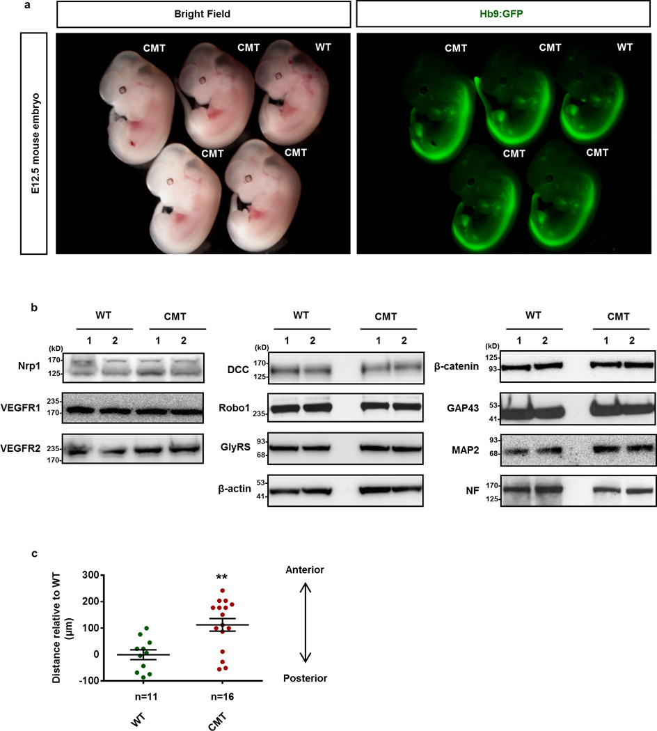
Selective neuronal loss is a hallmark of neurodegenerative diseases, which, counterintuitively, are often caused by mutations in widely expressed genes. Charcot-Marie-Tooth (CMT) diseases are the most common hereditary peripheral neuropathies, for which there are no effective therapies. A subtype of these diseases--CMT type 2D (CMT2D)--is caused by dominant mutations in GARS, encoding the ubiquitously expressed enzyme glycyl-transfer RNA (tRNA) synthetase (GlyRS). Despite the broad requirement of GlyRS for protein biosynthesis in all cells, mutations in this gene cause a selective degeneration of peripheral axons, leading to deficits in distal motor function. How mutations in GlyRS (GlyRS(CMT2D)) are linked to motor neuron vulnerability has remained elusive. Here we report that GlyRS(CMT2D) acquires a neomorphic binding activity that directly antagonizes an essential signalling pathway for motor neuron survival. We find that CMT2D mutations alter the conformation of GlyRS, enabling GlyRS(CMT2D) to bind the neuropilin 1 (Nrp1) receptor. This aberrant interaction competitively interferes with the binding of the cognate ligand vascular endothelial growth factor (VEGF) to Nrp1. Genetic reduction of Nrp1 in mice worsens CMT2D symptoms, whereas enhanced expression of VEGF improves motor function. These findings link the selective pathology of CMT2D to the neomorphic binding activity of GlyRS(CMT2D) that antagonizes the VEGF-Nrp1 interaction, and indicate that the VEGF-Nrp1 signalling axis is an actionable target for treating CMT2D.


Figures

Extended Data Figure 1
Extended Data Figure 1. Hydrogen-deuterium exchange analysis to compare P234KY-GlyRSCMT2D and GlyRSWT in solution
A global increase (15%) in deuterium incorporation for the mutant GlyRS was observed indicating overall structural opening. The regions having significant changes (>10%) in deuterium incorporation are highlighted under the human cytosolic GlyRS sequence with different color codes (see box).
Extended Data Figure 2
Extended Data Figure 2. Characterization of the binding activity of GlyRSCMT2D
a, In vitro pull-down of P234KY-GlyRSCMT2D proteins with the ectodomains of Nrp1, TrkB, DCC, Robo1 and Unc5C proteins. Note the much stronger binding of GlyRSCMT2D with Nrp1 compared to other receptors. GlyRS was detected by immunoblot with anti-GlyRS antibody; similar amounts of input receptors were visualized by Coomassie Brilliant Blue staining. b, In vitro pull-down of GlyRSCMT2D proteins with the ectodomain of Nrp1. In addition to L129P and P234KY, direct binding to Nrp1 was detected for E71G and G240R GlyRSCMT2D. c, GST pull-down to confirm that b1 domain of Nrp1 is the main binding site of GlyRSCMT2D. The amount of GST and GST fusion proteins used for GlyRSCMT2D binding was visualized by Ponceau staining. d,e, In vitro pull-down assay showing the mutual competition between L129P-GlyRSCMT2D and VEGF-A165 for Nrp1 binding.
Extended Data Figure 3
Extended Data Figure 3. Detection of GlyRS proteins in the cell medium
a, c, e, Western-blot analysis of the GlyRS protein levels in NSC34 motor neurons (a), C2C12 cell-differentiated myotubes (c) and undifferentiated C2C12 myoblasts (e). The level of GlyRS proteins in cell medium is diminished by application of the exosome-pathway inhibitor GW4869, but not by Brefeldin A (BFA), an inhibitor of the classical endoplasmic reticulum (ER) to Golgi secretory pathway. GAPDH (cytoplasmic protein), vWF (secretory protein through ER-Golgi pathway) and TSG101 (Exosomal protein) are used as controls. b, d, Quantification of GlyRS protein level indicated in a c. Data are presented as the mean ± SEM of three independent experiments (*p < 0.05, t-test). f, Western-blot analysis of the GlyRS protein level in NSC34 motor neurons. The level of GlyRS proteins in the cell medium is increased by the treatment of monensin (MON), an activator for microvesicle release by regulating the intracellular calcium level,. Vehicle-treated cells were used as control (Ctrl). g, Western-blot analysis of the GlyRS protein level in Cos7 cells transfected with plasmids encoding GlyRSWT and P234KY-GlyRSCMT2D. The expression of GlyRS proteins was detected by immuno-blot with antibody to V5 epitope tag. GAPDH was used as control. Note the similar level of GlyRSCMT2D and GlyRSWT in the media of transfected Cos7 cells. The observation that differentiated myotubes also secret GlyRS raises the possibility that muscles, which are directly innervated by the peripheral motor neurons, might contribute to the disease pathology.
Extended Data Figure 4
Extended Data Figure 4. Detection of GlyRS proteins in Exosome-enriched fractions
a, Diagram showing the procedure of “exosome” separation from the cell medium of NSC34 cells by differential centrifugation. See Materials and Methods for details. b, Western-blot analysis of proteins associated with various fractions. GlyRS proteins were detected in the “exosome”-enriched fractions but not in supernatant fractions. The quality of the “exosome” preparation was controlled by detection of TSG101 (exosomal protein), Bip (ER-associated protein), GAPDH (cytoplasmic protein), and vWF (secretory protein through ER-Golgi pathway).
Extended Data Figure 5
Extended Data Figure 5. CMT2D mutant embryos have overall normal morphology but exhibit facial motor neuron migration defects
a, Lateral view of WT and CMT2D mutant embryos at E12.5. Motor neurons are specifically labeled by a transgenic fluorescence reporter, Hb9:GFP (green). Note overall normal morphology of CMT2D mutant embryos (CMT) compared to their littermate controls (WT). b, Western-bolt analysis of protein expression in E12.5 mouse neural tissues. The expression levels of various neuronal proteins appear normal in CMT2D mutants compared to their littermate controls. c, The facial motor neuron migration phenotype is quantified by measuring the relative distance of the facial motor nucleus between WT and CMT littermate embryos (Each dot represents one facial motor nucleus, n=6 embryos for WT; n=8 embryos for CMT2D). We find the migration of facial motor neurons is significantly disrupted in CMT embryos. Data are presented as the mean ± SEM. **p < 0.01 (t-test).
Extended Data Figure 6
Extended Data Figure 6. Genetic-interaction between Gars and Nrp1 in the early stage of CMT2D
a, b, Hindlimb extension test of wild-type and mutant animals at 2 weeks. Note that 2 out of 9 GarsCMT2D;Nrp1+/− (CMT;Nrp1+/−) mutants exhibit hindlimb weakness with significantly lower scores compared to GarsCMT2D (CMT), Nrp1+/− (Nrp1+/−) and wild-type (WT) littermate controls. c, Comparison of stride lengths in different CMT2D mutant mice at 4-week-old: GarsCMT2D (CMT), GarsCMT2D;TrkB+/− (CMT;TrkB+/−), GarsCMT2D;DCC+/− (CMT;DCC+/−), GarsCMT2D;Robo1+/− (CMT;Robo1+/−), and GarsCMT2D;Unc5C+/− (CMT;Unc5C+/−). No significant differences were observed between compound heterozygotes and their littermate controls (CMT).
Extended Data Figure 7
Extended Data Figure 7. Axonal dystrophy in CMT2D mice
Histogram showing the axonal diameter frequencies in the sciatic nerves of 4-week-old wild-type (WT, a), Nrp1 heterozygous (Nrp1+/−, b), GarsCMT2D (CMT, c), and (CMT;Nrp1+/−, d) mutant mice. n=3 mice per group. Note the decreased numbers of larger-diameter axons in CMT;Nrp1+/− mutants compared to CMT, Nrp1 heterozygous, and wild-type controls.
Extended Data Figure 8
Extended Data Figure 8. Expression level of VEGF in mouse muscles
The expression level of VEGF proteins in muscle fibers of mice injected with lentivirus expressing LV-VEGF165-ires-GFP versus LV-GFP was determined by immunostaining with anti-VEGF antibodies. Note the expression level of VEGF in LV-VEGF infected muscles is significantly higher than in LV-GFP infected control groups.
Extended Data Figure 9
Extended Data Figure 9. VEGF treatment retains limb strength in CMT2D mice
a, Diagram showing that lentiviral vectors encoding GFP (LV-GFP) or VEGF-A165 (LV-VEGF165) are injected unilaterally into each hindlimb of the same GlyRSCMT2D mutant mouse at P5. c, e, At 5 weeks, LV-GFP-injected legs (L, left) of CMT2D animals have largely lost their ability to extend, while LV-VEGF165-treated legs (R, right) retained more limb strength with significantly higher scores in the hindlimb extension test (3 out of 7 animals). p<0.05 (Permutation test). No significant difference was observed between both injected legs of wild-type animals in the hindlimb extension test (b, d). f, g, GDNF and VEGF-A121 treatments fail to improve stride length in CMT2D mice. Walking strides of 2-month-old CMT2D mice bilaterally injected with lentiviral vectors (LV) encoding GFP, GDNF or VEGF-A121. No significant difference of hindlimb stride length was observed between animals treated with LV-GDNF, LV-VEGF-A121, and LV-GFP controls.
Extended Data Figure 10
Extended Data Figure 10. A simplified model for the neomorphic binding activity of GlyRSCMT2D
Left panel, GlyRSWT is a multifunctional protein with both intracellular and extracellular distributions. VEGF/Nrp1 signaling is an essential pathway for survival and function of motor neurons. (Note that VEGF may also act synergistically with other trophic factors, and/or maintains motor-function indirectly by acting on Nrp1 receptors on non-motor neurons.) Right panel, CMT2D mutations alter the conformation of GlyRS, enabling GlyRSCMT2D to bind Nrp1. This aberrant interaction antagonizes the binding of VEGF to Nrp1, contributing to motor defects in CMT2D. Our results do not exclude the possibility that GlyRSCMT2D may also interact with other extracellular and/or intracellular targets, related to CMT2D pathology.
Figure 1
Figure 1. Dispersed CMT2D mutations consistently cause neomorphic structural opening at the dimer interface of GlyRS
a, Distribution of 15 CMT2D-associated dominant mutations in the three domains of the cytosolic human GlyRS. The 3 strongest pathogenic mutations are highlighted in green. Two mutations identified in mice (*) are labeled with their corresponding residue numbers in the human protein. b, Human GlyRS structure (monomeric subunit) viewed from dimer interface. Consensus opened-up areas caused by 5 CMT2D mutations are labeled in red. c, Opened-up areas (red) by the P234KY mutation (>10 % increase in deuterium incorporation relative to WT GlyRS.)
Figure 2
Figure 2. GlyRSCMT2D specifically binds Nrp1 and antagonizes VEGF-Nrp1 interaction
a, In vitro pull-down (PD) of GlyRSCMT2D proteins by the ectodomain of Nrp1 but not TrkB. b, Co-immunoprecipitation (IP) to detect GlyRS-Nrp1 interaction in neural tissues of wild-type (WT) and P234KY-GarsCMT2D mice (CMT). c, Co-immunoprecipitation (IP) to detect GlyRS-Nrp1 interaction in lymphocytes from CMT2D patients carrying the L129P mutation (n=5) and from healthy individuals (n=3). d, Domain mapping using in vitro IP identifies the b1 domain of Nrp1 as the main binding site of GlyRSCMT2D. e, f, In vitro PD assay showing the competition between P234KY-GlyRSCMT2D and VEGF-A165 proteins for Nrp1 (b domains) binding. g, j, Schematic of facial motor neuron migration (g) and facial motor nucleus (j) in open-book preparations of WT (left half) and VEGF/Nrp1-deficient mouse hindbrains at E13.5 (right half). h, i, Fluorescence labeling of facial motor neuron somata and axons by ISLMN:GFP-F on one side of E13.5 mouse hindbrain of open-book preparation. k, l, Immunostaining of Isl-positive facial nucleus on one side of the E13.5 mouse hindbrain of open-book preparation. Scale bar represents 200µm in h, i, k, l.
Figure 3
Figure 3. Nrp1 is a genetic modifier of CMT2D
a, b, Hindlimb extension test at 4 weeks. ***p < 0.001 (Mann-Whitney test). c, Hindlimb footprints of WT and mutant animals at 4 weeks. GarsCMT2D/Nrp1+/− mutant mice exhibit disrupted gait patterns of different degrees (mild, severe). Note that severe cases show inability to walk. d, Stride length of WT and mutant animals at 4 weeks. e, f, Neuromuscular junction (NMJ) immunostaining in the gastrocnemius muscles of 4-week-old mice with the motor nerve terminal labeled in green and acetylcholine receptors on the muscle labeled in red. Data are presented as mean values ± SEM. n= 3 mice per group. Scale bar represents 50µm. g, h, Myelinated axons from sciatic nerves of 4-week-old mice. Scale bar represents 20µm. Histogram showing the quantification of axon numbers with the diameter larger than 2µm (h). n= 3 mice per group. *p < 0.05, **p < 0.01 (t-test) for d, f and h.
Figure 4
Figure 4. VEGF treatment improves motor function in CMT2D mice
a, Diagram showing bilateral intramuscular injection of lentivirus (LV) into mouse hindlimbs at P5. b, Inclined plane test of 4-week-old animals. c, Walking strides of 7-week-old animals. d, Rotarod test of 2-month-old animals. *p < 0.05, **p < 0.01, ***p < 0.001 (t-test) for b, c and d.

References

    1. Saxena S, Caroni P. Selective neuronal vulnerability in neurodegenerative diseases: from stressor thresholds to degeneration. Neuron. 2011;71:35–48. - PubMed
    1. Skre H. Genetic and clinical aspects of Charcot-Marie-Tooth's disease. Clin Genet. 1974;6:98–118. - PubMed
    1. Patzko A, Shy ME. Update on Charcot-Marie-Tooth disease. Curr Neurol Neurosci Rep. 2011;11:78–88. - PMC - PubMed
    1. Antonellis A, et al. Glycyl tRNA synthetase mutations in Charcot-Marie-Tooth disease type 2D and distal spinal muscular atrophy type V. Am J Hum Genet. 2003;72:1293–1299. - PMC - PubMed
    1. Wakasugi K, Schimmel P. Two distinct cytokines released from a human aminoacyl-tRNA synthetase. Science. 1999;284:147–151. - PubMed

Publication types

MeSH terms

Supplementary concepts