. 2015 Oct 27:5:15763.
doi: 10.1038/srep15763.

Retinal oxygen extraction in humans

Affiliations

Retinal oxygen extraction in humans

René M Werkmeister et al. Sci Rep. .

Abstract

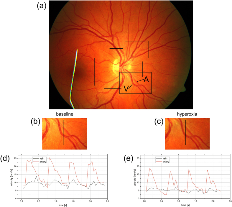
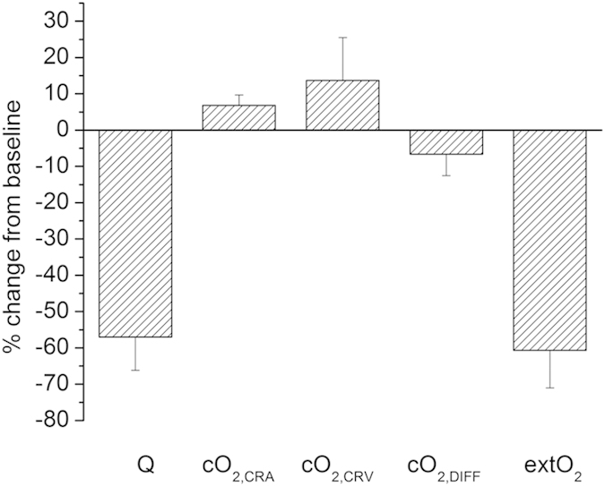
Adequate function of the retina is dependent on proper oxygen supply. In humans, the inner retina is oxygenated via the retinal circulation. We present a method to calculate total retinal oxygen extraction based on measurement of total retinal blood flow using dual-beam bidirectional Doppler optical coherence tomography and measurement of oxygen saturation by spectrophotometry. These measurements were done on 8 healthy subjects while breathing ambient room air and 100% oxygen. Total retinal blood flow was 44.3 ± 9.0 μl/min during baseline and decreased to 18.7 ± 4.2 μl/min during 100% oxygen breathing (P < 0.001) resulting in a pronounced decrease in retinal oxygen extraction from 2.33 ± 0.51 μl(O2)/min to 0.88 ± 0.14 μl(O2)/min during breathing of 100% oxygen. The method presented in this paper may have significant potential to study oxygen metabolism in hypoxic retinal diseases such as diabetic retinopathy.


Figures

Figure 1
Figure 1. Sample measurement in a healthy subject.
(a) Fundus image and OCT scanning patterns used to measure all retinal vessels entering the optic nerve head to measure total retinal blood flow. Black bars indicate the measurement locations. (b,c) Magnification of the fundus image during breathing room air and 100% oxygen, respectively. Vasoconstriction of retinal artery and vein can clearly be seen. (d,e) Time course of retinal arterial and venous blood velocity during breathing room air and breathing 100% oxygen respectively over approximately 4 cardiac cycles.
Figure 2
Figure 2. % change in total retinal blood flow (Q), arterial oxygen content (cO2,CRA), venous oxygen content (cO2,CRV), arteriovenous difference in oxygen content (cO2,DIFF) and retinal oxygen extraction (extO2) in response to 100% oxygen breathing (n = 8).
Data are presented as means ± SD.

References

    1. Pascolini D. & Mariotti S. P. Global estimates of visual impairment: 2010. Br J Ophthalmol 96, 614–618, 10.1136/bjophthalmol-2011-300539 (2012). - DOI - PubMed
    1. Pemp B. & Schmetterer L. Ocular blood flow in diabetes and age-related macular degeneration. Can J Ophthalmol 43, 295–301, 10.3129/i08-049 (2008). - DOI - PubMed
    1. Cherecheanu A. P., Garhofer G., Schmidl D., Werkmeister R. & Schmetterer L. Ocular perfusion pressure and ocular blood flow in glaucoma. Curr Opin Pharmacol 13, 36–42, 10.1016/j.coph.2012.09.003 (2013). - DOI - PMC - PubMed
    1. Pournaras C. J., Rungger-Brandle E., Riva C. E., Hardarson S. H. & Stefansson E. Regulation of retinal blood flow in health and disease. Prog Retin Eye Res 27, 284–330, 10.1016/j.preteyeres.2008.02.002 (2008). - DOI - PubMed
    1. Wangsa-Wirawan N. D. & Linsenmeier R. A. Retinal oxygen: fundamental and clinical aspects. Arch Ophthalmol 121, 547–557, 10.1001/archopht.121.4.547 (2003). - DOI - PubMed

Publication types

LinkOut - more resources