. 2016 Feb;26(2):181-92.
doi: 10.1093/glycob/cwv093. Epub 2015 Oct 26.

Single-chain antibody-fragment M6P-1 possesses a mannose 6-phosphate monosaccharide-specific binding pocket that distinguishes N-glycan phosphorylation in a branch-specific manner†

Affiliations

Single-chain antibody-fragment M6P-1 possesses a mannose 6-phosphate monosaccharide-specific binding pocket that distinguishes N-glycan phosphorylation in a branch-specific manner†

Ryan J Blackler et al. Glycobiology. 2016 Feb.

Abstract

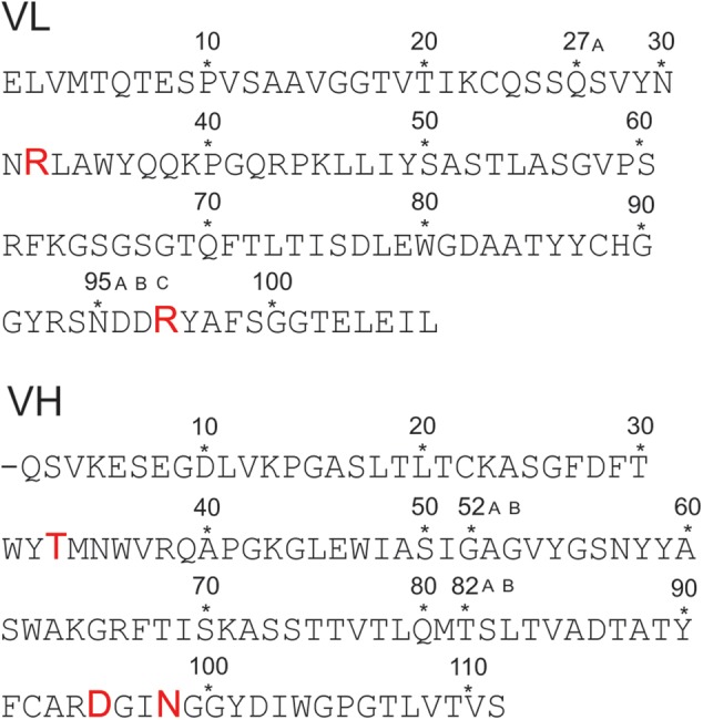
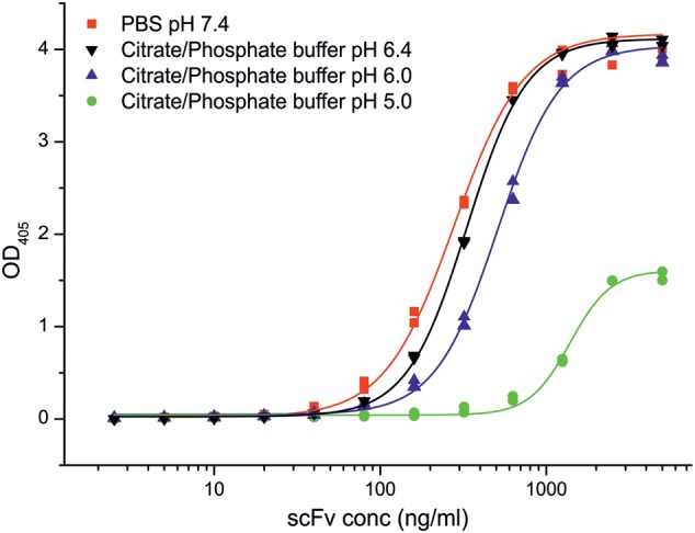
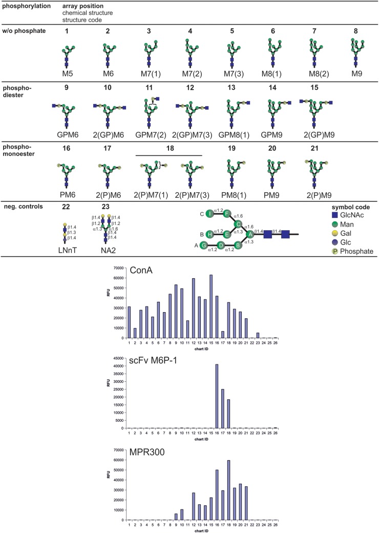
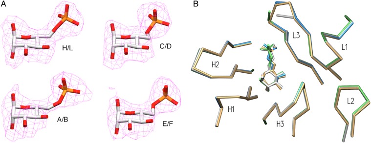
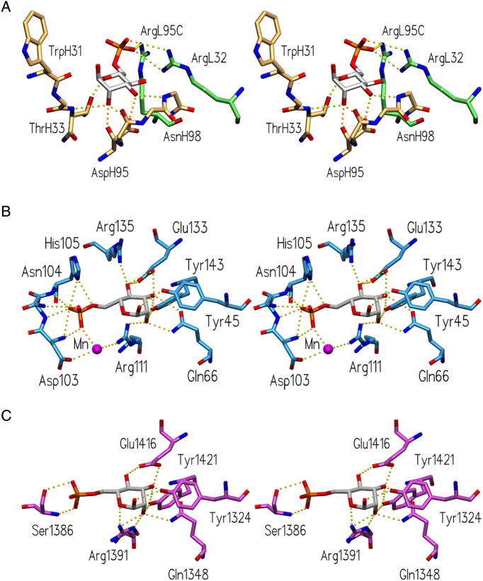
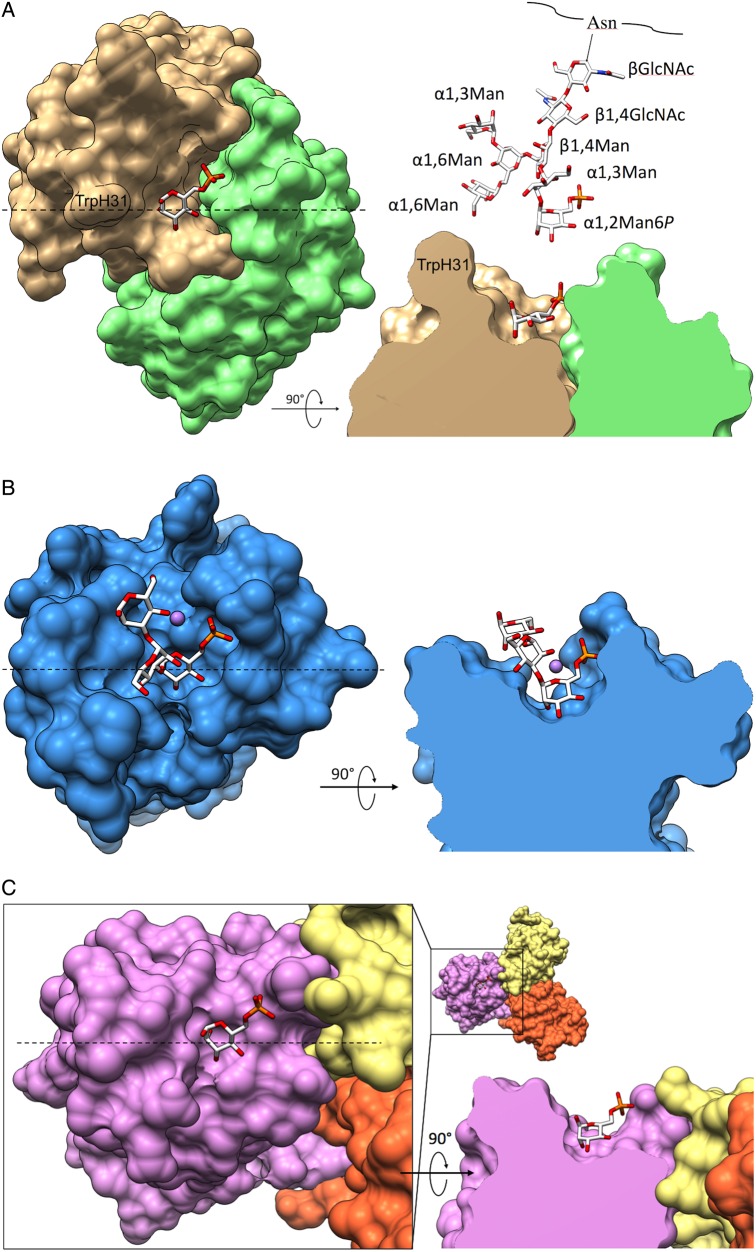
The acquisition of mannose 6-phosphate (Man6P) on N-linked glycans of lysosomal enzymes is a structural requirement for their transport from the Golgi apparatus to lysosomes mediated by the mannose 6-phosphate receptors, 300 kDa cation-independent mannose 6-phosphate receptor (MPR300) and 46 kDa cation-dependent mannose 6-phosphate receptor (MPR46). Here we report that the single-chain variable domain (scFv) M6P-1 is a unique antibody fragment with specificity for Man6P monosaccharide that, through an array-screening approach against a number of phosphorylated N-glycans, is shown to bind mono- and diphosphorylated Man6 and Man7 glycans that contain terminal αMan6P(1 → 2)αMan(1 → 3)αMan. In contrast to MPR300, scFv M6P-1 does not bind phosphodiesters, monophosphorylated Man8 or mono- or diphosphorylated Man9 structures. Single crystal X-ray diffraction analysis to 2.7 Å resolution of Fv M6P-1 in complex with Man6P reveals that specificity and affinity is achieved via multiple hydrogen bonds to the mannose ring and two salt bridges to the phosphate moiety. In common with both MPRs, loss of binding was observed for scFv M6P-1 at pH values below the second pKa of Man6P (pKa = 6.1). The structures of Fv M6P-1 and the MPRs suggest that the change of the ionization state of Man6P is the main driving force for the loss of binding at acidic lysosomal pH (e.g. lysosome pH ∼ 4.6), which provides justification for the evolution of a lysosomal enzyme transport pathway based on Man6P recognition.

Keywords: N-glycosylation; crystal structure; glycan array; lysosome; scFv.


Figures

Fig. 1.
Fig. 1.
Primary structure of the variable domains of the light (VL) and heavy chain (VH) scFv M6P-1 obtained by phage display from an immunized rabbit. The sequence determination has been published (Müller-Loennies et al. 2010) and residues were numbered using a world wide web interface (http://www.bioinf.org.uk/abs/) applying the Kabat convention (Martin 1996). Amino acids involved in interactions with the ligand in the crystal structure are shown with increased font size. This figure is available in black and white in print and in color at Glycobiology online.
Fig. 2.
Fig. 2.
Binding of scFv M6P-1 to immobilized PMP-BSA at different pH in ELISA. ScFv M6P-1 (Mw 28,228 Da) dissolved in the indicated buffers was titrated starting from 5 µg/mL (177 nM) on PMP-BSA neoglycoconjugate immobilized on polystyrene ELISA plates (85 ng/cup ≈ 24 pmol of PMP). The data were fitted by non-linear regression to a logistic function using Origin v. 7.0 SR4 (OriginLab Corp., Northampton). Displayed are the duplicate data points and the fitted curve. This figure is available in black and white in print and in color at Glycobiology online.
Fig. 3.
Fig. 3.
Binding of scFv M6P-1 to glycans immobilized in an array on glass slides. The glycan microarray used in this analysis contained the schematically depicted glycans that were printed in replicates of four at a concentration of 100 µM with the exception of glycans 3, 7, 11, 12, 15, 18 and 21, which were only available at lower concentrations (<100 µM). Below a structure code is given where G = GlcNAc (blue squares), P = phosphate (light yellow circles) and M = Man (green circles). Glucose and Galactose in controls are depicted as blue and yellow circles, respectively. Numbers indicate the number of residues and isomers are distinguished by numbers in brackets. N-Glycan residues are designated as schematically depicted in the lower panel following the nomenclature as published (Castonguay et al. 2012). The array was interrogated with biotinylated scFv M6P-1 and Con A and affinity purified fetal bovine serum MPR300. Bound scFv M6P-1 and Con A were quantified after incubation with Cy5-labeled streptavidin and MPR300 was assayed using a rabbit polyclonal antibody and a Cy5-labeled anti-rabbit IgG as described (Castonguay et al. 2012). Con A and MPR300 served as positive controls to validate the array. Relative fluorescence units were determined as the average of four replicates, and the glycan numbers correspond to the glycans above. Biotinylated Con A at 0.5 µg/mL (left) was detected with Cy5 Streptavidin (5 µg/mL); biotinylated scFv M6P-1 at 20 µg/mL (middle) detected with Cy5 Streptavidin (5 µg/mL); MPR300 at 5 µg/mL (right) detected with a rabbit polyclonal antibody (1:250 dilution) and Cy5-labeled goat, anti-rabbit IgG at 5 µg/mL. Slides were scanned for Cy5 in a ProScaArray scanner (PerkinElmer) and average fluorescence units were determined using the corresponding ProScanArray software.
Fig. 4.
Fig. 4.
Crystal structure of scFv M6P-1 (A). FoFc omit electron density maps contoured to 2σ for Man6P in each of the four Fv molecules in the AU. The refined coordinates of the corresponding Man6P are superposed for clarity. (B) Superposition of M6P-1 binding sites of the four molecules in the asymmetric unit, displaying α-carbon backbones of CDR loops and the Man6P molecule of each unit. Chains H/L in green, A/B white, C/D tan and E/F blue.
Fig. 5.
Fig. 5.
Comparison of Man6P-binding sites of Fv M6P-1 and bovine MPR. Stereoviews of the binding sites of (A) Fv M6P-1 (light chain green, heavy chain tan), (B) the CRD of MPR46 (pdb code 1M6P; Roberts et al. 1998) and (C) the CRD formed by the N-terminal domains 1–3 of MPR300 (pdb code 1SYO; Olson et al. 2004), showing hydrogen bonds and salt bridges as dashed yellow spheres.
Fig. 6.
Fig. 6.
Binding surfaces of Fv M6P-1 and MPRs. Top-down and side-view surface representations of Fv M6P-1, MPR46 (PDB code 1C39) and MPR300 domains 1–3 (PDB code 1SZ0). In each side-view, the model is clipped through the binding pocket. The dashed line in the top-down view indicates the location of the clipping plane. (A) Fv Man6P in complex with Man6P. Displayed above the side-view is a model of array glycan #16 for size comparison. (B) MPR46 in complex with PMP, showing only αMan6P(1 → 3)αMan(1 → 3)αMan as observed in that structure. Manganese ion is displayed as a mauve sphere. (C) MPR300 domains 1–3, sequentially colored yellow, orange, and mauve, in complex with Man6P.

References

    1. Adams PD, Afonine PV, Bunkoczi G, Chen VB, Davis IW, Echols N, Headd JJ, Hung LW, Kapral GJ, Grosse-Kunstleve RW et al. . 2010. PHENIX: A comprehensive Python-based system for macromolecular structure solution. Acta Crystallogr D Biol Crystallogr. 66:213–221. - PMC - PubMed
    1. Arakatsu Y, Ashwell G, Kabat EA. 1966. Immunochemical studies on dextrans. V. Specificity and cross-reactivity with dextrans of the antibodies formed in rabbits to isomaltonic and isomaltotrionic acids coupled to bovine serum albumin. J Immunol. 97:858–866. - PubMed
    1. Balaji PV, Qasba PK, Rao VS. 1994. Molecular dynamics simulations of high-mannose oligosaccharides. Glycobiology. 4:497–515. - PubMed
    1. Berman HM, Westbrook J, Feng Z, Gilliland G, Bhat TN, Weissig H, Shindyalov IN, Bourne PE. 2000. The Protein Data Bank. Nucleic Acids Res. 28:235–242. - PMC - PubMed
    1. Blackler RJ, Müller-Loennies S, Brade L, Kosma P, Brade H, Evans SV. 2012. Antibody recognition of chlamydia LPS: Structural insights of inherited immune responses. In: Kosma P, Müller-Loennies S, editors. Anticarbohydrate Antibodies. Vienna: Springer; p. 75–120.

Publication types