Review
. 2015 Oct 26:22:99.
doi: 10.1186/s12929-015-0210-7.

The HRASLS (PLA/AT) subfamily of enzymes

Affiliations
Review

The HRASLS (PLA/AT) subfamily of enzymes

Emily B Mardian et al. J Biomed Sci. .

Abstract

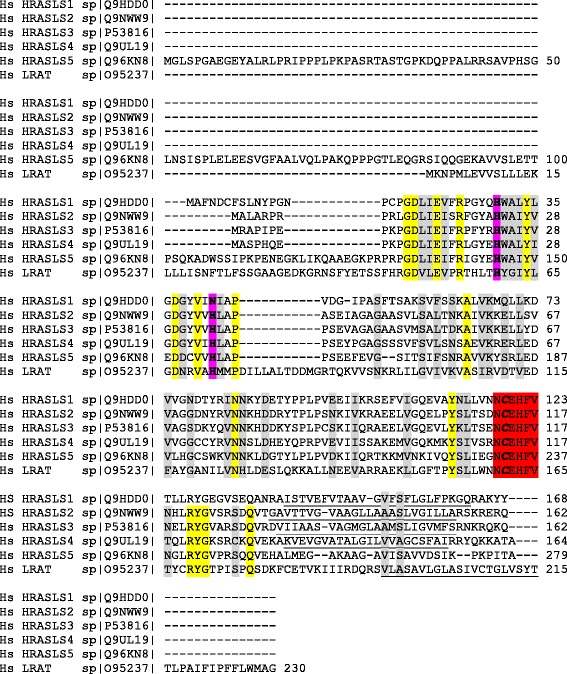
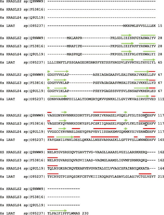
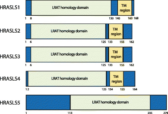
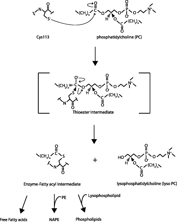
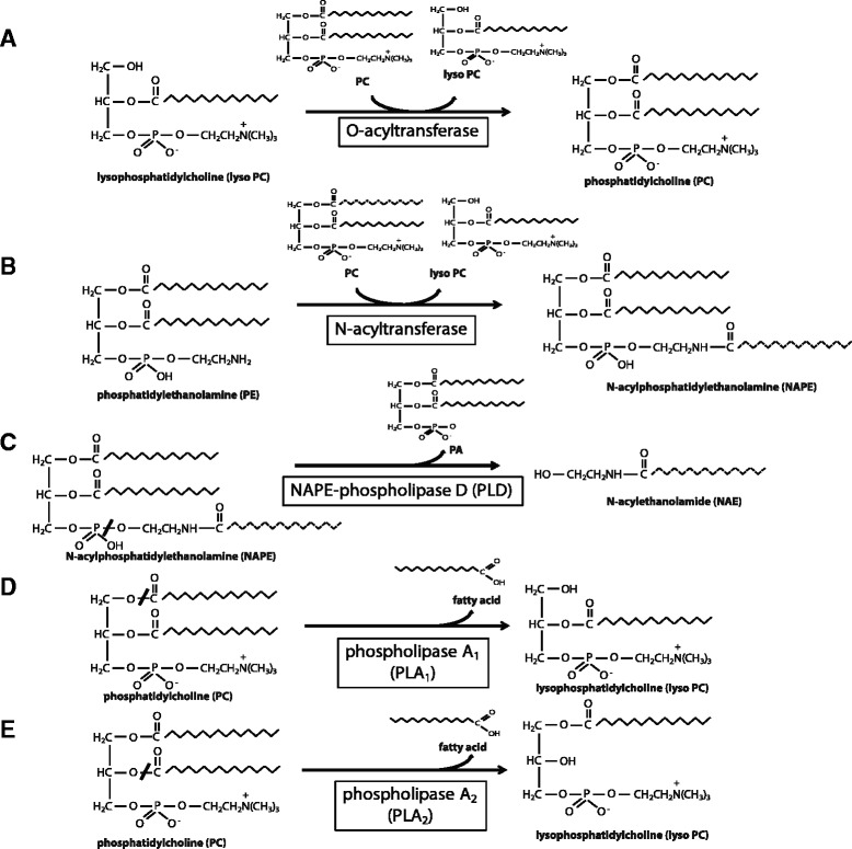
The H-RAS-like suppressor (HRASLS) subfamily consists of five enzymes (1-5) in humans and three (1, 3, and 5) in mice and rats that share sequence homology with lecithin:retinol acyltransferase (LRAT). All HRASLS family members possess in vitro phospholipid metabolizing abilities including phospholipase A1/2 (PLA1/2) activities and O-acyltransferase activities for the remodeling of glycerophospholipid acyl chains, as well as N-acyltransferase activities for the production of N-acylphosphatidylethanolamines. The in vivo biological activities of the HRASLS enzymes have not yet been fully investigated. Research to date indicates involvement of this subfamily in a wide array of biological processes and, as a consequence, these five enzymes have undergone extensive rediscovery and renaming within different fields of research. This review briefly describes the discovery of each of the HRASLS enzymes and their role in cancer, and discusses the biochemical function of each enzyme, as well as the biological role, if known. Gaps in current understanding are highlighted and suggestions for future research directions are discussed.


Figures

Fig. 1
Fig. 1
Sequence alignment of human HRASLS enzymes. Sequence alignment was performed using ClustalW2 [51]. The C-terminal transmembrane-spanning hydrophobic region is underlined. The NCEHFV motif is highlighted in red with the active site cysteine bolded and italicized. The remaining two active sites in the catalytic triad are also bolded, and are highlighted in purple. Conserved residues are highlighted in yellow, and homologous residues are highlighted in grey
Fig. 2
Fig. 2
Structural alignment of human HRASLS enzymes. Sequence alignment was performed using ClustalW2 [51] and structural alignment was performed for HRASLS2, HRASLS3, and HRASLS4 as described by Golczak et al. in 2012 [3] and Wei et al. in 2015 [6]. The β-sheets are indicated by green arrows and α-helices are indicated by red lines
Fig 3
Fig 3
Homology domains within the five human HRASLS enzymes. LRAT (lecithin:retinol acyltransferase) homology domain and the predicted transmembrane region (TM region) are shown
Fig 4
Fig 4
Illustration of a portion of the catalytic mechanism of HRASLS enzymes including depiction of the thioester intermediate formed during phospholipid hydrolysis. Reaction demonstrating the formation of a thioester intermediate following nucleophilic attack by the anionic sulphur of the deprotonated active site cysteine side chain in HRASLS proteins on the carbonyl group of the sn-1 fatty acyl chain of phosphatidylcholine. Hydrolysis results in the generation of a free lysophospholipid and an enzyme-fatty acyl intermediate that releases the enzyme and a free fatty acid following addition of a hydroxyl group to the carbonyl of the fatty acyl chain, forming a carboxylic acid. All HRASLS enzymes can catalyze complete phospholipase reactions in vitro, resulting in liberation of a free fatty acid as well as a lysophospholipid. All HRASLS enyzmes also show in vitro N- and O-acyltransferase activities, where the enzyme-fatty acyl intermediate binds a glycerolphospholipid leading to production of N-acylphosphatidylethanolamines (NAPEs) or phospholipids, respectively
Fig 5
Fig 5
Examples of phospholipid metabolism reactions involving HRASLS enzymes. A, O-acyltransferase (acyl-CoA independent transacylase) reaction demonstrating the conversion of lyso PC to PC, using the sn-1 acyl chain of PC as the acyl donor. B, N-acyltransferase reaction demonstrating the acylation of the amino group of phosphatidylethanolamine using the sn-1 fatty acyl chain donated by PC, resulting in the generation of an N-acylphosphatidylethanolamine (NAPE) and a lyso PC. C, Conversion of NAPE to N-acylethanolamine (NAE) by NAPE-phospholipase D-mediated hydrolysis. D, Phospholipase A1 (PLA1) reaction demonstrating the cleavage of a fatty acyl chain from the sn-1 position of PC, resulting in the liberation of a free fatty acid and a 1-hydroxy-2-acyl-lyso PC. E, Phospholipase A2 (PLA2)-mediated conversion of PC to lyso PC by hydrolysis of a fatty acyl chain at the sn-2 position

References

    1. Uyama T, Ikematsu N, Inoue M, Shinohara N, Jin XH, Tsuboi K, et al. Generation of N-acylphosphatidylethanolamine by members of the phospholipase A/acyltransferase (PLA/AT) family. J. Biol. Chem. 2012;287(38):31905–19. doi: 10.1074/jbc.M112.368712. - DOI - PMC - PubMed
    1. Jin XH, Uyama T, Wang J, Okamoto Y, Tonai T, Ueda N. cDNA cloning and characterization of human and mouse Ca(2+)-independent phosphatidylethanolamine N-acyltransferases. Biochim. Biophys. Acta. 2009;1791(1):32–8. doi: 10.1016/j.bbalip.2008.09.006. - DOI - PubMed
    1. Golczak M, Kiser PD, Sears AE, Lodowski DT, Blaner WS, Palczewski K. Structural basis for the acyltransferase activity of lecithin:retinol acyltransferase-like proteins. J. Biol. Chem. 2012;287(28):23790–807. doi: 10.1074/jbc.M112.361550. - DOI - PMC - PubMed
    1. Anantharaman V, Aravind L. Evolutionary history, structural features and biochemical diversity of the NlpC/P60 superfamily of enzymes. Genome Biol. 2003;4(2):R11. doi: 10.1186/gb-2003-4-2-r11. - DOI - PMC - PubMed
    1. Shyu RY, Hsieh YC, Tsai FM, Wu CC, Jiang SY. Cloning and functional characterization of the HRASLS2 gene. Amino acids. 2008;35(1):129–37. doi: 10.1007/s00726-007-0612-2. - DOI - PubMed

Publication types