. 2016 Jan;34(1):93-101.
doi: 10.1002/stem.2230. Epub 2015 Dec 6.

Induced Pluripotent Stem Cell-Derived Natural Killer Cells for Treatment of Ovarian Cancer

Affiliations

Induced Pluripotent Stem Cell-Derived Natural Killer Cells for Treatment of Ovarian Cancer

David L Hermanson et al. Stem Cells. 2016 Jan.

Abstract

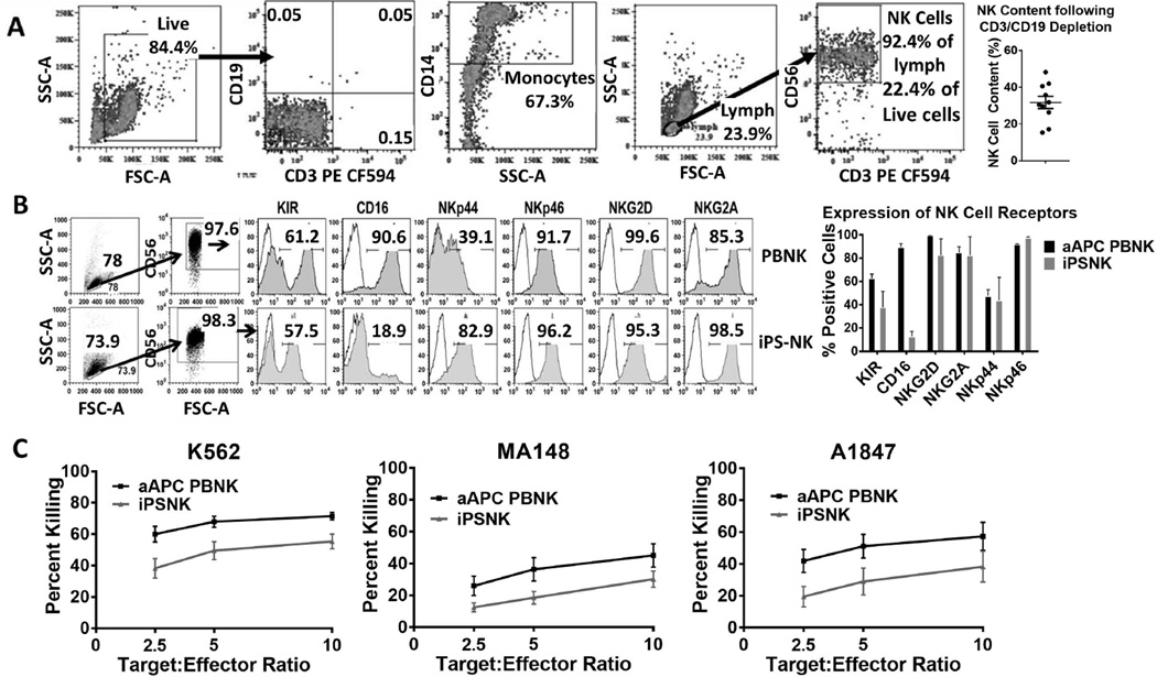
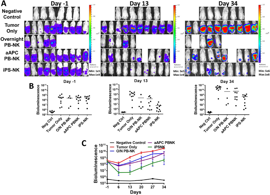
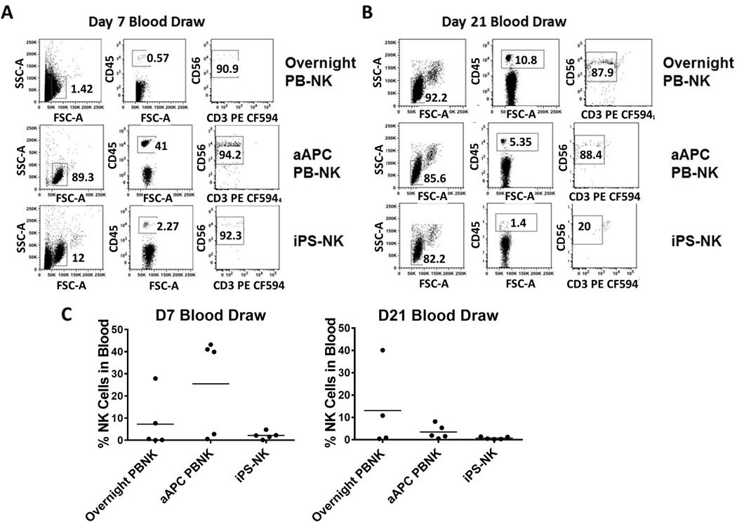
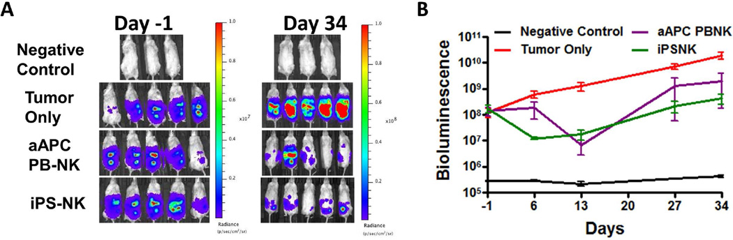
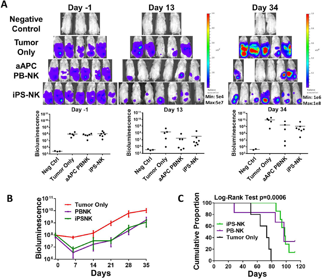
Natural killer (NK) cells can provide effective immunotherapy for ovarian cancer. Here, we evaluated the ability of NK cells isolated from peripheral blood (PB) and NK cells derived from induced pluripotent stem cell (iPSC) to mediate killing of ovarian cancer cells in a mouse xenograft model. A mouse xenograft model was used to evaluate the intraperitoneal delivery of three different NK cell populations: iPSC-derived NK cells, PB-NK cells that had been activated and expanded in long-term culture, and overnight activated PB-NK cells that were isolated through CD3/CD19 depletion of PB B and T cells. Bioluminescent imaging was used to monitor tumor burden of luciferase expressing tumor lines. Tumors were allowed to establish prior to administering NK cells via intraperitoneal injection. These studies demonstrate a single dose of any of the three NK cell populations significantly reduced tumor burden. When mice were given three doses of either iPSC-NK cells or expanded PB-NK cells, the median survival improved from 73 days in mice untreated to 98 and 97 days for treated mice, respectively. From these studies, we conclude iPSC-derived NK cells mediate antiovarian cancer killing at least as well as PB-NK cells, making these cells a viable resource for immunotherapy for ovarian cancer. Due to their ability to be easily differentiated into NK cells and their long-term expansion potential, iPSCs can be used to produce large numbers of well-defined NK cells that can be banked and used to treat a large number of patients including treatment with multiple doses if necessary.

Keywords: Immunotherapy; Induced pluripotent stem cells; Natural killer cells; Ovarian cancer.


Conflict of interest statement

Conflict of Interest: The authors declare no conflict of interest.

Disclosure of Potential Conflicts of Interest:

Dr. Kaufman consults for Fate Therapeutics. This relationship has been reviewed and managed by the University of Minnesota in accordance with its conflicts of interest policies.

Figures

Figure 1
Figure 1
Flow cytometric analysis of NK cell products. A) Phenotype of CD3/CD19 depleted PB-NK cell product with no aAPC expansion. Cells were gated based on live gate for CD3/CD19 and monocyte percentages. A lymphocyte gate based off the live gate was used to determine percent CD56+ NK cells. Final percentage of NK cells is percentage of NK cells in live gate. Dot plot represents the NK cell content of 10 CD3/CD19 depletions with SEM displayed. B) Phenotypic analysis of aAPC expanded PB-NK and iPSC-derived NK cells. A lymphocyte gate and a CD56+ gate were applied for individual NK cell receptors. Flow plots are representative of at least 3 independent experiments. The bar graph is the average + SEM for n=3. C) In vitro killing ability of NK cells was assessed using a standard 4-hour 51-Cr release assay. Lines shown are the average tumor lysis at the indicated effector:target ratios for at least 3 independent experiments.
Figure 2
Figure 2
PB-NK cells and iPSC-derived NK cells mediate in vivo killing of ovarian cancer cells in a MA-148 tumor model. Mice were inoculated with 2×10⁵ MA-148 GFP:Luc tumor cells 4 days prior to NK cell injection. Bioluminescent imaging was used to monitor tumor establishment and growth. Day -1 images were used to group mice prior to NK cell injection. A) Bioluminescent imaging of mice treated with overnight PB-NK, aAPC PB-NK, or iPS-NK cells as indicated in figures. B) Dot plots representing each group at shown time points. Bars are plotted at the mean. C) Means + SEM plotted for each group as shown. All treatment groups were statistically significantly different over the treatment period (p<0.0001) compared to tumor only, and iPSC-NK were significantly better than aAPC PB-NK (p=0.014).
Figure 3
Figure 3
Analysis of NK cells in blood of mice treated with NK cells. A representative mouse blood draw for either overnight activated PB-NK, aAPC PB-NK, or iPS-NK cells at (A) Day 7 or (B) Day 21. Cells were first gated based on FSC/SSC, then by human CD45+ cells, and finally CD56+ cells. C) Dot plots demonstrating the percentage of human NK cells in mice.
Figure 4
Figure 4
PB-NK cells and iPSC-derived NK cells mediate in vivo killing of ovarian cancer cells in a A1847 tumor model. Mice were inoculated with 2×10⁵ A1847 GFP:Luc tumor cells 4 days prior to NK cell injection. Bioluminescent imagining was used to monitor tumor establishment and growth. Day -1 images were used to group mice prior to NK cell injection. A&B) Bioluminescent imaging data for mice treated with aAPC PB-NK, or iPS-NK cells as indicated in figures. Line graph represents mean + SEM. Both treatment groups were statistically significantly better than tumor alone over the treatment period (both p<0.0001).
Figure 5
Figure 5
Anti-ovarian cancer activity using multiple doses of NK cells. Mice were inoculated with 2×10⁵ MA-148 GFP:Luc tumor cells 4 days prior to NK cell injection. Bioluminescent imagining was used to monitor tumor establishment and growth. NK cells were administered on D0, D7, and D14. A&B) Bioluminescent imaging data for mice treated with aAPC PB-NK, or iPS-NK cells as indicated in figures. Line graph represents mean + SEM. Both treatment groups were significantly better than tumor alone over the treatment period (both p<0.0001) C) Kaplan-Meier curve for overall survival of mice. Tumor only n=5 (5 failed), aAPC PB-NK n=6 (4 failed), and iPS-NK n=7 (6 failed). Treatment with iPS-NK and aAPC PB-NK statistically significantly improved overall survival compared to tumor only (p=0.008 and p=0.048, respectively).

References

    1. Armstrong DK. Relapsed ovarian cancer: challenges and management strategies for a chronic disease. Oncologist. 2002;7(Suppl 5):20–28. - PubMed
    1. Geller MA, Cooley S, Judson PL, et al. A phase II study of allogeneic natural killer cell therapy to treat patients with recurrent ovarian and breast cancer. Cytotherapy. 2011;13:98–107. - PMC - PubMed
    1. Miller JS, Soignier Y, Panoskaltsis-Mortari A, et al. Successful adoptive transfer and in vivo expansion of human haploidentical NK cells in patients with cancer. Blood. 2005;105:3051–3057. - PubMed
    1. Geller MA, Knorr DA, Hermanson DA, et al. Intraperitoneal delivery of human natural killer cells for treatment of ovarian cancer in a mouse xenograft model. Cytotherapy. 2013;15:1297–1306. - PMC - PubMed
    1. Koepsell SA, Miller JS, McKenna DH. Natural killer cells: a review of manufacturing and clinical utility. Transfusion. 2013;53:404–410. - PubMed

Publication types