. 2016 Feb;95(2):188-95.
doi: 10.1177/0022034515612022. Epub 2015 Oct 26.

Conditional TNF-α Overexpression in the Tooth and Alveolar Bone Results in Painful Pulpitis and Osteitis

Affiliations

Conditional TNF-α Overexpression in the Tooth and Alveolar Bone Results in Painful Pulpitis and Osteitis

B E Hall et al. J Dent Res. 2016 Feb.

Abstract

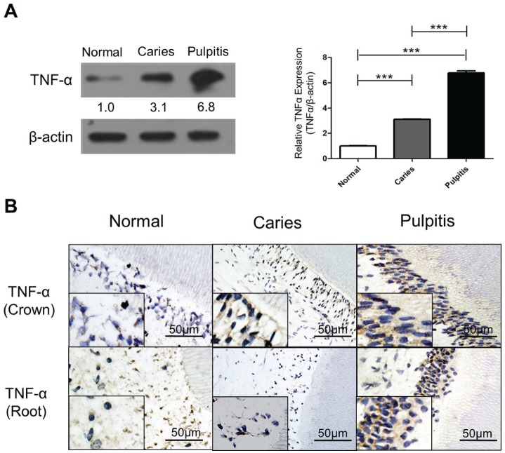
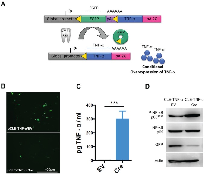
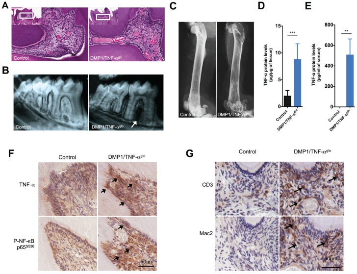
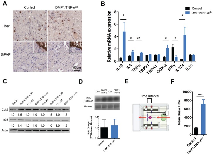
Tumor necrosis factor-α (TNF-α) is a proalgesic cytokine that is commonly expressed following tissue injury. TNF-α expression not only promotes inflammation but can also lead to pain hypersensitivity in nociceptors. With the established link between TNF-α and inflammatory pain, we identified its increased expression in the teeth of patients affected with caries and pulpitis. We generated a transgenic mouse model (TNF-α(glo)) that could be used to conditionally overexpress TNF-α. These mice were bred with a dentin matrix protein 1 (DMP1)-Cre line for overexpression of TNF-α in both the tooth pulp and bone to study oral pain that would result from subsequent development of pulpitis and bone loss. The resulting DMP1/TNF-α(glo) mice show inflammation in the tooth pulp that resembles pulpitis while also displaying periodontal bone loss. Inflammatory infiltrates and enlarged blood vessels were observed in the tooth pulp. Pulpitis and osteitis affected the nociceptive neurons innervating the orofacial region by causing increased expression of inflammatory cytokines within the trigeminal ganglia. With this new mouse model morphologically mimicking pulpitis and osteitis, we tested it for signs of oral pain with an oral function assay (dolognawmeter). This assay/device records the time required by a mouse to complete a discrete gnawing task. The duration of gnawing required by the DMP1/TNF-α(glo) mice to complete the task was greater than that for the controls; extended gnaw time in a dolognawmeter indicates reduced orofacial function. With the DMP1/TNF-α(glo) mice, we have shown that TNF-α expression alone can produce inflammation similar to pulpitis and osteitis and that this mouse model can be used to study dental inflammatory pain.

Keywords: Cdk5; animal model; cytokine(s); facial pain; inflammation; toothache.


Conflict of interest statement

The authors declare no potential conflicts of interest with respect to the authorship and/or publication of this article.

Figures

Figure 1.
Figure 1.
Tumor necrosis factor–α (TNF-α) expression is elevated by bacterial infection of the human tooth. (A) TNF-α (25 kD) is significantly upregulated during carious progression. A bar graph shows the quantification of the relative TNF-α expression (TNF-α/β-actin) between control, caries, and pulpitis groups (***P < 0.001). (B) Immunohistochemistry staining shows a similar result as the Western blot. Expression of TNF-α is increased in the tooth crown of the caries group and the pulpitis group. However, in the region of the tooth root, the pulpitis group displays a much stronger positive signal of TNF-α than the caries group. Lower inserts show higher magnification in each panel. Bar represents 50 µm.
Figure 2.
Figure 2.
Transgenic vector for Cre-mediated overexpression of tumor necrosis factor–α (TNF-α). (A) Schematic for conditional overexpression of TNF-α in the odontoblasts of transgenic mice. A transgenic mouse line designated TNF-αglo was generated, which requires Cre-mediated recombination to overexpress TNF-α. These TNF-αglo mice were bred with a dentin matrix protein 1 (DMP1)–Cre line for overexpression of TNF-α in both the tooth pulp and bone. (B) The transgenic vector was first tested in the MO6-G3 odontoblast cell line. The MO6-G3 cells show decreased green fluorescent protein (GFP) expression when cotransfected with Cre versus an empty vector. (C) Supernatants from MO6-G3 cells cotransfected with Cre show higher levels of TNF-α by enzyme-linked immunosorbent assay (***P < 0.001). (D) When cotransfected with Cre, MO6-G3 cells show increased active TNF-α signaling via nuclear factor (NF)–κB phosphorylation and a concomitant decrease in GFP expression. β-Actin was used as loading control. EV, empty vector.
Figure 3.
Figure 3.
Dentin matrix protein 1 (DMP1)/tumor necrosis factor–αglo (TNF-αglo) mice show inflammatory infiltrates within the tooth pulp. (A) Hematoxylin and eosin staining of the tooth pulp shows inflammation that is similar to pulpitis with infiltrating cells and enlarged blood vessels. (B) Radiographs also show alveolar bone loss around the teeth of the DMP1/TNF-αglo mice. (C) Femurs from the DMP1/TNF-αglo mice display reduced opacity, suggesting overall systemic bone loss due to overexpression of TNF-α by osteocytes. (D) Increased amounts of TNF-α were detected per µg of mandibular protein within the DMP1/TNF-αglo mice. (E) Serum levels of TNF-α were also increased in the DMP1/TNF-αglo mice as detected by a mouse TNF-α enzyme-linked immunosorbent assay (**P ≤ 0.01). (F) Immunohistochemical staining shows overexpression of TNF-α in the tooth pulp. Increased TNF-α cell signaling was also detected via phospho–NF-κB. Arrows denote sites of increased TNF-α expression and subsequent cell signaling via phosphorylation of NF-κB. (G) CD3 and Mac2 staining shows recruitment of lymphocytes and macrophages, respectively, in the tooth pulp. Arrows identify infiltrating lymphocytes and macrophages.
Figure 4.
Figure 4.
Dentin matrix protein 1 (DMP1)/tumor necrosis factor–αglo (TNF-αglo) mice exhibit orofacial pain due to the TNF-α–induced pulpitis and osteitis. (A) Inflammation initiated by TNF-α is primarily restricted to the tooth pulp and bone and does not cause neuronal injury or glial activation within the trigeminal ganglia (TG) as determined by Iba1 and GFAP staining (lower inserts positive staining for central root astrocytes). (B) The expression of proinflammatory markers within the TG was evaluated by quantitative real-time polymerase chain reaction (*P ≤ 0.05; **P ≤ 0.01). (C) Cdk5 is a protein kinase; its activity can be upregulated by inflammation, so levels of both Cdk5 and its activator p35 were examined within the TG to determine if chronic tooth inflammation increased their expression. In most cases, the levels of both Cdk5 and p35 were higher in the DMP1/TNF-αglo mice versus the littermate control (fold differences were measured as the ratio over β-actin). (D) A Cdk5 kinase assay was performed using protein from the TG. Graph and representative blot showing modest increase in Cdk5 kinase activity. (E) Schematic of the dolognawmeter (Dolan et al. 2010), an assay and device used to quantify nociception by measuring masticatory function. (F) DMP1/TNF-αglo mice require more time to gnaw through a hard dowel compared to Cre controls, signifying masticatory dysfunction and probable orofacial pain (****P ≤ 0.0001).

References

    1. Boettger MK, Hensellek S, Richter F, Gajda M, Stöckigt R, von Banchet GS, Bräuer R, Schaible HG. 2008. Antinociceptive effects of tumor necrosis factor alpha neutralization in a rat model of antigen-induced arthritis: evidence of a neuronal target. Arthritis Rheum. 58(8):2368–2378. - PubMed
    1. Bradley JR. 2008. TNF-mediated inflammatory disease. J Pathol. 214(2):149–160. - PubMed
    1. Bradley SV, Hyun TS, Oravecz-Wilson KI, Li L, Waldorff EI, Ermilov AN, Goldstein SA, Zhang CX, Drubin DG, Varela K, et al. 2007. Degenerative phenotypes caused by the combined deficiency of murine HIP1 and HIP1r are rescued by human HIP1. Hum Mol Genet. 16(11):1279–1292. - PubMed
    1. Byers MR, Närhi MV. 1999. Dental injury models: experimental tools for understanding neuroinflammatory interactions and polymodal nociceptor functions. Crit Rev Oral Biol Med. 10(1):4–39. - PubMed
    1. Chu CQ, Swart D, Alcorn D, Tocker J, Elkon KB. 2007. Interferon-gamma regulates susceptibility to collagen-induced arthritis through suppression of interleukin-17. Arthritis Rheum. 56(4):1145–1151. - PubMed

Publication types

MeSH terms

Substances