. 2015 Dec 1;108(3):324-34.
doi: 10.1093/cvr/cvv238. Epub 2015 Oct 26.

Pivotal role of miR-448 in the development of ROS-induced cardiomyopathy

Affiliations

Pivotal role of miR-448 in the development of ROS-induced cardiomyopathy

Sergii Kyrychenko et al. Cardiovasc Res. .

Abstract

Aims: Nicotinamide adenine dinucleotide oxidases (NOXs) are important contributors to cellular oxidative stress in the cardiovascular system. The NOX2 isoform is upregulated in numerous disorders, including dystrophic cardiomyopathy, where it drives the progression of the disease. However, mechanisms underlying NOX2 overexpression are still unknown. We investigated the role of microRNAs (miRs) in the regulation of NOX2 expression.

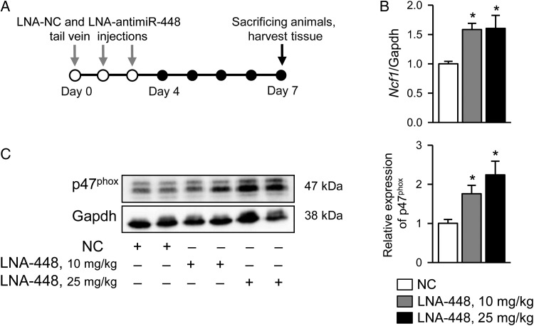
Methods and results: Duchenne muscular dystrophy (DMD) was used as a model of cardiomyopathy. After screening with miRNA target prediction databases and following qRT-PCR analysis, we found drastic downregulation of miR-448-3p in hearts of mdx mice, an animal model of DMD. The downregulation correlated with overexpression of the Ncf1 gene, encoding the NOX2 regulatory subunit p47(phox). Specificity of Ncf1 targeting by miR-448-3p was validated by luciferase reporter assay. Silencing of miR-448-3p in wild-type mice had a dramatic effect on cellular and functional properties of cardiac muscle as assessed by western blotting, qRT-PCR, confocal imaging, echocardiography, and histology. Acute treatment of mice with LNA-miR-448 inhibitors led to increased Ncf1 expression, abnormally elevated reactive oxygen species (ROS) production and exacerbated Ca(2+) signalling in cardiomyocytes, reminiscent of features previously observed in dystrophic cardiac cells. In addition, chronic inhibition of miR-448-3p resulted in dilated cardiomyopathy and arrhythmia, hallmarks of dystrophic cardiomyopathy.

Conclusions: Our studies suggest that downregulation of miR-448-3p leads to the increase in the expression of Ncf1 gene and p47(phox) protein, as well as to the substantial increase in NOX2-derived ROS production. Cellular oxidative stress subsequently triggers events that finally culminate in cardiac tissue damage and development of cardiomyopathy.

Keywords: Cardiomyopathy; Dystrophin; NOX; Oxidative stress; microRNA.


Figures

Figure 1
Figure 1
Increased level of NOX2 results in oxidative stress in dystrophic heart. (A) Representative images of DCF fluorescence in cardiomyocytes isolated from young WT and mdx mice. Graph in the middle illustrates time changes in the average DCF signals. Bar graph on the right shows the rate of DCF oxidation (slope) in WT cells (N = 3, n = 9) and mdx without (N = 3, n = 12) and with (N = 3, n = 8) incubation with DPI (10 µM). (B) Quantitative real-time PCR analysis of mRNA of Cybb, Ncf1, Cyba, Ncf4, Ncf2, and Rac1 genes in cardiac tissues of young, adult, and senescent WT and mdx mice. N = 5–6. (C) Western blot analysis indicates increase in expression of gp91phox and p47phox subunits in dystrophic heart compared with WT cardiac tissue, N = 5. *P < 0.05, **P < 0.001, t-test.
Figure 2
Figure 2
Changes in miRs expression in dystrophic heart. (A) Real-time PCR analysis of cardio-specific miRs and miRs predicted to target Cybb and Ncf1 genes. Graphs show changes in miR levels in mdx hearts relative to their levels in WT samples (indicated by dashed line). N = 5–6, *P < 0.05, t-test. (B) Genomic localization of mmu-miR-448 on intron 4 of the Htr2C gene located on X-chromosome of mouse genome. It is further processed to pre-miR-448 and later to the mature miR-448, where miR-448-3p is conserved among species. (C) Left, schematic representation of miR-448-3p binding region (‘seed region’) in mouse Ncf1 gene. On the bottom is the map of quadruple mutant (mutated residuals is in light grey). Right, luciferase activity assay shows the dose-dependent decrease of luciferase activity in cells transfected with miR-448 mimic and NCF1-3′UTR plasmid, but not with construct with the mutated seed region. For each transfection, a total of six wells for each condition were collected and measured individually, and then averaged (n = 6). A total of three separate transfections were assayed and averaged (N = 3). *P < 0.05, t-test.
Figure 3
Figure 3
Targeting of miR-448 by LNA-antimiR-448 inhibitor in WT mice results in an increase in Ncf1 gene and p47phox protein expression. (A) Protocol for LNA-antimiR-448 transfer. (B) Real-time PCR analysis of Ncf1 mRNA expression in cardiac tissue form WT mice treated with LNA-antimiR-448 inhibitor at two different concentrations. N = 5, *P < 0.05, t-test. (C) Expression of p47phox subunit of NOX2 in hearts of WT mice treated with LNA-NC and LNA-antimiR 448 inhibitors. N = 5, *P < 0.05, t-test.
Figure 4
Figure 4
Systemic inhibition of miR-448 in WT mice results in an increase in NOX2-dependent ROS production and exacerbated Ca2+ responses in ventricular cardiomyocytes. (A) Representative images of DCF fluorescence in myocytes isolated from mice treated with LNA-NC and LNA-antimiR-448 inhibitor at two different concentrations. Graph in the middle illustrates changes in average DCF signals. Bar graph on the right illustrates the rate of DCF oxidation in six different groups of experiments: LNA-NC (N = 5, n = 17; N = 4, n = 22), LNA-antimiR-448, 10 mg/kg (N = 3, n = 25; N = 3, n = 13), and LNA-antimiR-448, 25 mg/kg (N = 3, n = 22; N = 3, n = 15) without and with incubation with DPI, respectively. (B) Intracellular Ca2+ responses to mild hypo-osmotic shock in ventricular cardiomyocytes from WT mice treated with LNA-NC and LNA-antimiR-448. Left panels are line-scan representations of series of images acquired from cells and converted to a two-dimensional X,t image. Middle panel represents time course of normalized fluo-4 fluorescence in WT cells treated with LNA-NC and LNA-antimiR-448. Right panel shows pooled data of mean values of normalized fluorescence during 60 s after the osmotic shock. Number of cells studied was LNA-NC (N = 4, n = 26), LNA-antimiR-448, 10 mg/kg (N = 4 n = 27; N = 3 n = 10), and LNA-antimiR-448, 25 mg/kg (N = 4 n = 27; N = 3 n = 12) without and with incubation with DPI, respectively. *P < 0.05, **P < 0.001, t-test (for ROS and Ca2+ measurements) and #P < 0.05, ##P < 0.001, t-test (for ROS and Ca2+ measurements after DPI treatment).
Figure 5
Figure 5
Chronic inhibition of miR-448 in WT mice impairs cardiac functions. (A) Protocol for LNA-antimiR-448 injection. (B) Left, real-time PCR analysis of Ncf1 mRNA expression in heart of WT mice injected with LNA-antimiR inhibitor of miR-448 (10 mg/kg) compared with the corresponding NC (N = 5). Middle, immunoblot and right, summary of p47phox subunit expression under the same experimental conditions. N = 5, *P < 0.05, t-test. (C) Left, M-mode echocardiographic images of left ventricles (LVs) of LNA-treated adult WT mice. Right, bar graphs illustrate changes in FS and ventricle ejection fraction in animals treated with LNA-NC (N = 7) and LNA-antimiR-448 (N = 4), respectively. *P < 0.05, t-test.
Figure 6
Figure 6
Chronic inhibition of miR-448 in WT mice induces cardiac remodelling. (A) Morphological changes in hearts following chronic LNA treatment. (B) Morphological changes in cardiac myocytes. Left, representative images of LV myocardium of LNA-treated mice stained with haematoxylin-eosin. Right, averaged cross-sectional area of the myocytes in hearts of animals treated with either LNA-NC (N = 3) or LNA-antimiR-448 (N = 3). *P < 0.05, t-test. (C and D) Left, patterns of perivascular and cardiac fibrosis in LV remote myocardium stained with Masson trichrome (C) or picric acid sirius red (D). Right, relative amount of fibrotic patches in hearts of animals treated with either LNA-NC (N = 3) or LNA-antimiR-448 (N = 3). **P < 0.001, t-test. (E) Real-time PCR analysis shows overexpression of pro-fibrotic genes mRNA in cardiac tissues after acute (grey bars) and chronic (black bars) inhibition of miR-448. Dashed line indicates gene expression in LNA-NC samples. N = 4–6. *P < 0.05, **P < 0.001, t-test.

References

    1. Paravicini TM, Touyz RM. NADPH oxidases, reactive oxygen species, and hypertension: clinical implications and therapeutic possibilities. Diabetes Care 2008;31:S170–S180. - PubMed
    1. Nabeebaccus A, Zhang M, Shah AM. NADPH oxidases and cardiac remodelling. Heart Fail Rev 2010;16:5–12. - PubMed
    1. Zhang M, Perino A, Ghigo A, Hirsch E, Shah AM. NADPH oxidases in heart failure: poachers or gamekeepers? Antioxid Redox Signal 2013;18:1024–1041. - PMC - PubMed
    1. Bedard K, Krause K-H. The NOX family of ROS-generating NADPH oxidases: physiology and pathophysiology. Physiol Rev 2007;87:245–313. - PubMed
    1. Shirokova N, Niggli E. Cardiac phenotype of Duchenne muscular dystrophy: insights from cellular studies. J Mol Cell Cardiol 2013;58:217–224. - PMC - PubMed

Publication types

MeSH terms