. 2016 Jan 29;291(5):2087-106.
doi: 10.1074/jbc.M115.696419. Epub 2015 Oct 26.

Mutant B-Raf(V600E) Promotes Melanoma Paracellular Transmigration by Inducing Thrombin-mediated Endothelial Junction Breakdown

Affiliations

Mutant B-Raf(V600E) Promotes Melanoma Paracellular Transmigration by Inducing Thrombin-mediated Endothelial Junction Breakdown

Pu Zhang et al. J Biol Chem. .

Abstract

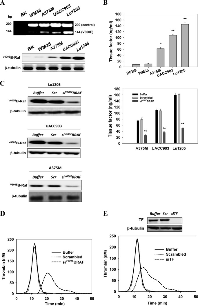
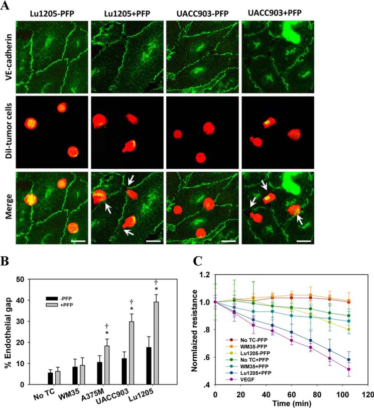
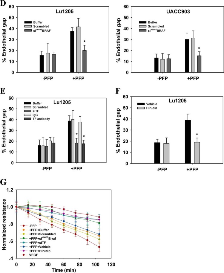
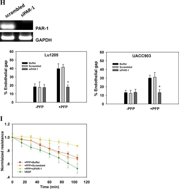
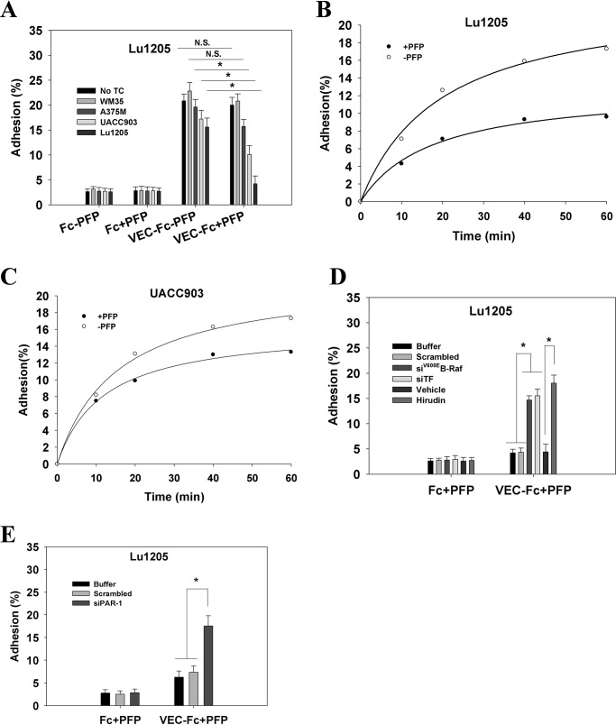
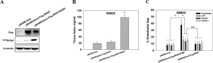
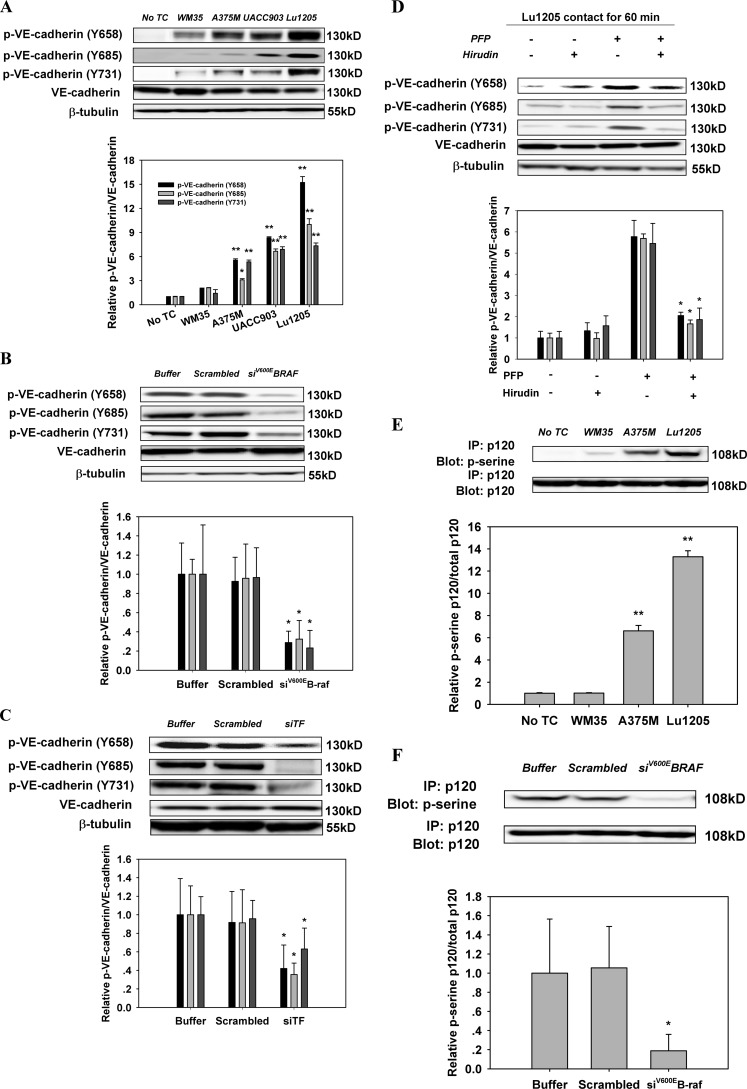
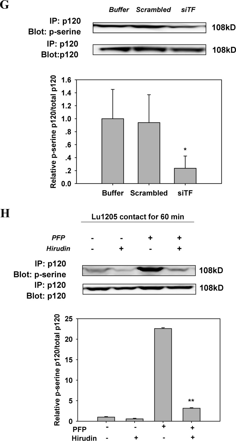
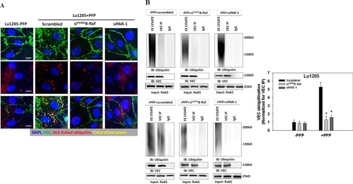
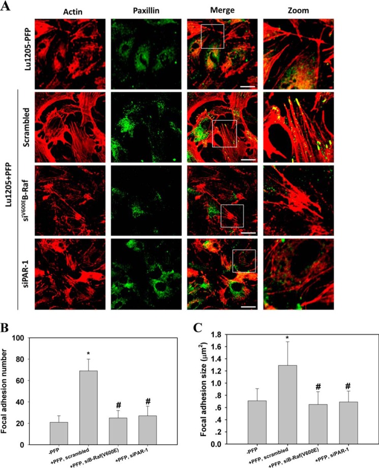
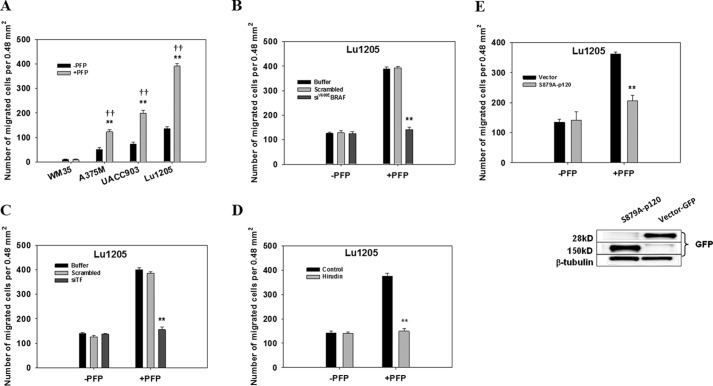
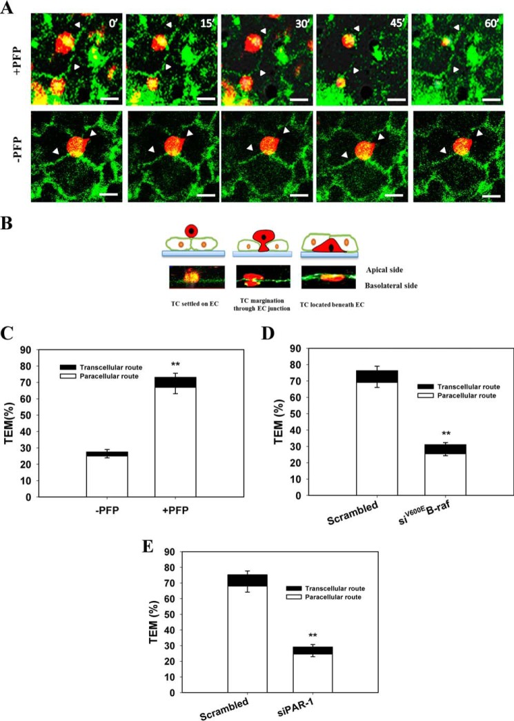
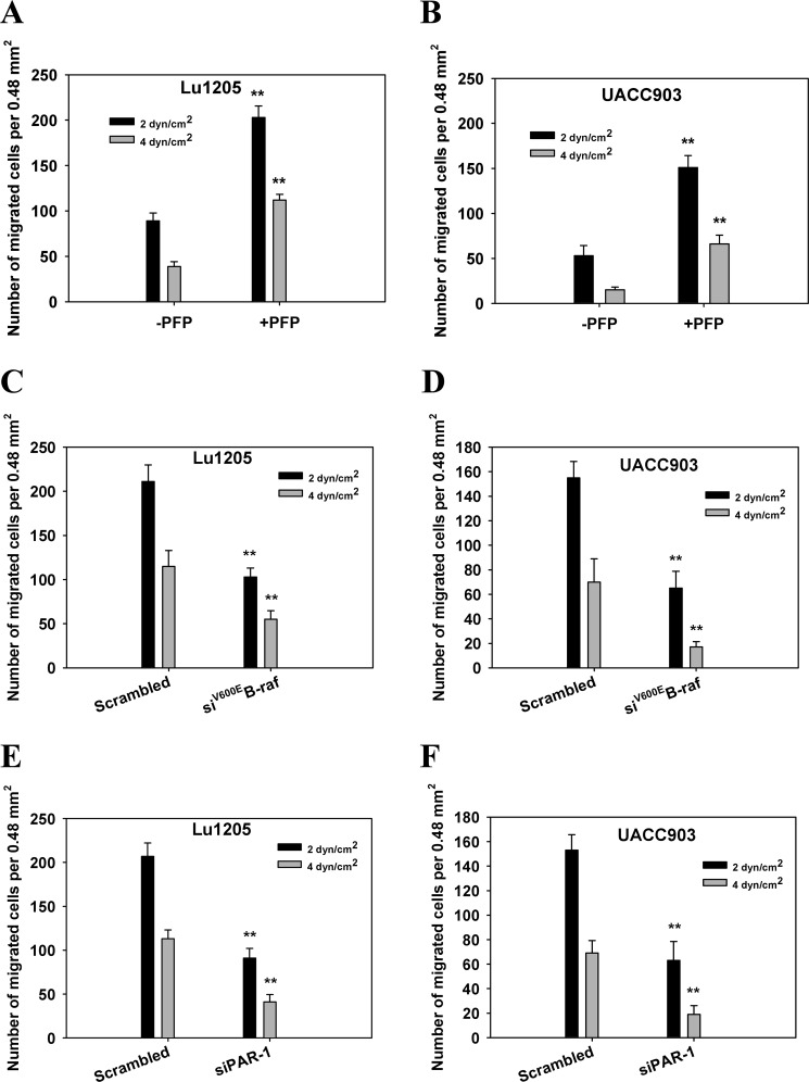
Tumor invasiveness depends on the ability of tumor cells to breach endothelial barriers. In this study, we investigated the mechanism by which the adhesion of melanoma cells to endothelium regulates adherens junction integrity and modulates tumor transendothelial migration (TEM) by initiating thrombin generation. We found that the B-Raf(V600E) mutation in metastatic melanoma cells up-regulated tissue factor (TF) expression on cell membranes and promoted thrombin production. Co-culture of endothelial monolayers with metastatic melanoma cells mediated the opening of inter-endothelial spaces near melanoma cell contact sites in the presence of platelet-free plasma (PFP). By using small interfering RNA (siRNA), we demonstrated that B-Raf(V600E) and TF silencing attenuated the focal disassembly of adherens junction induced by tumor contact. Vascular endothelial-cadherin (VE-cadherin) disassembly was dependent on phosphorylation of p120-catenin on Ser-879 and VE-cadherin on Tyr-658, Tyr-685, and Tyr-731, which can be prevented by treatment with the thrombin inhibitor, hirudin, or by silencing the thrombin receptor, protease-activated receptor-1, in endothelial cells. We also provided strong evidence that tumor-derived thrombin enhanced melanoma TEM by inducing ubiquitination-coupled VE-cadherin internalization, focal adhesion formation, and actin assembly in endothelium. Confocal microscopic analysis of tumor TEM revealed that junctions transiently opened and resealed as tumor cells accomplished TEM. In addition, in the presence of PFP, tumor cells preferentially transmigrated via paracellular routes. PFP supported melanoma transmigration under shear conditions via a B-Raf(V600E)-thrombin-dependent mechanism. We concluded that the activation of thrombin generation by cancer cells in plasma is an important process regulating melanoma extravasation by disrupting endothelial junction integrity.

Keywords: V600EB-raf; VE-cadherin; adherens junction; focal adhesion; thrombin; tissue factor; tumor metastasis.


Figures

FIGURE 1.
FIGURE 1.
B-Raf(V600E) mutation conferred on melanoma a thrombogenic potential by inducing TF expression. A, ARMS-PCR and Western blotting were used to detect the presence of B-Raf(V600E) in the WM35, A375M, UACC903, and Lu1205 melanoma cell lines. The Fo-Ro primer pair generated a common fragment of 200 bp flanking the mutation site. The Fo-Rimut primer pair generated the 144-bp fragment specific for the B-Raf(V600E) gene. BK, blank no DNA (control). Anti-B-Raf(V600E) (VE1) antibody was used to detect B-Raf(V600E) protein levels. B, cell membrane fractions from four melanoma cell lines were isolated with octyl β-d-glucopyranoside, and TF expression was measured by ELISA. TF levels (nanograms/ml) are shown as means of three independent experiments ± S.E. *, p < 0.05; **, p < 0.01 compared with control cases. C, knockdown of B-Raf(V600E) with siRNA in A375M, UACC903 and Lu1205 cells significantly reduced TF expression. A375M, UACC903, and Lu1205 cells transfected with buffer, scrambled siRNA, or B-Raf(V600E) siRNA were subjected to TF measurement. TF levels (nanograms/ml) are shown as means of three independent experiments ± S.E. **, p < 0.01 compared with control cases. Knockdown efficiency was assessed by Western blotting. Scr, scrambled; siV600EBRAF, B-Raf(V600E) siRNA. D, knockdown of B-Raf(V600E) with siRNA in Lu1205-impaired thrombin generation. Thrombin activities were measured with a calibrated automated thrombogram. E, knockdown of TF with siRNA in Lu1205 cells suppressed thrombin generation. Thrombin activities were measured with a calibrated automated thrombogram. Knockdown efficiency was assessed by Western blotting. siTF, TF siRNA.
FIGURE 2.
FIGURE 2.
Melanoma contacts increased endothelial gap formation and permeability in a B-Raf(V600E)- and PAR-1-dependent manner. A, gaps in AJ were formed at the sites of melanoma adhesion in the presence of PFP. 1 × 10⁶ Lu1205 or UACC903 cells that were stained with DiI were co-cultured with a HUVEC monolayer for 60 min in the presence (+PFP) or absence of PFP (−PFP) before the VE-cadherin was stained. With PFP, Lu1205 or UACC903 contacts induced gap formation and reduced VE-cadherin staining at the cell periphery. White arrows indicate the locations of junction dissociation and tumor pseudopod protrusion. Bar, 5 μm. B, percent endothelial gaps significantly increased upon melanoma-HUVEC co-culture in the presence of plasma. 1 × 10⁶ WM35, A375M, UACC903, and Lu1205 melanoma cells were co-cultured with a HUVEC monolayer for 60 min in the presence or absence of PFP before the endothelial gap area was determined. *, p < 0.05 compared with −PFP; †, p < 0.05 compared with no TC control. C, HUVECs were grown on transwell inserts with 0.4-μm pores before being left alone or co-cultured with WM35 or Lu1205 melanoma cells with or without PFP for the indicated time periods. HUVEC monolayer TER was measured as described under “Experimental Procedures.” The time-dependent change of the electrical resistance of HUVEC monolayer stimulated with 80 ng/ml VEGF served as a positive control. D, silencing B-Raf(V600E) with siRNA in late-stage melanoma cells, Lu1205 and UACC903, resulted in a dramatic decrease in gap formation in the presence of, but not in the absence of, PFP. *, p < 0.05 compared with control cases. siV600EBRAF, B-Raf(V600E) siRNA. E, depletion of TF with siRNA or blocking of TF with antibody-attenuated Lu1205/PFP-induced gap formation. *, p < 0.05 compared with control cases (buffer, scrambled, or isotype control). F, anti-coagulant, hirudin, reduced Lu1205/PFP-induced gap formation. *, p < 0.05 compared with vehicle control. G, HUVECs were grown on transwell inserts with 0.4-μm pores before co-culturing with Lu1205 melanoma cells transfected with buffer, scrambled siRNA, B-Raf(V600E) siRNA (siV600EBRAF), or siTF, or treated with vehicle control or 40 units/ml hirudin with PFP for indicated time periods. HUVEC monolayer TER was measured as described in “Experimental Procedures.” The time-dependent changes in the electrical resistance of the HUVEC monolayer, which was left alone or stimulated with 80 ng/ml VEGF, served as controls. H, depletion of PAR-1 with siRNA attenuated Lu1205- or UACC903/PFP-induced gap formation. *, p < 0.05 compared with control cases (buffer or scrambled). Knockdown efficiency was assessed by PCR. siPAR-1, PAR-1 siRNA. I, HUVECs transfected with buffer, scrambled siRNA, or siPAR-1 were grown on transwell inserts with 0.4-μm pores. Then they were co-cultured with Lu1205 melanoma cells for indicated time periods. HUVEC monolayer TER was measured as described under “Experimental Procedures.” The time-dependent change in the electrical resistance of the HUVEC monolayer, which was stimulated with 80 ng/ml VEGF, served as a control. Values are mean ± S.E. from at least three independent experiments.
FIGURE 2.
FIGURE 2.
Melanoma contacts increased endothelial gap formation and permeability in a B-Raf(V600E)- and PAR-1-dependent manner. A, gaps in AJ were formed at the sites of melanoma adhesion in the presence of PFP. 1 × 10⁶ Lu1205 or UACC903 cells that were stained with DiI were co-cultured with a HUVEC monolayer for 60 min in the presence (+PFP) or absence of PFP (−PFP) before the VE-cadherin was stained. With PFP, Lu1205 or UACC903 contacts induced gap formation and reduced VE-cadherin staining at the cell periphery. White arrows indicate the locations of junction dissociation and tumor pseudopod protrusion. Bar, 5 μm. B, percent endothelial gaps significantly increased upon melanoma-HUVEC co-culture in the presence of plasma. 1 × 10⁶ WM35, A375M, UACC903, and Lu1205 melanoma cells were co-cultured with a HUVEC monolayer for 60 min in the presence or absence of PFP before the endothelial gap area was determined. *, p < 0.05 compared with −PFP; †, p < 0.05 compared with no TC control. C, HUVECs were grown on transwell inserts with 0.4-μm pores before being left alone or co-cultured with WM35 or Lu1205 melanoma cells with or without PFP for the indicated time periods. HUVEC monolayer TER was measured as described under “Experimental Procedures.” The time-dependent change of the electrical resistance of HUVEC monolayer stimulated with 80 ng/ml VEGF served as a positive control. D, silencing B-Raf(V600E) with siRNA in late-stage melanoma cells, Lu1205 and UACC903, resulted in a dramatic decrease in gap formation in the presence of, but not in the absence of, PFP. *, p < 0.05 compared with control cases. siV600EBRAF, B-Raf(V600E) siRNA. E, depletion of TF with siRNA or blocking of TF with antibody-attenuated Lu1205/PFP-induced gap formation. *, p < 0.05 compared with control cases (buffer, scrambled, or isotype control). F, anti-coagulant, hirudin, reduced Lu1205/PFP-induced gap formation. *, p < 0.05 compared with vehicle control. G, HUVECs were grown on transwell inserts with 0.4-μm pores before co-culturing with Lu1205 melanoma cells transfected with buffer, scrambled siRNA, B-Raf(V600E) siRNA (siV600EBRAF), or siTF, or treated with vehicle control or 40 units/ml hirudin with PFP for indicated time periods. HUVEC monolayer TER was measured as described in “Experimental Procedures.” The time-dependent changes in the electrical resistance of the HUVEC monolayer, which was left alone or stimulated with 80 ng/ml VEGF, served as controls. H, depletion of PAR-1 with siRNA attenuated Lu1205- or UACC903/PFP-induced gap formation. *, p < 0.05 compared with control cases (buffer or scrambled). Knockdown efficiency was assessed by PCR. siPAR-1, PAR-1 siRNA. I, HUVECs transfected with buffer, scrambled siRNA, or siPAR-1 were grown on transwell inserts with 0.4-μm pores. Then they were co-cultured with Lu1205 melanoma cells for indicated time periods. HUVEC monolayer TER was measured as described under “Experimental Procedures.” The time-dependent change in the electrical resistance of the HUVEC monolayer, which was stimulated with 80 ng/ml VEGF, served as a control. Values are mean ± S.E. from at least three independent experiments.
FIGURE 3.
FIGURE 3.
Melanoma in the presence of PFP reduced VE-dependent HUVEC adhesion in a B-Raf(V600E)- and thrombin-dependent manner. A, HUVEC monolayers were left alone or co-cultured with WM35, A375M, UACC903, or Lu1205 cells in the presence or absence of PFP. After being detached, HUVECs were plated onto a VEC-Fc- or Fc-coated dish in the presence of respective melanoma-HUVEC co-culture medium for 60 min. Cell adhesion was measured as described under “Experimental Procedures” and is shown as the percentage of adherent cells. Values are mean ± S.E. from three independent experiments. B and C, after co-culture with Lu1205 and UACC903 cells in the presence or absence of PFP, HUVECs were plated onto a VEC-Fc-coated dish for the time indicated. Cell adhesion is shown as the percentage of adherent cells. D, HUVECs were co-cultured with Lu1205 cells transfected with buffer, scrambled siRNA, siB-Raf(V600E), or siTF, or treated with vehicle or hirudin in the presence of PFP. Then they were assessed for adhesion activity as described in the legend to A. E, HUVECs transfected with buffer, scrambled siRNA, or siPAR-1 were co-cultured with Lu1205 cells in the presence of PFP. Then they were assessed for adhesion activity as described in the legend to A. *, p < 0.05. N.S., not significant.
FIGURE 4.
FIGURE 4.
Ectopic expression of B-Raf(V600E) in WM35 enhanced gap formation by increasing TF expression and thrombin generation. A, WM35 cells were transfected with pBABE-bleo, pBABE(bleo)-FLAG-BRAF, or pBABE(bleo)-FLAG-BRAF(V600E) packed in retrovirus. Transfection efficiency was assessed by Western blotting with anti-FLAG and anti-B-Raf(V600E) antibodies. β-Tubulin serves as a loading control. B, WM35 cells transfected with pBABE-bleo, pBABE(bleo)-FLAG-BRAF, or pBABE(bleo)-FLAG-BRAF(V600E) were assessed for membrane TF expression. TF measurement is described in legend to Fig. 1B. *, p < 0.05 compared with pBABE(bleo)-FLAG-BRAF. C, WM35 cells transfected with pBABE-bleo, pBABE(bleo)-FLAG-BRAF, or pBABE(bleo)-FLAG-BRAF(V600E) were co-transfected with scrambled siRNA or siTF or treated with hirudin. They were co-cultured with HUVECs transfected with scrambled siRNA or siPAR-1 for 60 min in the presence of PFP before endothelial gap formation was measured. Values are mean ± S.E. from three independent experiments. *, p < 0.05. N.S., not significant.
FIGURE 5.
FIGURE 5.
Inhibition of the activities of B-Raf(V600E), TF, or thrombin reversed the increase of phosphorylation of VE-cadherin and p120-catenin in endothelium caused by melanoma contacts in plasma. A, co-culture between B-Raf(V600E)-positive melanoma and HUVECs promoted phosphorylation of VE-cadherin at tyrosine 658, 685, and 731. Confluent HUVEC monolayers were serum-starved for 12 h. They were either left alone (no TC) or co-cultured with 1 × 10⁶ melanoma cells (WM35, A375M, UACC903, or Lu1205) in the presence of PFP for 60 min before Western blot analysis. B, silencing B-Raf(V600E) suppressed co-culture-induced tyrosine phosphorylation of VE-cadherin. HUVECs were subjected to Western blot analysis after being co-cultured with 1 × 10⁶ Lu1205 melanoma cells transfected with buffer, scrambled siRNA, or B-Raf(V600E) siRNA in the presence of PFP for 60 min. C, silencing TF suppressed co-culture-induced tyrosine phosphorylation of VE-cadherin. HUVECs were subjected to Western blot analysis after being co-cultured with 1 × 10⁶ Lu1205 melanoma cells transfected with buffer, scrambled siRNA, or TF siRNA in the presence of PFP for 60 min. D, hirudin suppressed co-culture-induced tyrosine phosphorylation of VE-cadherin. HUVECs were subjected to Western blot analysis after being co-cultured with 1 × 10⁶ Lu1205 melanoma cells in the presence or absence of PFP with or without 40 units/ml hirudin for 60 min. E, serine phosphorylation of p120-catenin was triggered by melanoma contacts in the presence of PFP. HUVECs were left alone (no TC) or co-cultured with 1 × 10⁶ melanoma cells (WM35, A375M, or Lu1205) in the presence of PFP for 60 min. HUVECs were subjected to immunoprecipitation (IP). F, serine phosphorylation of p120-catenin was suppressed by B-Raf(V600E) silencing. HUVECs were co-cultured with buffer, scrambled siRNA, or B-Raf(V600E) siRNA-transfected Lu1205 cells in the presence of PFP. HUVECs were subjected to immunoprecipitation. G, serine phosphorylation of p120-catenin was suppressed by TF silencing. HUVECs were co-cultured with buffer, scrambled siRNA, or TF siRNA-transfected Lu1205 cells in the presence of PFP. HUVECs were subjected to immunoprecipitation. H, serine phosphorylation of p120-catenin was abolished by hirudin treatment. HUVECs were subjected to immunoprecipitation after being co-cultured with 1 × 10⁶ Lu1205 melanoma cells in the presence or absence of PFP with or without 40 units/ml hirudin for 60 min. For Western blot analysis, HUVECs were lysed and subjected to blotting using phospho-VE-cadherin-specific antibodies (Tyr(P)-658, Tyr(P)-685, and Tyr(P)-731). VE-cadherin and β-tubulin served as loading controls. For immunoprecipitation, precipitated proteins were analyzed by immunoblotting with an antibody against phosphoserine. The same blot was stripped and then reprobed with an antibody to p120-catenin. Relative VE-cadherin phosphorylation and p120 phosphorylation for densitometric analysis are shown below each blot. Values are mean ± S.E. *, p < 0.05; **, p < 0.01 compared with control. All results are representative of at least three independent experiments.
FIGURE 5.
FIGURE 5.
Inhibition of the activities of B-Raf(V600E), TF, or thrombin reversed the increase of phosphorylation of VE-cadherin and p120-catenin in endothelium caused by melanoma contacts in plasma. A, co-culture between B-Raf(V600E)-positive melanoma and HUVECs promoted phosphorylation of VE-cadherin at tyrosine 658, 685, and 731. Confluent HUVEC monolayers were serum-starved for 12 h. They were either left alone (no TC) or co-cultured with 1 × 10⁶ melanoma cells (WM35, A375M, UACC903, or Lu1205) in the presence of PFP for 60 min before Western blot analysis. B, silencing B-Raf(V600E) suppressed co-culture-induced tyrosine phosphorylation of VE-cadherin. HUVECs were subjected to Western blot analysis after being co-cultured with 1 × 10⁶ Lu1205 melanoma cells transfected with buffer, scrambled siRNA, or B-Raf(V600E) siRNA in the presence of PFP for 60 min. C, silencing TF suppressed co-culture-induced tyrosine phosphorylation of VE-cadherin. HUVECs were subjected to Western blot analysis after being co-cultured with 1 × 10⁶ Lu1205 melanoma cells transfected with buffer, scrambled siRNA, or TF siRNA in the presence of PFP for 60 min. D, hirudin suppressed co-culture-induced tyrosine phosphorylation of VE-cadherin. HUVECs were subjected to Western blot analysis after being co-cultured with 1 × 10⁶ Lu1205 melanoma cells in the presence or absence of PFP with or without 40 units/ml hirudin for 60 min. E, serine phosphorylation of p120-catenin was triggered by melanoma contacts in the presence of PFP. HUVECs were left alone (no TC) or co-cultured with 1 × 10⁶ melanoma cells (WM35, A375M, or Lu1205) in the presence of PFP for 60 min. HUVECs were subjected to immunoprecipitation (IP). F, serine phosphorylation of p120-catenin was suppressed by B-Raf(V600E) silencing. HUVECs were co-cultured with buffer, scrambled siRNA, or B-Raf(V600E) siRNA-transfected Lu1205 cells in the presence of PFP. HUVECs were subjected to immunoprecipitation. G, serine phosphorylation of p120-catenin was suppressed by TF silencing. HUVECs were co-cultured with buffer, scrambled siRNA, or TF siRNA-transfected Lu1205 cells in the presence of PFP. HUVECs were subjected to immunoprecipitation. H, serine phosphorylation of p120-catenin was abolished by hirudin treatment. HUVECs were subjected to immunoprecipitation after being co-cultured with 1 × 10⁶ Lu1205 melanoma cells in the presence or absence of PFP with or without 40 units/ml hirudin for 60 min. For Western blot analysis, HUVECs were lysed and subjected to blotting using phospho-VE-cadherin-specific antibodies (Tyr(P)-658, Tyr(P)-685, and Tyr(P)-731). VE-cadherin and β-tubulin served as loading controls. For immunoprecipitation, precipitated proteins were analyzed by immunoblotting with an antibody against phosphoserine. The same blot was stripped and then reprobed with an antibody to p120-catenin. Relative VE-cadherin phosphorylation and p120 phosphorylation for densitometric analysis are shown below each blot. Values are mean ± S.E. *, p < 0.05; **, p < 0.01 compared with control. All results are representative of at least three independent experiments.
FIGURE 6.
FIGURE 6.
Knockdown of B-Raf(V600E) or PAR-1 reduced melanoma-induced endothelial VE-cadherin ubiquitination and endocytosis in plasma. HUVECs transfected with scrambled siRNA or siRNA against PAR-1 were incubated with 300 μm chloroquine for 3 h. Then they were co-cultured with Lu1205 cells transfected with scrambled siRNA or siRNA against B-Raf(V600E) in the presence or absence of PFP for 60 min. A, Lu1205/PFP-induced VE-cadherin internalization (red staining) and co-localization in intracellular vesicles with K63-linked ubiquitin (green staining) in HUVECs were abrogated following siB-Raf(V600E) or siPAR-1 transfection. Pixels representing ubiquitin and VE-cadherin colocalization are highlighted in yellow. Red arrowheads point to internalized VE-cadherin. Data are representative of three independent experiments. Bar, 10 μm. B, VE-cadherin (VEC) was immunoprecipitated (IP) from the early endosome fraction, and ubiquitinated VE-cadherin was detected with Western blotting (IB) using an anti-ubiquitin antibody (FK2, Millipore). EE LYSATE, lysate from early endosome preparation. VEC IP, immunoprecipitated VE-cadherin. IgG, immunoprecipitation using an IgG isotype control. Rab5 protein was probed to validate the status of early endosomes and ensure equal loading across samples. Representative blots from three independent experiments are shown. Bottom panel is a quantitation of ubiquitinated VE-cadherin in different cases. Data were normalized against the amount of VE-cadherin immunoprecipitated from early endosome preparation. Values are mean ± S.E. *, p < 0.05 compared with Lu1205/PFP/Scrambled.
FIGURE 7.
FIGURE 7.
Endothelial stress fiber formation and focal adhesion assembly induced by melanoma contacts were suppressed after knockdown of B-Raf(V600E) or PAR-1. A, HUVEC untransfected or transfected with siRNA against PAR-1 were co-cultured with melanoma cells transfected with scrambled siRNA or siRNA against B-Raf(V600E) in the presence or absence of PFP. HUVECs were stained with rhodamine-phalloidin and paxillin antibody. The right panel shows magnified views of the boxed area in the merged images. Bar = 10 μm. (Green = paxillin and red = F-actin.) B and C, quantification of the average number and size (μm2) of paxillin-containing focal adhesions in HUVECs using ImageJ software. 12 cells were analyzed per condition in each experiment. Values are mean ± S.E. *, p < 0.05 compared with −PFP; #, p < 0.05 compared with scrambled, +PFP.
FIGURE 8.
FIGURE 8.
Inhibition of B-Raf(V600E)-mediated thrombin generation attenuated melanoma TEM. A, melanoma TEM was significantly increased by plasma treatment. The migration of 1 × 10⁶ WM35, A375M, UACC903, or Lu1205 melanoma cells across the HUVEC monolayer was assessed in a Boyden chamber in the presence or absence of PFP. **, p < 0.01 compared with −PFP; ††, p < 0.01 compared with no TC control. Values are mean ± S.E. B–D, silencing B-Raf(V600E) (B) or TF (C) or inhibition of thrombin with hirudin (D) dramatically reduced PFP-induced Lu1205 cell TEM. **, p < 0.01 compared with control cases. Values are mean ± S.E. E, p120-catenin S879A mutation attenuated PFP-induced Lu1205 cell TEM. **, p < 0.01 compared with control cases. Values are mean ± S.E. Immunoblots show the expression of S879A-p120 (150 kDa) in HUVECs. β-Tubulin served as a loading control.
FIGURE 9.
FIGURE 9.
Melanoma cells underwent TEM in plasma via paracellular routes. A, confocal images show the migration of melanoma cells from the apical side to the basolateral side of HUVECs in the presence or absence of PFP. The locations of the same Lu1205 cells were captured at 0, 15, 30, 45, and 60 min of TEM. Lu1205 cells were stained with DiI before being seeded on a HUVEC monolayer, which was transfected with GFP-VE-cadherin. B, X/Z cross-sections of images of transmigrating Lu1205 cells. Micrographs show the stages of TEM as follows: TC settlement on EC; TC margination through EC; and TC localization beneath EC. The schematic drawing above each image represents the stage of TEM shown in the micrograph. Bar, 10 μm. White arrowheads indicate junctional sites. C, percentage of adherent Lu1205 cells taking transcellular and paracellular routes for TEM in the presence or absence of PFP. D, percentage of buffer, scrambled siRNA, or siB-Raf(V600E)-transfected Lu1205 cells taking transcellular and paracellular routes for TEM in the presence of PFP. E, percentage of Lu1205 cells taking transcellular and paracellular routes for migrating across buffer, scrambled siRNA, or siPAR-1-transfected HUVEC monolayer in the presence of PFP. Values are mean ± S.E. from three independent experiments. **, p < 0.01 compared with control cases.
FIGURE 10.
FIGURE 10.
Knockdown of B-Raf(V600E) or PAR-1 reduced melanoma TEM under shear conditions. A, Lu1205; B; UACC903 TEMs were measured at a shear stress of 2 or 4 dynes/cm2 in the presence or absence of PFP. C and D, scrambled siRNA or siB-Raf(V600E)-transfected Lu1205 (C) and UACC903 (D) TEMs were measured at a shear stress of 2 or 4 dynes/cm2 in the presence of PFP. E and F, transmigrations of Lu1205 (E) or UACC903 (F) across scrambled siRNA or siPAR-1-transfected HUVECs were measured at a shear stress of 2 or 4 dynes/cm2 in the presence of PFP. Values are mean ± S.E. from three independent experiments. **, p < 0.01 compared with control.

Similar articles

Cited by

References

    1. Konstantopoulos K., and Thomas S. N. (2009) Cancer cells in transit: the vascular interactions of tumor cells. Annu. Rev. Biomed. Eng. 11, 177–202 - PubMed
    1. Wirtz D., Konstantopoulos K., and Searson P. C. (2011) The physics of cancer: the role of physical interactions and mechanical forces in metastasis. Nat. Rev. Cancer 11, 512–522 - PMC - PubMed
    1. Biggerstaff J. P., Seth N., Amirkhosravi A., Amaya M., Fogarty S., Meyer T. V., Siddiqui F., and Francis J. L. (1999) Soluble fibrin augments platelet/tumor cell adherence in vitro and in vivo, and enhances experimental metastasis. Clin. Exp. Metastasis 17, 723–730 - PubMed
    1. Ruf W., and Edgington T. S. (1994) Structural biology of tissue factor, the initiator of thrombogenesis in vivo. FASEB J. 8, 385–390 - PubMed
    1. Brummel K. E., Paradis S. G., Butenas S., and Mann K. G. (2002) Thrombin functions during tissue factor-induced blood coagulation. Blood 100, 148–152 - PubMed

Publication types

MeSH terms