. 2015 Nov 10;112(45):E6148-57.
doi: 10.1073/pnas.1509416112. Epub 2015 Oct 26.

Tether mutations that restore function and suppress pleiotropic phenotypes of the C. elegans isp-1(qm150) Rieske iron-sulfur protein

Affiliations

Tether mutations that restore function and suppress pleiotropic phenotypes of the C. elegans isp-1(qm150) Rieske iron-sulfur protein

Gholamali Jafari et al. Proc Natl Acad Sci U S A. .

Abstract

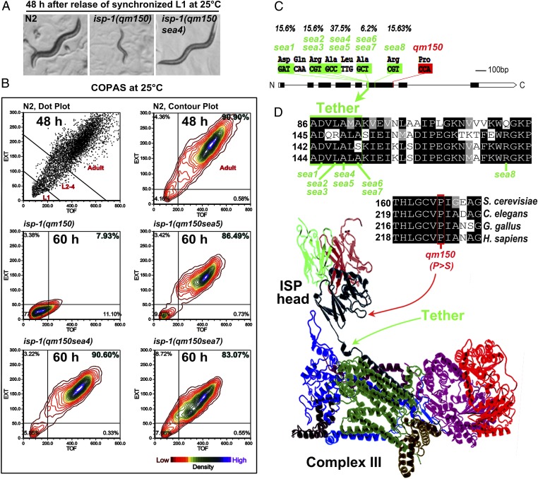
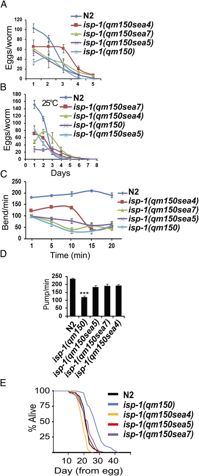
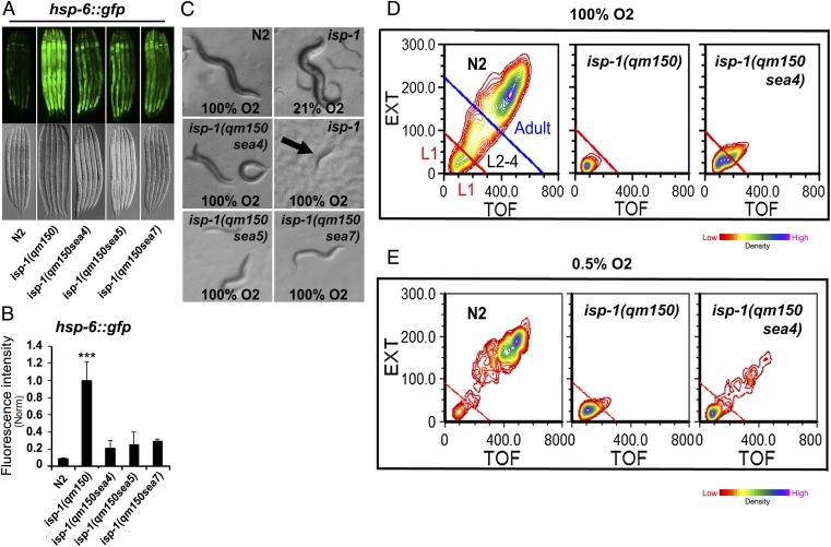
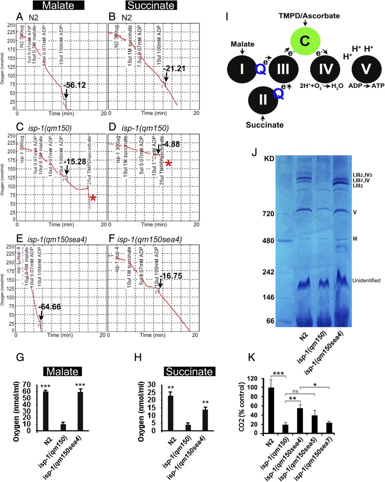
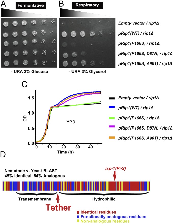
Mitochondria play an important role in numerous diseases as well as normative aging. Severe reduction in mitochondrial function contributes to childhood disorders such as Leigh Syndrome, whereas mild disruption can extend the lifespan of model organisms. The Caenorhabditis elegans isp-1 gene encodes the Rieske iron-sulfur protein subunit of cytochrome c oxidoreductase (complex III of the electron transport chain). The partial loss of function allele, isp-1(qm150), leads to several pleiotropic phenotypes. To better understand the molecular mechanisms of ISP-1 function, we sought to identify genetic suppressors of the delayed development of isp-1(qm150) animals. Here we report a series of intragenic suppressors, all located within a highly conserved six amino acid tether region of ISP-1. These intragenic mutations suppress all of the evaluated isp-1(qm150) phenotypes, including developmental rate, pharyngeal pumping rate, brood size, body movement, activation of the mitochondrial unfolded protein response reporter, CO2 production, mitochondrial oxidative phosphorylation, and lifespan extension. Furthermore, analogous mutations show a similar effect when engineered into the budding yeast Rieske iron-sulfur protein Rip1, revealing remarkable conservation of the structure-function relationship of these residues across highly divergent species. The focus on a single subunit as causal both in generation and in suppression of diverse pleiotropic phenotypes points to a common underlying molecular mechanism, for which we propose a "spring-loaded" model. These observations provide insights into how gating and control processes influence the function of ISP-1 in mediating pleiotropic phenotypes including developmental rate, movement, sensitivity to stress, and longevity.

Keywords: Rip1; aging; complex III; isp-1; mitochondrial iron–sulfur protein.


Conflict of interest statement

The authors declare no conflict of interest.

Figures

Fig. 1.
Fig. 1.
Intragenic suppressors of isp-1(qm150) suppress the slow development and are located in the tether region. (A) Development of N2, isp-1(qm150), and one of the suppressors, 48 h after release of synchronized L1 at 25 °C. (B) Complex Object Parametric Analyzer and Sorter (COPAS) assessment of the size of the individuals in the population cultivated at 25 °C for 48 h for N2, or 60 h for isp-1(qm150), and the suppressor strains (n = 6,000 per strain). N2 were assayed at 48 h to prevent substantial contamination with progeny. (C) Structure of the isp-1 gene showing the positions, residues, and the incidence of the suppressor mutations in green and the P > S mutation in isp-1(qm150) in red. (D) Protein sequences of ISP from S. cerevisiae, C. elegans, Gallus gallus, and Homo sapiens, were aligned using ClustalW and represented with Boxshade. Amino acid numbers corresponding to the first shown residue are to the left of each segment. Functionally identical residues are indicated with black background. The proline that is mutated to serine in isp-1(qm150) is highlighted in red, and suppressor mutations within the tether region are in green background. Arrows in the ribbon structure of the cytochrome bc1 complex show the tether and P > S mutation locations.
Fig. 2.
Fig. 2.
Physiological parameters of the isp-1(qm150) suppressors. (A and B) Brood size of N2, isp-1(qm150), and one of the suppressors at two different temperatures (20 °C and 25 °C), n = 25 for each strain/condition. (C) Thrashing in the course of 20 min. (D) Pharyngeal pumping rates, n = 30 per strain, the two-tailed heteroscedastic t test was performed on two strains at a time in all 10 combinations to establish which differences in pumping rate were statistically significant. (E) Lifespan of the suppressors, at 25 °C. The depicted result is a single typical trial. N2 (n = 90) isp-1(qm150) (n = 65), isp-1(qm150sea4) (n = 53), isp-1(qm150sea5) (n = 158), isp-1(qm150sea7) (n = 198). The graph indicates animals that died during the course of the experiment. Animals that left the agar surface are not considered. No other censoring was performed. Error bars: SEM for highlighted experiments, *P < 0.05; **P < 0.01; ***P < 0.001.
Fig. 3.
Fig. 3.
Effects of intragenic suppressors on isp-1(qm150) induction of the mitochondrial unfolded protein response, sensitivity to hyperoxia, and sensitivity to hypoxia. (A) Suppressors attenuated the hsp-6p::gfp expression (mtUPR response). Epifluorescence and DIC Nomarski images of the worms expressing hsp-6p::gfp. (B) hsp-6p::gfp fluorescence intensity of the strains were compared by ImageJ software (50). (C) Intragenic suppressors partially rescued isp-1(qm150) hyperoxia lethality. N2 at 100% O2 produced next generation. isp-1(qm150) at 21% O2 produced next generation. isp-1(qm150) died at L1 stage at 100% O2. Intragenic suppressors reached to L3/4 stage under 100% O2. Images are from day eight from synchronized L1 seeded onto NGM-OP50 at 25 °C. (D and E) COPAS sorted worms in hyperoxia and hypoxia. N2 worms were sorted at day two and other strains at day five at 25 °C. Please note that L1 in N2 are from the next generation. Error bars: SEM for highlighted experiments, *P < 0.05 **P < 0.01 ***P < 0.001.
Fig. 4.
Fig. 4.
ADP-stimulated respiration of intact isolated mitochondria. Oxygen consumption traces are shown in N2, isp-1(qm150), and isp-1(qm150sea4). (A) State 3 of ADP-stimulation rates (arrow) in N2 is measured by using malate substrate as an upstream (complex I) electron donor. State 3 respiration with 2 mM ADP represents the maximum capacity for oxidative phosphorylation of the mitochondria in N2. (B) Oxygen consumption traces in N2 by using succinate substrate as an upstream (complex II) electron donor. (C–F) comparison of state 3 of ADP-stimulation rates (arrows) in isp-1(qm150) and isp-1(qm150sea4) by using complex III upstream substrates (malate and succinate). In C and D, asterisks (*) show the oxygen consumption in isp-1(qm150) after adding TMPD/ascorbate (electron donor to cytochrome c, downstream of complex III). (G and H) Quantitative results for state 3 measurements of 3 biological replicates are depicted. P values are compared with isp-1(qm150) triplicates. P values in H: N2: 0.000204, isp-1(qm150sea4): 0.000707. P values in I: N2: 0.002405, isp-1(qm150sea4): 0.009303. Respiration of intact isolated mitochondria was measured with a Clark electrode. The traditional unit for the respiration rate, AO/min/mgprotein is equivalent to nmol 1/2O2/min/mgprotein (52). (I) A schematic cartoon depicting electron flow from the electron donor substrates (malate, succinate, or TMPD/ascorbate) through the mitochondrial ETC complexes. ADP stimulates respiration (state 3) because its phosphorylation to ATP by complex V allows proton flow, through complexes I, III, IV (out), and V (in). Note that electron flow from TMPD/ascorbate is independent of functional complex III. (J) Blue Native Gel. Complex I is present in supercomplex bands of N2 and isp-1(qm150sea4) but not in isp-1(qm150). (K) isp-1(qm150) CO2 output compared with the suppressors and N2 (for each strain: n = 1,000 COPAS sorted synchronized young adult animals of identical size (and likely weight) in three technical replicates (3000 worms/strain/condition). Error bars: SEM for highlighted experiments, *P < 0.05 **P < 0.01 ***P < 0.001.
Fig. 5.
Fig. 5.
Tether mutations rescue the respiratory defect of S. cerevisiae Rip1(P166S). Yeast lacking the entire RIP1 gene ORF (rip1Δ) were transformed with a plasmid containing empty vector, or plasmids expressing wild-type Rip1(WT), Rip1(P166S), or Rip1(P166S) with D87N (Sea1), or Rip1(P166S) with A90T(Sea4). Respiratory growth of yeast expressing Rip1(P166S) was dramatically impaired, which is rescued by the secondary mutations within the tether region, D87N or A90T. (A and B) Growth was assessed on solid synthetic media containing 2% (wt/vol) glucose (fermentative conditions; A) or 3% (wt/vol) glycerol (respiratory conditions; B). (C) Growth curves were generated from Bioscreen liquid culture experiments in rich media containing 2% (wt/vol) glucose. (D) Comparison of Rip1 conservation with isp-1.
Fig. 6.
Fig. 6.
Three configurations of the extrinsic domain of ISP show the spring-loaded mechanism in action. (Top) ISP docked at cyt b interface, H-bonded to the Qo-site occupant [Protein Data Bank (PDB) ID 3H1K, avian complex, stigmatellin bound]. The main features of interest are labeled. (Middle) ISP in relaxed configuration [PDB ID 3L71, avian complex, azoxystrobin (MOA-inhibitor) bound]. (Bottom) ISP H-bonding with heme c1 at cyt c1 interface (PDB ID 1BE3, bovine complex, no Qo-site occupant indicated). In the relaxed configuration (Middle), the cluster ligand (H161 in bovine) makes no H-bonding contacts, so the configuration in the tether region is not under tension. When the H161 H-bonds to the Qo-site occupant (Top), the tether is stretched due to application of a force (mainly from the strength of the H-bond to the occupant) to lengthen the tether and rotate the head. The helical region is pulled into an extended configuration, leading to the breaking of several intra- and interspan H-bonds. When H161 H-bonds to heme c1, the displacements are much smaller, and the tether region retains a H-bonding configuration similar to that of the relaxed state. SI Appendix, Fig. S6 and Table S6 shows details of H-bonding changes between Qo-site docked (stigmatellin) and relaxed (azoxystrobin) configurations. Chains are colored as follows: chain c, cyt b, pink; chain d, cyt c1, yellow; chain e, ISP, orange; chain j, 7.2-kDa protein, cyan; chain p, cyt b, green (or blue-green in 1BE3); chain q, cyt c1, olive (or light green in 1BE3). Gray spans have the cartoon colored by the CPK coloring of the C-atom or C-C bond. Atoms within 7 Å of a neighboring chain are shown as space-filled spheres of 0.2-Å radius. Atoms within 3.5 Å of a neighboring chain are shown as space-filled spheres of 0.4-Å radius. Selected atoms are CPK colored (by atom type). The three structures are shown with the protein scaffold in the same orientation, so that the main difference comes from the configurational changes of the ISP tether and extrinsic domains. Stereoscopic images for crossed-eye viewing.

Comment in

Similar articles

Cited by

References

    1. Wallace DC. Mitochondrial DNA mutations in disease and aging. Environ Mol Mutagen. 2010;51(5):440–450. - PubMed
    1. Bratic A, Larsson NG. The role of mitochondria in aging. J Clin Invest. 2013;123(3):951–957. - PMC - PubMed
    1. Butler JA, Mishur RJ, Bhaskaran S, Rea SL. A metabolic signature for long life in the Caenorhabditis elegans Mit mutants. Aging Cell. 2013;12(1):130–138. - PMC - PubMed
    1. de Jong L, Meng Y, Dent J, Hekimi S. Thiamine pyrophosphate biosynthesis and transport in the nematode Caenorhabditis elegans. Genetics. 2004;168(2):845–854. - PMC - PubMed
    1. Felkai S, et al. CLK-1 controls respiration, behavior and aging in the nematode Caenorhabditis elegans. EMBO J. 1999;18(7):1783–1792. - PMC - PubMed

Publication types

MeSH terms

Substances

LinkOut - more resources