. 2015 Oct 13:2:150043.
doi: 10.1038/sdata.2015.43. eCollection 2015.

Test-retest resting-state fMRI in healthy elderly persons with a family history of Alzheimer's disease

Collaborators, Affiliations

Test-retest resting-state fMRI in healthy elderly persons with a family history of Alzheimer's disease

Pierre Orban et al. Sci Data. .

Abstract

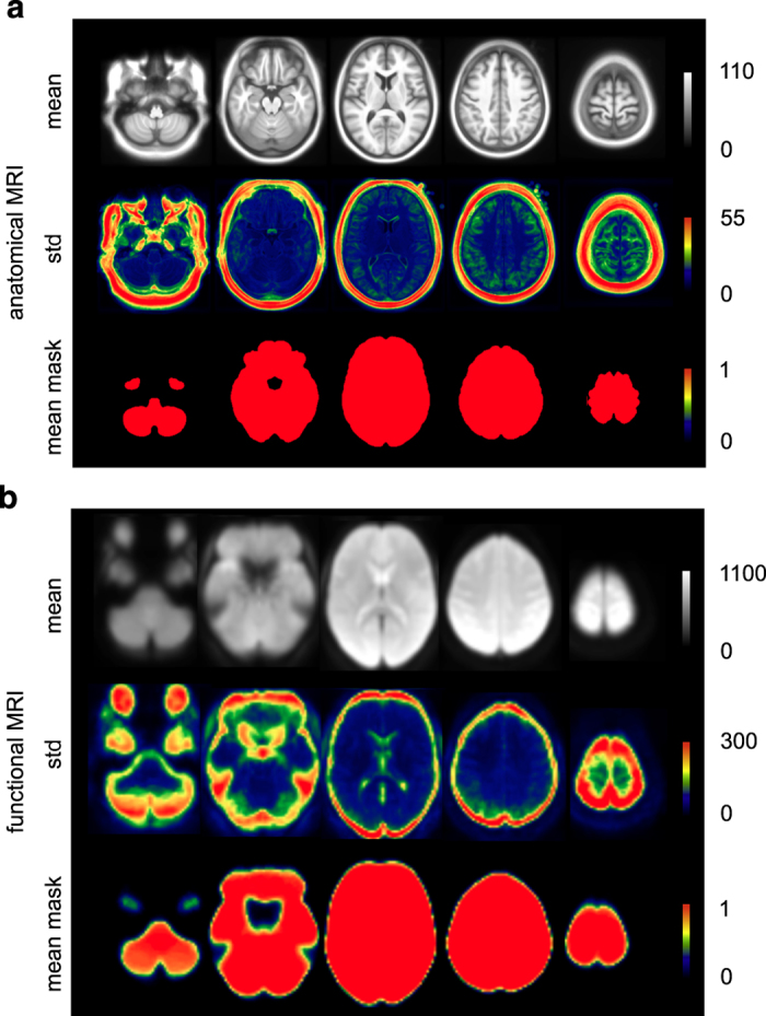
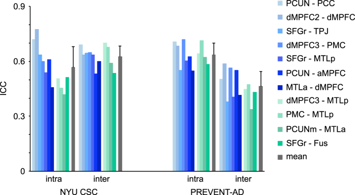
We present a test-retest dataset of resting-state fMRI data obtained in 80 cognitively normal elderly volunteers enrolled in the "Pre-symptomatic Evaluation of Novel or Experimental Treatments for Alzheimer's Disease" (PREVENT-AD) Cohort. Subjects with a family history of Alzheimer's disease in first-degree relatives were recruited as part of an on-going double blind randomized clinical trial of Naproxen or placebo. Two pairs of scans were acquired ~3 months apart, allowing the assessment of both intra- and inter-session reliability, with the possible caveat of treatment effects as a source of inter-session variation. Using the NeuroImaging Analysis Kit (NIAK), we report on the standard quality of co-registration and motion parameters of the data, and assess their validity based on the spatial distribution of seed-based connectivity maps as well as intra- and inter-session reliability metrics in the default-mode network. This resource, released publicly as sample UM1 of the Consortium for Reliability and Reproducibility (CoRR), will benefit future studies focusing on the preclinical period preceding the appearance of dementia in Alzheimer's disease.


Conflict of interest statement

The authors declare no competing financial interests.

Figures

Figure 1
Figure 1. Group-level summary statistics of co-registration procedures and motion parameters.
Averages over the two test and two retest runs are shown for all subjects, which are sorted in ascending order in terms of data quality. Spatial correlations between individual anatomical and functional scans are shown for each subject (a). Maximal rotation and translation movement parameters are shown for each subject (b). The mean frame displacement (FD) values are shown for each subject, before and after scrubbing of volumes with FD>0.5 (c). Only runs with >60 volumes left after scrubbing were included in the analyses.
Figure 2
Figure 2. Group-level summary maps of co-registration procedures.
Maps (mean across subjects, standard deviation across subjects, mean of the individual brain masks) are shown for both anatomical (a) and functional (b) MRI.
Figure 3
Figure 3. Output map of the meta-analysis.
BASC parcels with the highest number of coordinates showing a significant effect across contrasts in the selected studies, indicating the consistency of findings across studies. Maps are superimposed onto the anatomical ICBM 152 template. MNI coordinates are given for sagittal, coronal and axial slices.
Figure 4
Figure 4. Average connectivity (correlation) maps in the DMN.
Maps are shown for 3 seeds located in the anterior medial prefrontal cortex (aMPFC), the posterior cingulate cortex (PCC), and the temporo-parietal junction (TPJ). Correlation values indicate the correspondence for connectivity maps obtained in the NYU CSC and the PREVENT-AD datasets. Maps are superimposed onto the anatomical ICBM 152 template. MNI coordinates are given for representative slices.
Figure 5
Figure 5. Reliability analysis.
Run-averaged reliability was assessed intra- and inter-sessions with intra-class correlation coefficients (ICC). Reliability analyses were performed in the NYU CSC and PREVENT-AD datasets for 11 connections of interest, 7 within the DMN (blue) and 4 between the DMN and other networks (green). See Table 3 for abbreviations.

References

Data Citations

    1. Bellec P. 2014. Functional Connectomes Project International Neuroimaging Data-Sharing Initiative. http://dx.doi.org/10.15387/fcp_indi.corr.um1 - DOI

References

    1. Jacobs H. I. L., Radua J., Lückmann H. C. & Sack A. T. Meta-analysis of functional network alterations in Alzheimer's disease: toward a network biomarker. Neurosci Biobehav Rev 37, 753–765 (2013). - PubMed
    1. Sheline Y. I. & Raichle M. E. Resting State Functional Connectivity in Preclinical Alzheimer’s Disease. Biol. Psychiatry 74, 340–347 (2013). - PMC - PubMed
    1. Fox M. D. & Raichle M. E. Spontaneous fluctuations in brain activity observed with functional magnetic resonance imaging. Nat. Rev. Neurosci. 8, 700–711 (2007). - PubMed
    1. Damoiseaux J. S. et al. Consistent resting-state networks across healthy subjects. Proc. Natl Acad. Sci. USA 103, 13848–13853 (2006). - PMC - PubMed
    1. Greicius M. D., Srivastava G., Reiss A. L. & Menon V. Default-mode network activity distinguishes Alzheimer's disease from healthy aging: evidence from functional MRI. Proc. Natl Acad. Sci. USA 101, 4637–4642 (2004). - PMC - PubMed

Publication types