. 2015 Sep 3;6(10):3748-56.
doi: 10.1364/BOE.6.003748. eCollection 2015 Oct 1.

In vivo cell characteristic extraction and identification by photoacoustic flow cytography

Affiliations

In vivo cell characteristic extraction and identification by photoacoustic flow cytography

Guo He et al. Biomed Opt Express. .

Abstract

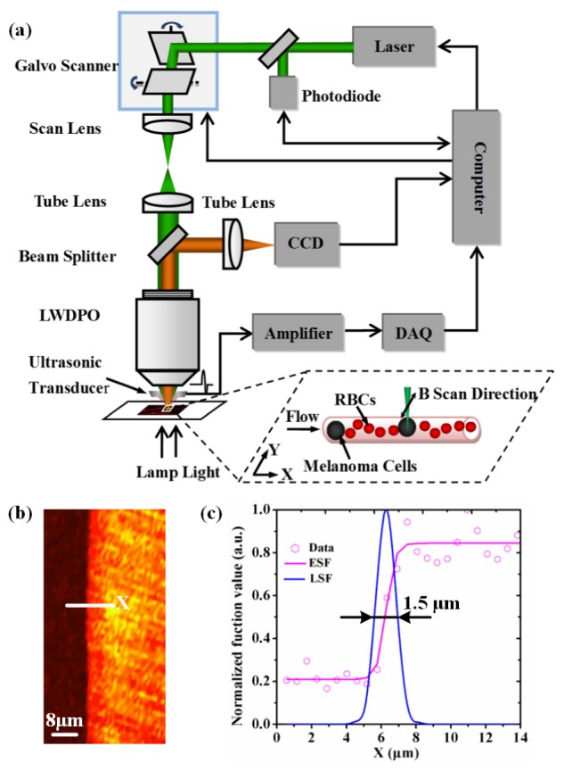
We present a photoacoustic flow cytography with fast cross-sectional (B-scan) imaging to precisely identify specific cells in vivo. The B-scan imaging speed of the system is up to 200 frame/s with a lateral resolution of 1.5 μm, which allows to dynamically image the flowing cells within the microvascular. The shape, size and photoacoustic intensity of the target cells are extracted from streaming images and integrated into a standard pattern to distinguish cell types. Circulating red blood cells and melanoma cells in blood vessels are simultaneously identified on melanoma-bearing mouse model. The results demonstrate that in vivo photoacoustic flow cytography can provide cells characteristics analysis and cell type's visual identification, which will be applied for noninvasively monitoring circulating tumor cells (CTCs) and analyzing hematologic diseases.

Keywords: (110.5120) Photoacoustic imaging; (170.0180) Microscopy; (170.1530) Cell analysis.


Figures

Fig. 1
Fig. 1
(a) Schematic of photoacoustic flow cytography. LWDPO, long working distance plan objective (20 × , NA = 0.55). DAQ, data acquisition. CCD, charge coupled device. (b) The PA image of a sharp edge. (c) Estimation of lateral resolution. ESF, edge spread function. LSF, line spread function.
Fig. 2
Fig. 2
(a) Absorption spectra of solutions with the mixture of WBCs and PLT, RBCs, B16F10 cells, plasma and PBS, respectively. (b) The corresponding PA signal intensities at 532 nm.
Fig. 3
Fig. 3
Morphology comparison. (a), (b) and (c) PA images of RBCs, adherent melanoma B16F10 cells, and suspended B16F10 cells in RBCs. The scanning interval was 1 μm. (d), (e) and (f) The corresponding optical microscope images.
Fig. 4
Fig. 4
Characteristic extraction and identification. (a) Processes of obtaining cell diameters after edge detection, filling holes and extraction. Dl and Ds were the long and short diameters. (b) The maximum PA signal intensity extracted from a B16F10 cell image. The bar shown in figures represents 3 μm. (c) Scatter plot of the long versus short diameters. (d) Scatter plot of the long diameter and PA intensity. (e) Scatter plot of the Γ parameters of cells. The hollow circles are RBCs and the filled circles are B16F10 cells.
Fig. 5
Fig. 5
In vivo recognition of RBCs and melanoma CTCs. (a) PA image of a capillary at the edge of a mouse ear. (b) B-scan x-t image of cells in vessels located in (a). (c) Melanoma tumor growth in mouse dorsum. (d) The superficial microvasculature PA image in a mouse ear. (e) B-scan x-t image of cells in a selected vessel labeled x3 in (d). The diameters of selected vessels were 10 μm, 18 μm and 15 μm respectively. The white dotted lines represented walls of vessels. And the recognized melanoma CTCs and RBCs were labeled with yellow and white squares respectively. (f) The number of melanoma CTCs recorded in one minute as a function of weeks after tumor inoculation.

References

    1. Nedosekin D. A., Verkhusha V. V., Melerzanov A. V., Zharov V. P., Galanzha E. I., “In vivo photoswitchable flow cytometry for direct tracking of single circulating tumor cells,” Chem. Biol. 21(6), 792–801 (2014).10.1016/j.chembiol.2014.03.012 - DOI - PMC - PubMed
    1. Liu R. R., Wang C., Hu C., Wang X. D., Wei X. B., “In vivo, label-free, and noninvasive detection of melanoma metastasis by photoacoustic flow cytometry,” Proc. SPIE 8944, 89440Q (2014).10.1117/12.2041022 - DOI
    1. Mahjoubfar A., Chen C., Niazi K. R., Rabizadeh S., Jalali B., “Label-free high-throughput cell screening in flow,” Biomed. Opt. Express 4(9), 1618–1625 (2013).10.1364/BOE.4.001618 - DOI - PMC - PubMed
    1. Novak J., Georgakoudi I., Wei X., Prossin A., Lin C. P., “In vivo flow cytometer for real-time detection and quantification of circulating cells,” Opt. Lett. 29(1), 77–79 (2004).10.1364/OL.29.000077 - DOI - PMC - PubMed
    1. Ding Y., Wang J., Fan Z., Wei D., Shi R., Luo Q., Zhu D., Wei X., “Signal and depth enhancement for in vivo flow cytometer measurement of ear skin by optical clearing agents,” Biomed. Opt. Express 4(11), 2518–2526 (2013).10.1364/BOE.4.002518 - DOI - PMC - PubMed

LinkOut - more resources