. 2015:2015:753210.
doi: 10.1155/2015/753210. Epub 2015 Oct 4.

Novel Model of Pulmonary Artery Banding Leading to Right Heart Failure in Rats

Affiliations

Novel Model of Pulmonary Artery Banding Leading to Right Heart Failure in Rats

Masataka Hirata et al. Biomed Res Int. 2015.

Abstract

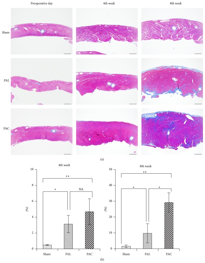
Background: Congenital heart diseases often involve chronic pressure overload of the right ventricle (RV) which is a major cause of RV dysfunction. Pulmonary artery (PA) banding has been used to produce animal models of RV dysfunction. We have devised a new and easier method of constricting the PA and compared it directly with the partial ligation method.

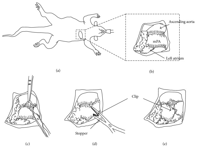
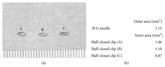
Methods: Eight-week-old male Sprague-Dawley rats (240-260 g) were divided into three groups: sham operation, partial pulmonary artery ligation (PAL) procedure, and pulmonary artery half-closed clip (PAC) procedure. RV function and remodeling were determined by echocardiography and histomorphometry.

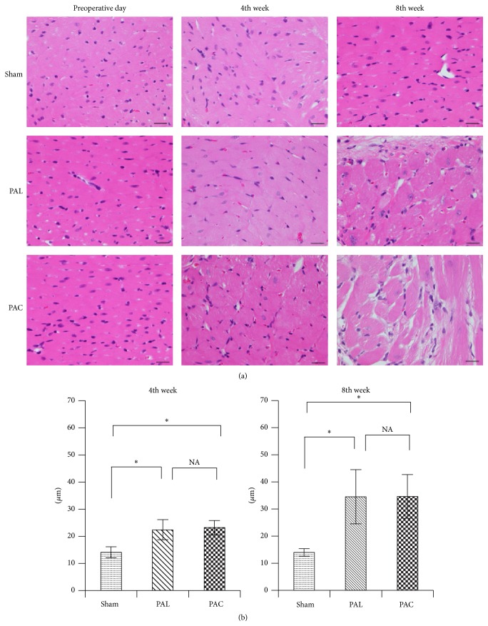
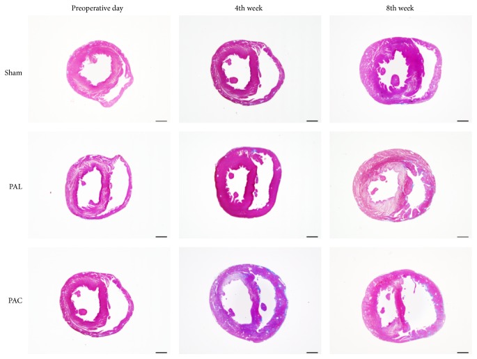
Results: Surgical mortality was significantly lower in the PAC group while echocardiography revealed significantly more signs of RV dysfunction. At the 8th week after surgery RV fibrosis rate was significantly higher in the PAC group.

Conclusions: This procedure of pulmonary artery banding in rats is easier and more efficient than partial ligation.


Figures

Figure 1
Figure 1
The PAC method. (a) Left thoracotomy was performed in the right semilateral decubitus position. (b) mPA was exposed through the left thoracotomy. ((c), (d)) Placement of a half-closed clip around mPA.
Figure 2
Figure 2
(a) Half-closed clips (A), (B), and (C) with different placed stopper. (b) Size of inner area of clips (A), (B), and (C), respectively, comparing with 18 G needle outer size. In this paper, we used (B) type for the PAC method.
Figure 3
Figure 3
Schemata of echocardiograms measurements. (a) The parasternal long-axis view. (b) Parasternal short-axis view. (c) The apical four-chamber view.
Figure 4
Figure 4
Photomicrographs of hematoxylin and eosin-stained sections showed significantly hypertrophied ventricular myocytes in the PAL and PAC groups. Scale bar: 20 μm. p < 0.01.
Figure 5
Figure 5
Macroscopic photographs of Masson's trichrome-stained cross sections showed RV wall thickening, cavity enlarging, and the interventricular septum shifting toward the left side in the PAL and PAC groups. Scale bar: 2 mm.
Figure 6
Figure 6
(a) Representative photomicrographs of Masson's trichrome-stained RV free wall (scale bar: 500 μm). (b) Evaluation of fibrosis area for RV free wall. p < 0.05, ∗∗ p < 0.01.

References

    1. Patel M. S., Kogon B. E. Care of the adult congenital heart disease patient in the United States: a summary of the current system. Pediatric Cardiology. 2010;31(4):511–514. doi: 10.1007/s00246-009-9629-5. - DOI - PubMed
    1. Bolger A. P., Coats A. J. S., Gatzoulis M. A. Congenital heart disease: the original heart failure syndrome. European Heart Journal. 2003;24(10):970–976. doi: 10.1016/s0195-668x(03)00005-8. - DOI - PubMed
    1. Murphy J. G., Gersh B. J., Mair D. D., et al. Long-term outcome in patients undergoing surgical repair of tetralogy of Fallot. The New England Journal of Medicine. 1993;329(9):593–599. doi: 10.1056/nejm199308263290901. - DOI - PubMed
    1. Gatzoulis M. A., Balaji S., Webber S. A., et al. Risk factors for arrhythmia and sudden cardiac death late after repair of tetralogy of Fallot: a multicentre study. The Lancet. 2000;356(9234):975–981. doi: 10.1016/s0140-6736(00)02714-8. - DOI - PubMed
    1. Faber M. J., Dalinghaus M., Lankhuizen I. M., et al. Right and left ventricular function after chronic pulmonary artery banding in rats assessed with biventricular pressure-volume loops. American Journal of Physiology—Heart and Circulatory Physiology. 2006;291(4):H1580–H1586. doi: 10.1152/ajpheart.00286.2006. - DOI - PubMed