

The Systemic Effect of Burn Injury and Trauma on Muscle and Bone Mass and Composition
- PMID: 26505718
- PMCID: PMC4876821
- DOI: 10.1097/PRS.0000000000001723
The Systemic Effect of Burn Injury and Trauma on Muscle and Bone Mass and Composition
Abstract
Background: By understanding the global inflammatory effects on distant myopathies, surgeons can better guide the rehabilitative process for burn patients. The authors tested the systemic effect of burn injury on distant injured muscle and native bone using immunohistochemistry and validated a new morphometric analytic modality to reproducibly quantify muscle atrophy using computed tomographic imaging.
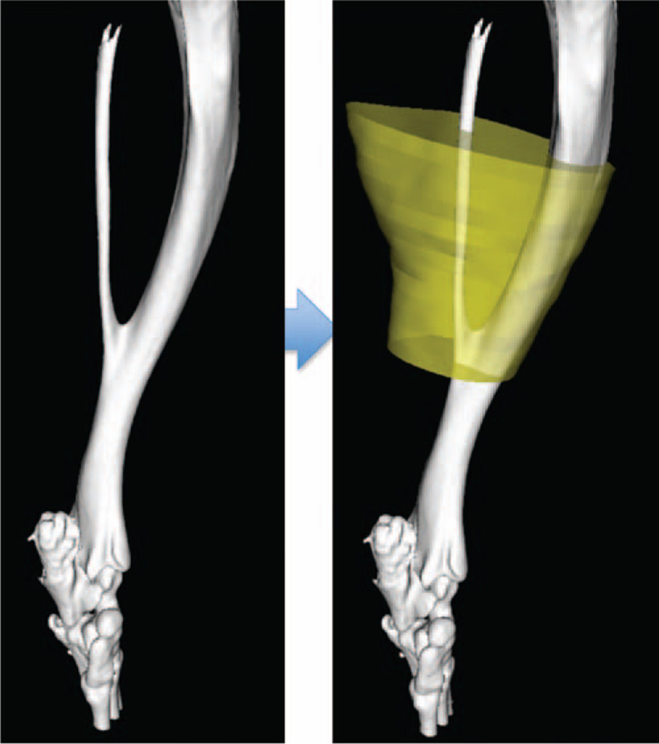
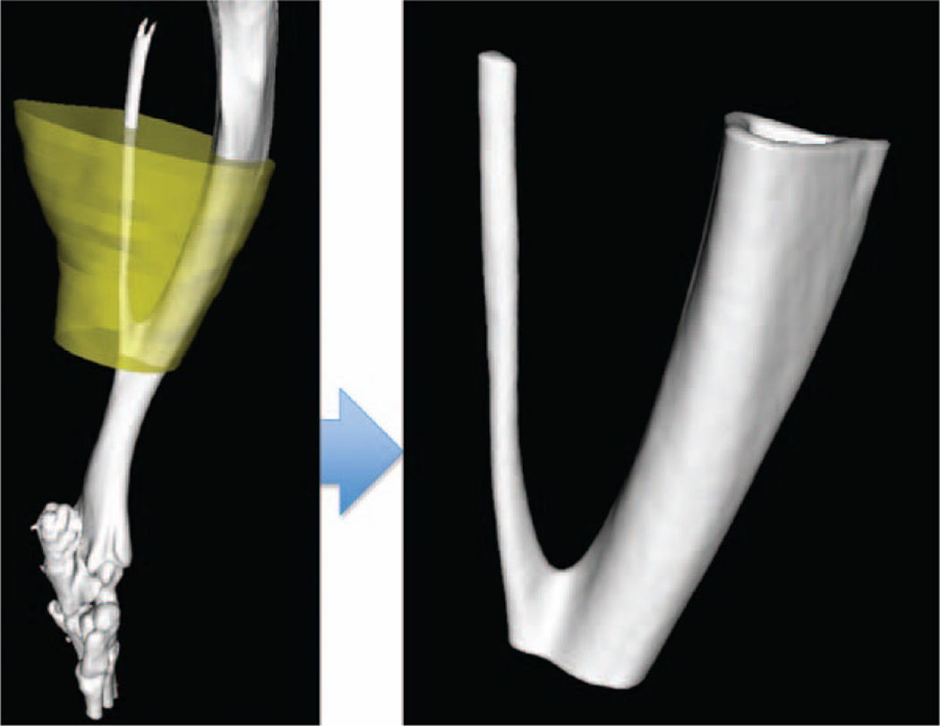
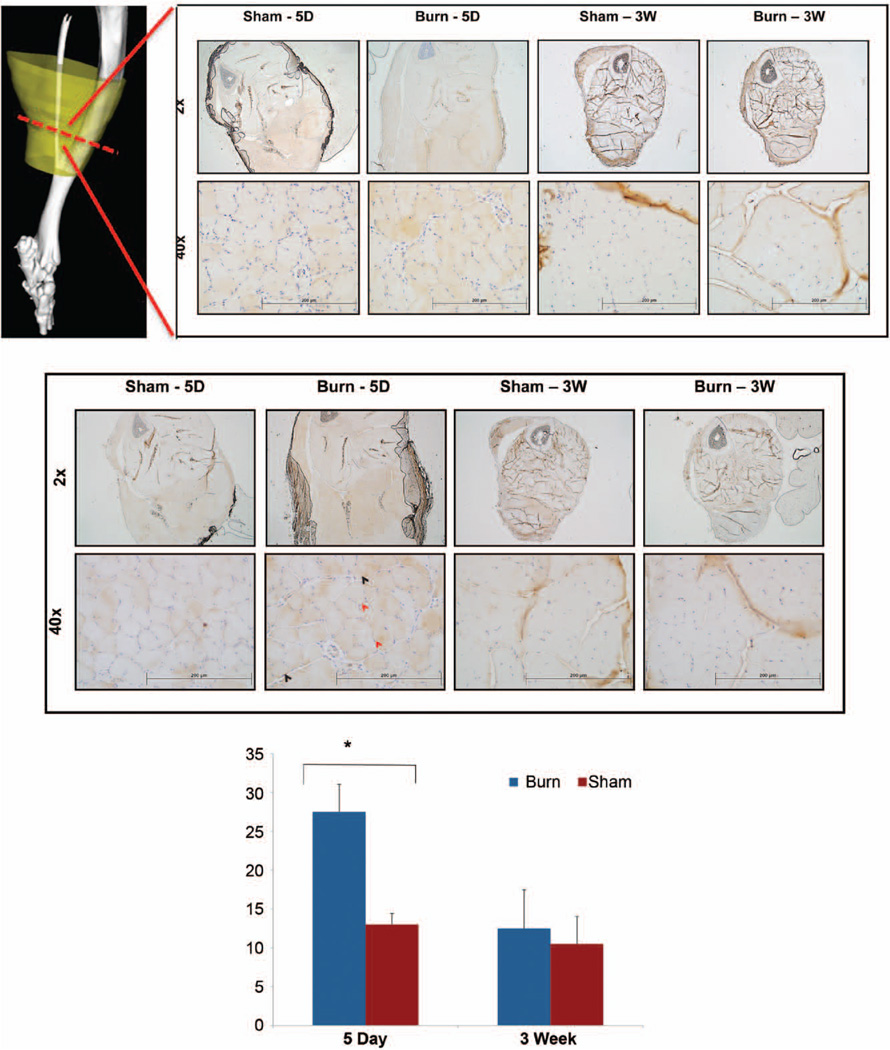
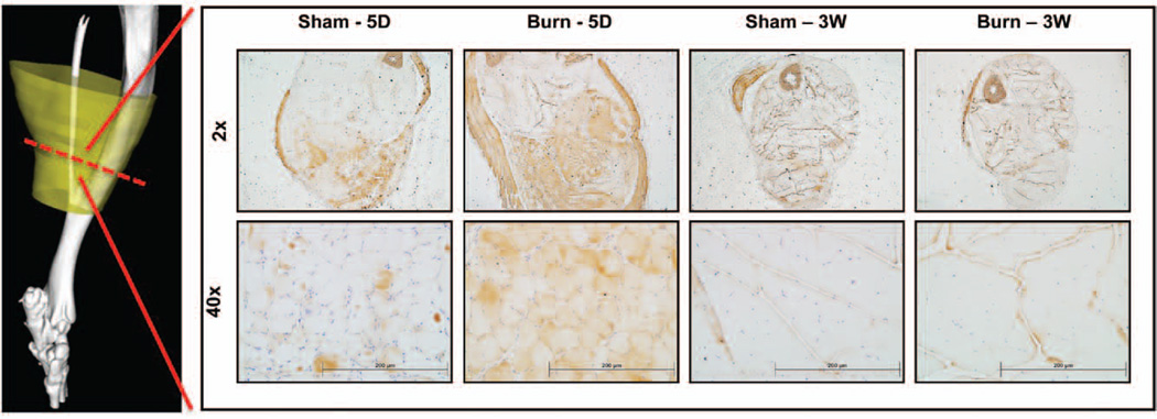
Methods: In vivo studies were performed on C57/BL6 mice using an Achilles tenotomy with concurrent burn injury model. Total muscle and bone (tibia and fibula) volume/density were quantified near the site of Achilles tenotomy using micro-computed tomography at 5, 7, and 9 to 12 weeks after surgery. The impact of burn injury on the inflammatory cascade [nuclear factor (NF)-κB, p-NF-κB] and the interconnected protein catabolism signaling pathway (Atrogin-1) was assessed by immunohistochemistry.
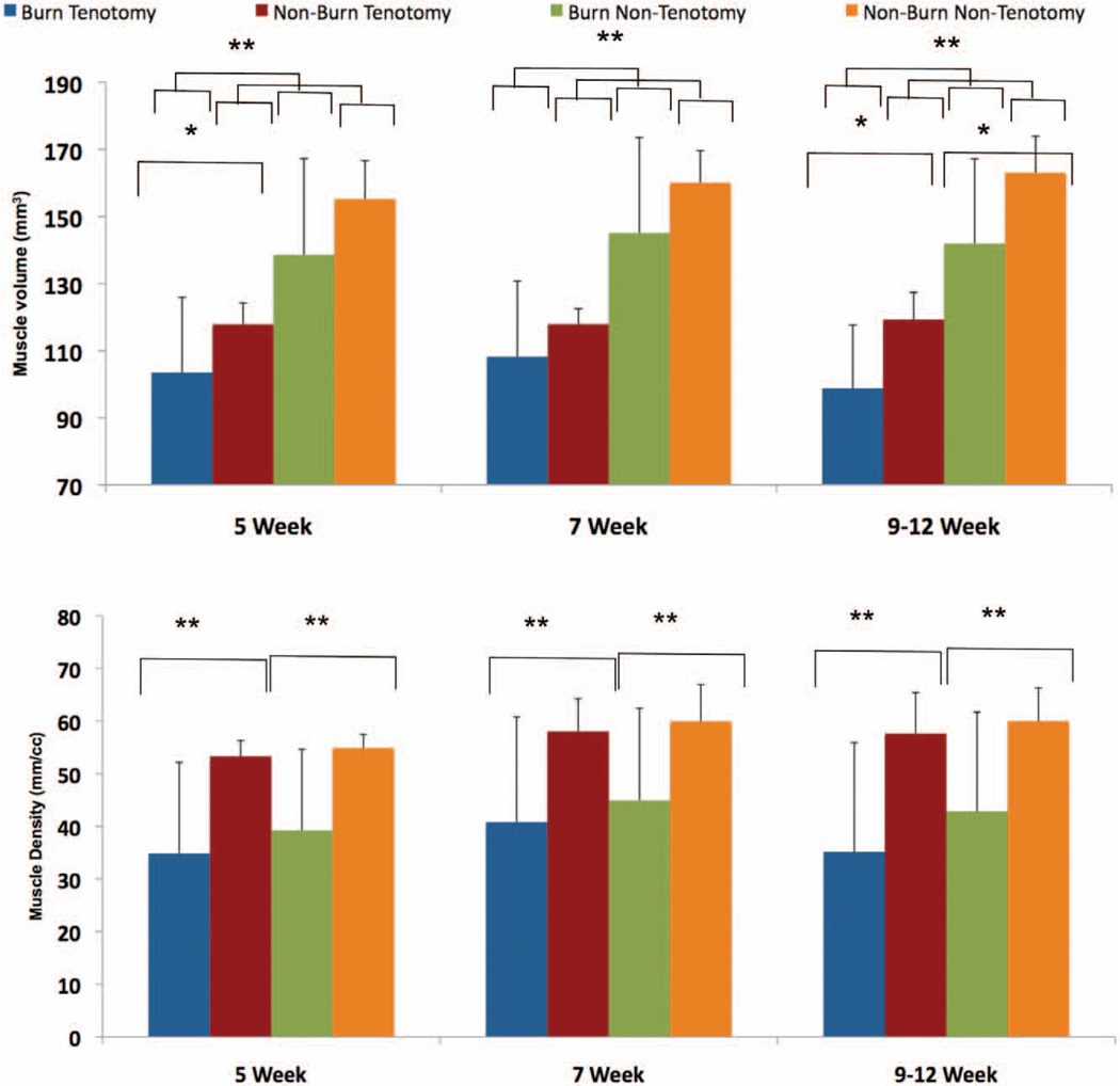
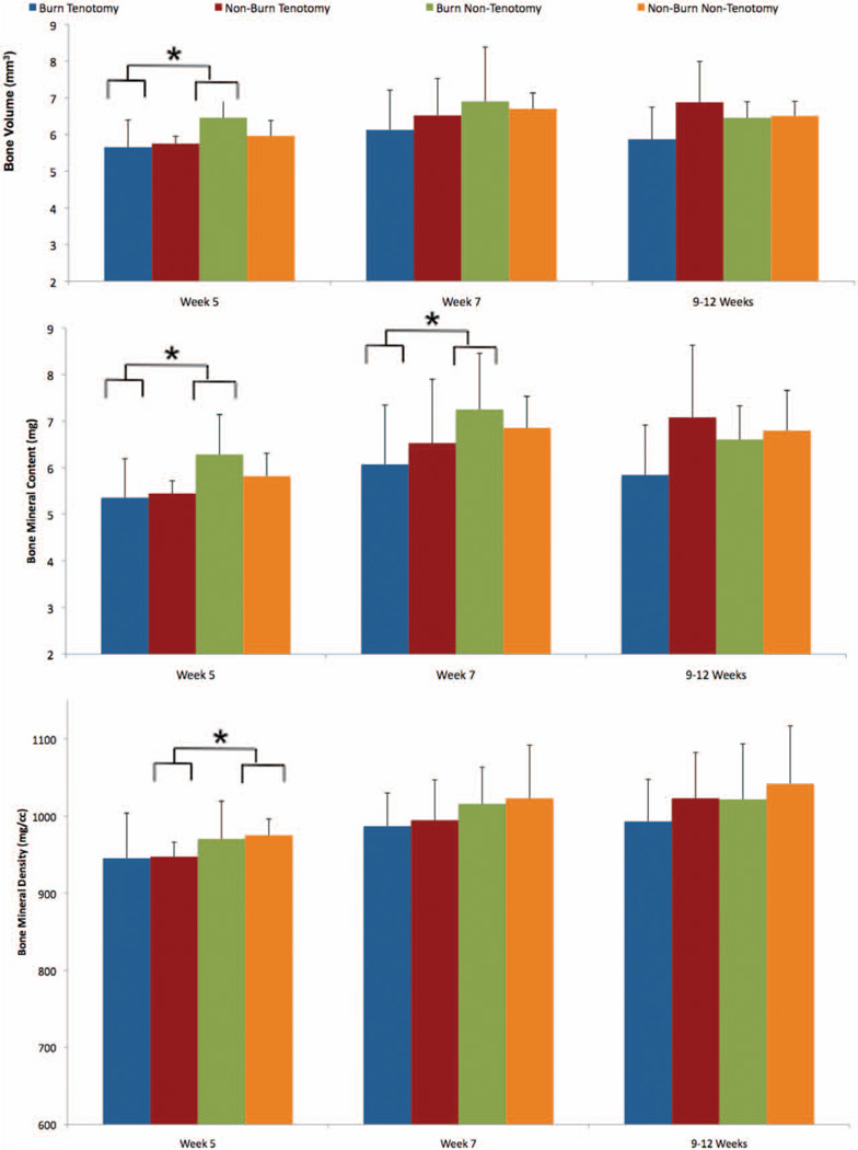
Results: Muscle volume and density at the site of Achilles tenotomy in burned mice were significantly diminished compared with nonburned mice at 5 weeks and 9 to 12 weeks. Similar decreases in muscle volume and density were observed when comparing tenotomy to no tenotomy. Cortical bone health remained stable in burn/tenotomy mice compared with tenotomy. Muscle atrophy was associated with up-regulation of p-NF-κB, NF-κB, and Atrogin-1 assessed by immunohistochemistry.
Conclusions: Burn injury significantly decreases muscle volume and density. Increased muscle atrophy using our computed tomographic morphometric analysis correlated with a significant increase in intramuscular inflammatory markers and proteolysis enzymes. This study demonstrates a unique characterization of how burn injuries may worsen local myopathy. Moreover, it provides a novel approach for quantifying muscle atrophy over an expanded period.
Figures
References
-
- Hart DW, Wolf SE, Mlcak R, et al. Persistence of muscle catabolism after severe burn. Surgery. 2000;128:312–319. - PubMed
-
- Chandra RK. Nutrition and immunology: From the clinic to cellular biology and back again. Proc Nutr Soc. 1999;58:681–683. - PubMed
-
- Wilmore DW. Catabolic illness: Strategies for enhancing recovery. N Engl J Med. 1991;325:695–702. - PubMed
-
- Ibebunjo C, Martyn J. Disparate dysfunction of skeletal muscles located near and distant from burn site in the rat. Muscle Nerve. 2001;24:1283–1294. - PubMed
Publication types
MeSH terms
Grants and funding
LinkOut - more resources
Full Text Sources
Medical
